Exciton energy transfer in nanotube bundles
Abstract
Photoluminescence is commonly used to identify the electronic structure of individual nanotubes. But, nanotubes naturally occur in bundles. Thus, we investigate photoluminescence of nanotube bundles. We show that their complex spectra are simply explained by exciton energy transfer between adjacent tubes, whereby excitation of large gap tubes induces emission from smaller gap ones via Frster interaction between excitons. The consequent relaxation rate is faster than non-radiative recombination, leading to enhanced photoluminescence of acceptor tubes. This fingerprints bundles with different compositions and opens opportunities to optimize them for opto-electronics.
pacs:
78.67.Ch, 71.35.-y, 78.55.-m, 71.35.Cc, 73.22.-fCarbon nanotubes are rolled graphene sheets rbook . One dimensional quantum confinement makes their band structure fundamentally different from graphene, with sub-bands and singularities in the density of states rbook . These are fully determined by their chiral indexes (n,m), which specify how graphene is folded into each nanotube. Thus, measuring the optical transitions allows in principle to determine the chiral indexes, and fully identify a nanotube sample. For this reason, a massive effort was put to measure photoluminescence in nanotubes since their discovery. However, it took more than ten years to unambiguously detect and identify photoluminescence emission from single wall nanotubes (SWNT)Connell ; Bachilo1 . Indeed, SWNT naturally occur in bundles, due to Van-der-Waals interactions Hertel , but a significant intensity was only measured when efficient de-bundling was achievedConnell ; Bachilo1 . This paved the way to the interpretation of their complex absorption and emission spectraConnell ; Bachilo1 . The discrepancy between single-particle theory and experiments, pointed to the major role of electron-electron and electron-hole interactions in shaping their band-structureWang1 ; Maultzsch . The exciton binding energies were recently measuredWang1 ; Maultzsch . These are very large, from tens meV to 1 eV, depending on diameter, chirality, and dielectric screening Wang1 ; Maultzsch ; Perebeinos2 . Thus, excitons dominate even at room temperature.
The investigation of the optical properties of nanotubes is now a most pursued research areaConnell ; Bachilo1 ; Misewich ; Chen ; Perebeinos2 ; Wang1 ; Maultzsch ; Ma , however this still focuses on individual tubes, in contrast with their natural occurrence in bundles. Furthermore, the luminescence quantum yield of individual SWNTs is very low and this hinders their applications in optoelectronics Bachilo1 ; Misewich ; Chen . Here we present a thorough investigation of absorption and emission spectra of nanotube bundles. We show that their apparently complex spectra can be simply interpreted considering exciton energy transfer between tubes. This is a well known phenomenon in biological systems, conjugated polymers, quantum wires, dots, and other low-dimensional systemsBecker ; Biju ; Forster ; Rizzo ; Kagan , which we now clearly identify in nanotubes. We find that energy transfer is a major non-radiative relaxation channel for large gap tubes, strongly enhancing the photoluminescence of the acceptor tubes. Thus, contrary to what usually assumed, nanotube bundles could be ideal for high yield optoelectronics applications, far surpassing the poor performance of individual tubes Misewich ; Chen . Furthermore, energy transfer fingerprints bundles with different nanotube concentrations, finally offering a quantitative means to monitor the composition of bundles in solution, a key requirement for research and applicationsConnell ; Bachilo1 .
We measure absorption on CoMoCAT SWNT(South West Nanotechnologies) Kitiyanan suspensions in D2O with sodium dodecylbenzene sulfonate (SDBS) surfactantConnell , using a Perkin-Elmer 950 spectrometer. A JY Fluorolog-3 is used for Photoluminescence Excitation (PLE).

Figure 1a plots PLE maps from the as-prepared solution. Each spot can be labeled as (, ), where , are, respectively, the excitation and emission wavelengths. Several high intensity peaks in Fig.1a are exciton-exciton resonances Bachilo1 ; Bachilo2 . In this case corresponds to the energy of the excitonic states associated with the th electronic interband transitions (=1, 2, 3, 4) in the single particle picture Bachilo1 ; Bachilo2 , while is the emission energy of the lowest exciton transition . Other spots in Fig.1a are related to exciton-phonon sidebands Perebeinos1 ; Plentz ; Miyauchi . The spectral features in Fig.1a are summarized in Fig.2. We identify 16 different SWNT species in the range 800nm-1300nm, and assign their chiral indexes in Fig. 2, as for Ref. Bachilo1, . The phonon sidebands for the and excitons are shown in Fig. 2 with open circles and diamonds. The wavelengths of most SWNT here are 3-10 nm larger than Ref. Bachilo1, . This red-shift is expected in the presence of bundling Reich3 ; Wang . Fig.2 has some interesting features compared with previous data on isolated SWNT suspensions Bachilo1 ; Bachilo2 : 1) the spectral profile of exciton resonances significantly elongates in the horizontal and vertical directions; 2) new peaks appear, such as, e.g., (645nm, 1265nm) and (568nm, 1250nm), with intensity much stronger than the (, ) peaks of (10, 5), (8, 7) and (9, 5) SWNTs; 3) a strong broad band near (980nm, 1118nm) is observed.
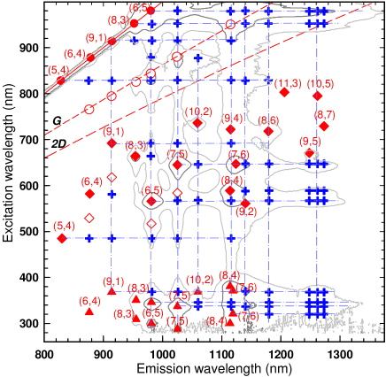
To clarify the origin of these bands, we check PLE from the same solution after two months (Fig. 1b). Figs. 1a,b have similar features. However, the emission wavelengths of most SWNTs in Fig. 1b redshift3-5 nm relative to Fig. 1a. This suggests further aggregation into bigger bundles. Also, almost all spots decrease in intensity. But a careful examination of Figs. 1a,b, shows that the (980nm, 1118nm) peak becomes stronger after two months. Also, two peaks near (568nm, 1118nm) and (346nm, 1118nm), indicated by circles in Figs. 1a,b, are identified more clearly, due to the lower intensity of the (, )(=2, 3, 4) bands of (8, 4) and (7, 6) tubes, which shadowed them in the pristine solution. Notably, these three peaks do not correspond to any of the known exciton-exciton resonances of SWNTs in this spectral range Bachilo1 ; Bachilo2 . The (980nm, 1118nm) peak is not assigned to a phonon sideband of (8, 4), (7, 6) or (9, 4) tubes, due to the lack of such sideband in previous investigations of these and other tubes Perebeinos1 ; Plentz ; Htoon ; Chou . Indeed, the excitation energies of the (980nm, 1118nm), (568nm, 1118nm) and (346nm, 1118nm) bands match, respectively, the , and transitions of (6, 5) tubesBachilo1 , whereas their emission around 1118 nm is consistent with (8, 4), (7, 6), (9, 4) . Thus, resonant excitation of large gap donors tubes induces emission from smaller gap acceptors. This implies energy transfer between SWNT in bundles. Due to the large exciton binding energiesWang1 ; Maultzsch ; Perebeinos2 , this happens mainly via excitons, not via intertube electron or hole migration Torrens .
A thorough examination of all peaks in Figs. 1a,b, allows us to identify several other exciton energy transfer (EET) features (solid crosses in Fig. 2). The peaks not attributable to known exciton-exciton resonances along each horizontal dashed-dotted line in Fig.2 are assigned to excitation of donor tubes, inducing emission from acceptors. Vice-versa, the crosses along each vertical dashed-dotted line are emission of an acceptor, following EET from excitation of donors. Broad/elongated patterns of Fig.1, shown by grey contours in Fig.2, contain overlapping peaks from tubes with similar excitation or emission energies. Size, concentration and distribution of nanotube species within a bundle will determine the EET-induced intensities. The higher the concentration of semiconducting tubes, the higher the probability of them being adjacent, the higher the chance of EET-induced emission. Thus, the strongest peaks will appear around transitions of semiconducting tubes with highest concentration, such as (6, 5), (7, 5), (8, 4) in our CoMoCAT solutions Kitiyanan ; Bachilo2 .
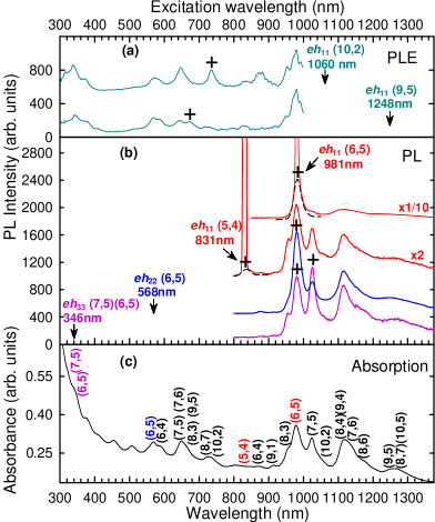
Fig.3 compares the absorption of the as-prepared solution with its photoluminescence emission and excitation spectra. The (, ) peaks are marked by crosses. We assign most of the other bands to energy transfer from donor to acceptors within bundles. The (, ) peak is the strongest amongst all possible (, ) for a given (n, m), as, e.g., in the (6, 5) tube of Fig. 3b. This is because excitons have higher density of states than , Spataru0 . Thus more photons are absorbed by states. Then, as shown in Fig. 3a, the excitation of large gap donors is a more efficient way to enhance emission of smaller gap acceptors than the direct and excitation of the acceptors, despite the low photoluminescence quantum efficiency of individual tubes Connell .
We can estimate the exciton energy transfer efficiency in bundles as follows. Let us consider the exciton relaxation of two adjacent tubes with different gaps, following the resonant excitation of the larger gap tube. The rate equations of the donor-acceptor system are:
| (1) |
| (2) |
where is the energy transfer lifetime between donor and acceptor, is the population of excitons in the donor and in the acceptor, , , and are the radiative (r) and non-radiative (nr) lifetimes, the exciton density in the donor created by photo-excitation. An estimation of the EET efficiency is the ratio of acceptor emission intensity (=) to that of the donor (=). Then, deriving from Eqs. (1,2) at steady state, we get:
| (3) |
The radiative lifetime is reported 20-180ps, dependent on temperature Hagen . For tube diameters 0.75-0.95 nm, it is about 20-30 ps at room temperatureHirori . This is much shorter than the theoretical radiative lifetime (10 ns)Spataru . Thus, the observed lifetimes are determined by non-radiative recombination. Eq. (3) can then be simplified as .
We measure a very high in bundles. E.g., under excitation of the (5, 4) tubes in Fig. 3b, the ratio of photoluminescence intensity of all acceptor tubes with emission above 900 nm [such as (6, 5), (7, 5), (8, 4),(7,6)] to that at831 nm of the (5, 4) donor tubes is at least75. This indicates that most resonantly-excited (5, 4) excitons transfer their energy to the acceptors, rather than recombine. Thus, in bundles exciton relaxation is comparable or even faster than non-radiative recombination. This fast relaxation suppresses emission from donors. But it significantly increases the acceptors luminescence. This suggests that small bundles entirely formed of semiconducting tubes can be ideal for opto-electronics, such as in light-emitting devicesMisewich ; Chen .
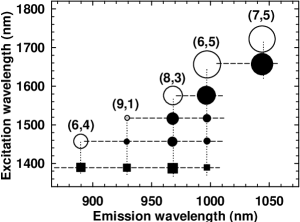
Two-photon excitation is used to derive exciton binding energiesWang1 ; Maultzsch . Fig.4 summarizes the two-photon map of Ref.Maultzsch, . The open circles are two-photon exciton resonances. These are slightly shifted with respect to those of Ref.Wang1, due to the presence of small bundlesMaultzsch . Below each two-photon band, Ref.Maultzsch reported a set of peaks, indicated by solid squares and circles in Fig.4. These appear like a Rydberg series of states, each matching the excitation energy of a larger gap tube, as indicated by horizonal dashed lines in Fig. 4. These are analogous to the energy transfer-induced peaks along each horizonal dashed-dotted line in Fig. 2. We attribute them to emission of small gap tubes due to exciton energy transfer from larger gap tubes in bundles. We assign the four features in Fig. 4 with 1390 nm excitation (solid squares) to EET from (5,4) donors to (6, 4), (9, 1), (8, 3) and (6, 5) acceptors. Since two-photon luminescence increases quadratically with excitation power, the energy transfer features show more distinct peaks compared to Fig.1.
In low-dimensional systems, exciton tunneling, photon-exchange and Frster Resonance Energy Transfer (FRET) are efficient exciton energy transfer mechanisms Alexander ; Forster ; Kagan ; Biju ; Becker ; Rizzo . We attribute energy transfer in bundles to FRET. Indeed, tunneling requires coupling of exciton wavefunctions. Its rate decays rapidly with donor-acceptor distance () and is very sensitive to the energy difference Alexander . The 16 tube species in our experiment have diameters0.65-1.05 nm, 0.06-0.5 eV, and chiral angle variation5-26oBachilo1 ; Bachilo2 . Therefore, the efficiency should strongly depend on specific donor and acceptor couples. However, the spectrum in Fig. 3b excited at (5, 4) , reproduces the profile of the absorption in Fig.3c above 850nm, with no (n,m) preference. This suggests that the factor dominating exciton energy transfer in bundles is tube concentration, not symmetry, diameter or bandgap difference, thus exciton tunneling is not the dominant mechanism.
Photon-exchange is exciton-photon coupling with no direct donor-acceptor interaction. It has a smaller dependence on than FRET, thus it can become significant for much longer distances than FRET. However, the lack of significant EET features in isolated tube solutions Bachilo1 ; Bachilo2 combined with the low quantum efficiency Connell suggest that even if photon-exchange might exist between bundles or between isolated SWNTs, it is not dominant between adjacent tubes in a given bundle.
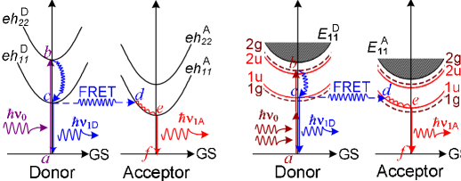
FRET is a very efficient exciton energy transfer mechanism via a resonant, near-field, dipole-dipole interactionForster ; Kagan ; Biju ; Becker ; Rizzo . It is commonly observed in biological systems, conjugated polymers, wires, dotsForster ; Kagan ; Biju ; Becker ; Rizzo , where it dominates at short and intermediate distances (0.5-10nm) Forster ; Kagan ; Biju ; Becker ; Rizzo . Its efficiency is determined by the spectral overlap of donor emission and acceptor absorption, by , and by the relative orientation of emission and absorption dipoles Forster . The rate of energy transfer is proportional to Forster . The FRET efficiency in bundles is expected to be high. Indeed, there is a large overlap between emission of large gap tubes and absorption of small gap tubes. SWNTs in bundles are parallel, giving a maximum dipole orientation factor. They aggregate with small wall-to-wall distance Hertel ; Reich3 . This makes higher multipolar contributions possible as well Forster ; Kagan . Indeed, considerable luminesce quenching of CdSe-ZnS dots conjugated to SWNTs was reported due to FRET from dots to tubes Biju . This further suggests FRET to be dominant in bundles. This process is schematized in Figs. 5a,b for both one and two-photon spectroscopies.
In summary, we presented a thorough investigation of photoluminescence in nanotube bundles. We have shown that the apparently complex absorption and emission spectra can be simply explained by exciton energy transfer between adjacent semiconducting tubes. By studying the spectral evolution for increasing bundle size, we assigned all the excition energy transfer peaks. We argue that Frster interaction between excitons dominates the transfer process. This is highly efficient in nanotube bundles, adding a major relaxation channel for excitons, explaining the low luminescence yield of large gap nanotubes. Thus, contrary to what usually assumed, bundles could be ideal for high yield optoelectronics applications, far surpassing the poor performance of individual tubes. Furthermore, energy transfer fingerprints bundles with different tube concentrations, finally offering a quantitative means to monitor the composition of solutions and films, a key requirement for research and applications.
We acknowledge D. Prezzi, A. Rubio, A. Hartschuh for useful discussions; funding from EPSRC GR/S97613 and Ministry of Information and Communication, Republic of Korea (No. A1100-0501-0073). PHT and ACF acknowledge funding from the Royal Society. ACF from the Leverhulme Trust.
References
- (1) S. Reich, C. Thomsen, J. Maultzsch, Carbon nanotubes (Wiley, Weinheim),(2004).
- (2) M. J. O’Connell et al., Science 297, 593 (2002).
- (3) S. M. Bachilo et al., Science 298, 2361 (2002).
- (4) T. Hertel, R. E. Walkup, P. Avouris, Phys. Rev. B 58, 13870 (1998).
- (5) F. Wang et al., Science 308, 838 (2005).
- (6) J. Maultzsch et al., Phys. Rev. B 72, 241402(R) (2005).
- (7) V. Perebeinos, J. Tersoff, P. Avouris, Phys. Rev. Lett. 92, 257402 (2004).
- (8) Y. Z. Ma et al., Phys. Rev. Lett. 94, 157402 (2005).
- (9) J. A. Misewich et al., Science 300, 783 (2003).
- (10) J. Chen et al., Science 301, 1171(2005).
- (11) T. Frster, Discuss. Faraday Soc. 27, 7 (1959).
- (12) C. R. Kagan et al., Phys. Rev. Lett. 76, 1517 (1996).
- (13) S. R. Adams et al, Nature 349, 694 (1991).
- (14) K. Becker et al., Nat. Mater. 5, 777 (2006).
- (15) V. Biju et al., J. Phys. Chem. B 110, 26068 (2006).
- (16) B. Kitiyanan et al., Chem. Phys. Lett. 317, 497(2000).
- (17) S. M. Bachilo et al., J. Am. Chem. Soc. 125, 11186 (2003).
- (18) V. Perebeinos, J. Tersoff,P. Avouris, Phys. Rev. Lett. 94, 027402 (2005).
- (19) F. Plentz et al., Phys. Rev. Lett. 95, 247401 (2005).
- (20) Y.Miyauchi,S.Maruyama,Phys. Rev. B 74,035415 (2006).
- (21) S. Reich, C. Thomsen, P. Ordejon, Phys. Rev. B 65,155411 (2002).
- (22) F. Wang et al., Phys. Rev. Lett. 96, 167401 (2006).
- (23) H. Htoon et al., Phys. Rev. Lett. 94, 127403(2005).
- (24) S. G. Chou, et al., Phys. Rev. Lett. 94, 127402(2005).
- (25) O. N. Torrens et al., Nano Lett. 6, 2864(2006).
- (26) C. D. Spataru et al., Phys. Rev. Lett. 92, 077402 (2004).
- (27) A. Hagen et al., Phys. Rev. Lett. 95, 197401 (2005).
- (28) H. Hirori et al., Phys. Rev. Lett. 97, 257401 (2006).
- (29) C. D. Spataru et al., Phys. Rev. Lett. 95, 247402 (2005).
- (30) M. G. W. Alexander et al. Phys. Rev. B 41, 12295 (1990).