Search for low lying dipole strength in the neutron rich nucleus 26Ne
Abstract
Coulomb excitation of the exotic neutron-rich nucleus 26Ne on a natPb target was measured at 58 A.MeV in order to search for low-lying E1 strength above the neutron emission threshold. Data were also taken on an natAl target to estimate the nuclear contribution. The radioactive beam was produced by fragmentation of a 95 A.MeV 40Ar beam delivered by the RIKEN Research Facility. The set-up included a NaI gamma-ray array, a charged fragment hodoscope and a neutron wall. Using the invariant mass method in the 25Ne+n channel, we observe a sizable amount of E1 strength between 6 and 10 MeV. The reconstructed 26Ne angular distribution confirms its E1 nature. A reduced dipole transition probability of is deduced. For the first time, the decay pattern of low-lying strength in a neutron-rich nucleus is obtained. The results are discussed in terms of a pygmy resonance centered around 9 MeV.
1 Introduction
Giant Resonances are a general feature of nuclei and their properties give us a handle on the macroscopic and microscopic behavior of nuclear matter. These modes have been extensively studied in stable nuclei over the last 50 years and the recent inception of Radioactive Ion Beam facilities opens the opportunity to extend these investigations to exotic nuclei. Far from stability new modes are expected to appear. In particular, the dipole response of neutron-rich nuclei may exhibit strength at energies lower than the standard Giant Dipole Resonance (GDR) often depicted as the oscillation of a deeply bound core against a neutron skin giving rise to a so-called pygmy resonance. Recent studies on Oxygen [1] and Tin [2] isotopes have given the first experimental indications for such an effect, while at the same time calling into question its nature.
The theoretical approach is often based on mean field calculations. Relativistic random phase approximation (RRPA) and quasi-particle RRPA (QRRPA) calculations have been carried out by Cao and Ma in 26Ne [3] which predict that below 10 MeV excitation energy, almost 5% of the Thomas-Reiche-Kuhn (TRK) energy weighted sum rule is exhausted by strength centered around 8 MeV. This region of energy is located between the one neutron and the two neutron emission thresholds. Thus, in order to investigate this prediction, we performed Coulomb excitation of 26Ne at intermediate energies on a lead target and used the invariant mass method to reconstruct the B(E1) strength from the 26Ne25Ne reaction.
2 Experimental details
The experiment was performed at the RIKEN Accelerator Research Facility. A secondary 26Ne beam was produced through fragmentation of a 95 A.MeV 40Ar primary beam on a 2-mm-thick 9Be target. The 26Ne was separated by the RIKEN Projectile Fragment Separator (RIPS) [4]. Particle identification was unambiguously performed by means of the time-of-flight (TOF) and the purity was 80%. The 26Ne beam of intensity pps and incident energy 58 A.MeV, was tracked with two parallel-plate avalanche counters providing incident angle and hit position onto the target. It then impinged alternatively on a 230 mg/cm2 natPb and a 130 mg/cm2 natAl target. Data obtained with aluminum target are used in the following to estimate the contribution of nuclear excitation to the data.
The outgoing charged fragments were measured using a set of telescopes placed at 1.2 m downstream of the target. They consisted of two layers (X and Y) of 500 m silicon strip detectors (SSD) with 5 mm strips which provided an total energy resolution of 1.5 MeV (FWHM). The last layer used 3-mm-thick Si(Li) from the charged-particle detector MUST [5], provided a 9 MeV (FWHM) resolution on the remaining energy (E). Unambiguous mass and charge identification of all projectile like fragments was obtained using the E-E method.
In-beam gamma rays were detected using a 4-gamma-detector, DALI2 [6], which consists of 152 NaI detectors placed around the target. For 1.3 MeV gamma-rays, its measured efficiency is approximately 15% with an energy resolution of 7% (FWHM). The Doppler corrected gamma energy distribution obtained in coincidence with the 25Ne isotope allows us to identify the gamma decay from the adopted 1702.7(7) keV and 3316.4(11) keV excited states. In addition we observed a keV gamma-ray which we related to the keV excited state, only seen up to now by transfer reaction [7].
The hodoscope for neutron detection was an array of 4 layers of 29 plastic rods each, placed 3.5 m downstream of the target. Each layer was composed of 13 [2.1 m66 cm2] and 16 [1.1 m66 cm2] rods, arranged in a shape of a cross. Its total intrinsic efficiency for the detection of one 60 MeV neutron was calculated to be %. Finally 29 thin plastic rods covered the front face of the wall in order to veto charged particles as well as to provide an active beam stopper. The neutron position is determined with an error of 3 cm and the energy, from TOF information, with a 2.5 MeV (FWHM) resolution for the neutrons of interest.
3 Results
We have performed a simulation of the experimental setup using the Geant 3 package [8]. In order to check the reliability of the analysis, we built the elastic scattering angular distribution of 26Ne on natPb at 55 A.MeV. It is in good agreement with a DWBA calculation based on a 20Ne+208Pb at 40 A.MeV optical potential from [9]. We empirically generated optical potential parameters for the 26Ne+27Al reaction and tested them by comparing with the experimental elastic scattering.
Using the invariant mass method, the excitation energy of an unbound state in the nucleus decaying to a state in can be expressed by: , where is the relative energy between neutron and and the one neutron emission threshold. The gamma detection efficiency was not high enough to apply an event-by event reconstruction technique. To extract the excitation energy spectrum, we hence used a method based on adequate subtraction of relative energy spectra built for these two kind of event.
The method can be illustrated in the schematic case where the daughter nucleus has only one excited state below its neutron threshold. The excitation energy spectrum can be decomposed in the sum of two contributions: the decay of to the ground-state (gs) of and the decay of to the excited state (I) of .
The first contribution can be obtained by subtracting from the inclusive relative energy spectrum the relative spectrum of gamma-coincidence events divided by the gamma detection efficiency , and shifted “” by the one neutron emission threshold i.e.: .
The second contribution is simply the relative energy spectrum of gamma-coincidence events shifted by the gamma energy: . Finally, the excitation energy spectrum for the 25Ne+n decay channel is given by: . The method was tested by simulation in this schematic case, and also in the realistic case taking into account the experimental decay scheme of 25Ne.
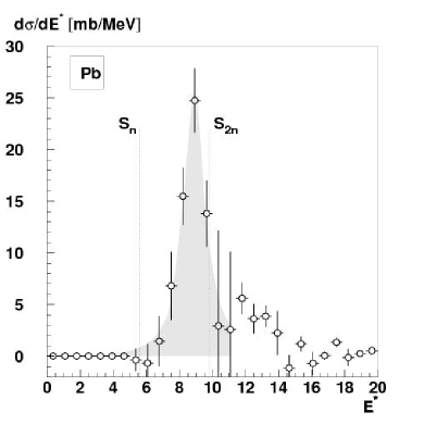
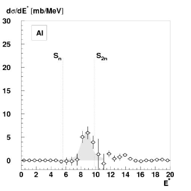
The excitation energy spectra reconstructed for the 25Ne+n decay channel obtained with the natPb and natAl targets are represented on Fig. 1. Note that above MeV, the decay of 26Ne is expected to occur mainly by 2 neutron emission. Between 8 and 10 MeV, a sizable amount of cross-section is observed for both targets. In intermediate energy inelastic scattering with a heavy target such as natPb, the Coulomb dominance of the E1 excitation is well-known. The contribution of possible E2 excitation to the spectrum obtained with the lead target has — in a first step — been determined using data taken with the aluminum target and the coupled channels ECIS 97 [10] code. Assuming simple collective vibration mode with equal nuclear and Coulomb deformation lengths, the E2 deformation parameters were extracted from the measured cross-section with the natAl target ( mb). The cross section in lead was then calculated using the deformation lengths extracted in the previous step. We obtained mb. After subtraction of the contribution, the resulting mb cross section corresponds, using ECIS 97, to a Coulomb deformation parameter of which led to a via the relation with the Coulomb radius. This value of reduced transition probability correspond to % of the Thomas-Reiche-Kuhn energy weighted sum rule for an excitation energy of 9 MeV. We estimate the error due to the choice of optical potential by performing the same analysis using now parameters from the 40Ar+208Pb reaction at 40 A.MeV [11]. We obtained i.e. % of the TRK, in good agreement with the values previously extracted, which demonstrates that we are not strongly sensitive to the choice of the optical potential for the reaction on lead.
Due to the high granularity and the good resolution of the present setup, it is possible to reconstruct the scattering angular distribution for 26Ne on the natPb target which is represented in Fig. 2. Hence, we have used a second method to extract the E1 excitation which relies on a multipole decomposition analysis of this angular distribution. The and angular distributions (dashed and dotted lines) were obtained from simulation based on ECIS 97 angular distribution calculated for MeV. The data were fitted with a linear combination of the two distributions. The results of the fit give us which corresponds to % of the TRK, again for an excitation energy of 9 MeV. If we suppose now that the remaining part of the contribution is due to excitation we can extracted a for this structure.
![[Uncaptioned image]](/html/0705.1753/assets/x3.png)
Figure 2: Result (solid line) of the multipole decomposition of the experimental angular distribution (circle) of the 26Ne angular scattering onto lead target, by (dashed line) and -type (dotted line) distributions.
The two results from the two different methods are in agreement but since the multipole decomposition of the angular distribution relies only on the data from lead target, the second value of will be retained. The results obtained from our experiment concerning the E1 transition are compared in the following to theoretical calculations.
4 Comparison with theory
In the introduction we already presented results from Cao and Ma [3]. Using the relativistic QRPA framework and the response function formalism they predicted a pygmy resonance centered around 8.4 MeV and which exhausts of the TRK, which is close to our experimental values. Another calculation has been performed by Khan et al. [12]. It is based on effective SGII Skyrme interactions and is performed in the spherical QRPA framework. It predicts a redistribution of the strength at low energy centered around MeV exhausting of the TRK. Two other preliminary calculations has been performed in the deformed QRPA framework using Gogny forces [13] and in the deformed relativistic QRPA framework [14]. Both predict a redistribution of the strength at low energy, centered around MeV and MeV respectively. The first calculation also predicts that only of the TRK should be exhausted. Preliminary shell-model calculations have also been performed by Nowackiet al. [15] who predict a state at 9.3 MeV exhausting of the TRK. All these theories agree on the presence of a structure a low excitation energy, compatible with our experimental result but they disagree on its nature: the amount of strength differs as well as as well as its collective or single particle nature.
5 Decay of pygmy resonances in 26Ne
| Final 25Ne state | Experiment | Statistical decay | ||||
|---|---|---|---|---|---|---|
| Label | Pb | Al | ||||
| (gs) | 40% | 28% | 22% | |||
| (I) | 55% | 67% | 75% | |||
| (II) | 5% | 4% | 3% | |||
Our reconstruction method for the excitation energy allows us to extract for the first time data on the decay of pygmy resonance of neutron–rich nuclei. We present the experimental branching ratios towards the various states of 25Ne in Tab. 1. The clear difference between the branching ratios obtained with lead and aluminum targets proves that states of different nature have been excited. For comparison, we also performed a statistical decay calculation for states using the CASCADE code[16], assuming spins and parities of 25Ne states as listed in Tab. 1. It clearly shows that the decay is not statistical, which is not surprising for a light nucleus. No predictions of the direct decay of pygmy states yet exist from the previously mentioned microscopic models. Future comparisons with our data should be a strong test for these models.
6 Conclusion
We performed the Coulomb excitation of 26Ne in order to measure its low lying dipole strength below 10 MeV excitation energy, using the invariant mass method. We extract an E1 strength value of between the one– and the two–neutron emission threshold as well as the corresponding decay pattern. Our results are compatible with various theoretical predictions. Future comparison of the extracted decay pattern with theoretical models may allow us to elucidate whether the strength is of single particle nature or rather due to a collective pygmy resonance.
References
- [1] A. Leistenschneider, et al., Phys. Rev. Lett. 86 (2001) 5442.
- [2] P. Adrich, et al., Phys. Rev. Lett. 95 (2005) 132501.
- [3] L.-G. Cao, Z.-Y. Ma, Phys. Rev. C 71 (2005) 034305.
- [4] T. Kubo, et al., Nucl. Ins. and Meth. B 70 (1992) 309.
- [5] Y. Blumenfeld, et al., Nucl. Ins. and Meth. A 421 (1999) 471.
- [6] S. Takeuchi, et al., RIKEN Accel. Prog. Rep. 36 (2002) 148.
- [7] R. H. Wilcox, et al., Phys. Rev. Lett. 30 (1973) 866.
- [8] R. Brun, et al., CERN DD/EE/84-1.
- [9] T. Suomijärvi, et al., Nucl. Phys. A 491 (1989) 314.
- [10] J. Raynal, ECIS-97, Unpublished.
- [11] T. Suomijärvi, et al., Nucl. Phys. A 509 (1990) 369.
- [12] E. Khan, et al., Private communication (2005).
- [13] S. Péru, et al., This proceedings (2006).
- [14] P. Ring, et al., This proceedings (2006).
- [15] F. Nowacki, et al., Private communication (2005).
- [16] F. Puhlhofer, Nucl. Phys. A 280 (1977) 267.