Single cell mechanics: stress stiffening and kinematic hardening
Abstract
Cell mechanical properties are fundamental to the organism but remain poorly understood. We report a comprehensive phenomenological framework for the nonlinear rheology of single fibroblast cells: a superposition of elastic stiffening and viscoplastic kinematic hardening. Our results show, that in spite of cell complexity its mechanical properties can be cast into simple, well-defined rules, which provide mechanical cell strength and robustness via control of crosslink slippage.
pacs:
87.15.La, 83.60.Df, 83.60.La, 87.16.KaIntracellular transport, cell locomotion, resistance to external mechanical stress and other vital biomechanical functions of eukaryotic cells are governed by the cytoskeleton, an active biopolymer gel Bray (2001). This gel is made of three types of biopolymers, actin, microtubules and intermediate filaments, crosslinked by a multitude of proteins with different properties in terms of connection angles, bond strengths and bond lifetimes. The actin cytoskeleton –the major force-sustaining structure in our experiments– is made of filaments of about micrometer length and presents a weak local structural order. The cytoskeleton also comprises molecular motors, proteins that move on actin or microtubule filaments driven by chemical energy. How the cytoskeleton in conjunction with biochemical regulatory circuits performs specific, active mechanical tasks is not understood. When cells attach to biological material they often biochemically recognize the binding partner. The cytoskeleton organizes accordingly and produces a mechanical response. Active cell responses such as contraction are well separated from passive rheological properties by their timescales Thoumine and Ott (1997). Passive rheological cell properties have been studied with various techniques on subcellular and supercellular scale Pullarkat et al. (2007). From the measurements with different techniques on different eukaryotic cell types a broad relaxation spectrum arises as a common feature of passive, linear cell rheology Pullarkat et al. (2007); Fabry et al. (2001). The description of the non-linear regime remains elusive; both stiffening Wang et al. (1993, 2002); Fernández et al. (2006); Wakatsuki et al. (2000); Fernández et al. (2007) and linear responses to large stretch Yang and Saif (2005); Wakatsuki et al. (2000); Fernández et al. (2007); Desprat et al. (2005) have been observed.
In the following we present microplate rheology experiments where individual cells are stretched between two plates (Fig. 1). The advantage of the setup is that the cells possess a well-controlled geometry and adhere via chosen biochemical linkers, which better define the cytoskeletal state. Quasi-differential cell deformations reveal an elastic stiffening response. The corresponding nonlinear elastic modulus depends on the cell prestress but is independent of cell length. Large deformations reveal an inelastic regime with a (counterintuitive) linear force–length relation. Both relations simply superpose to generate the response to more complex deformations. The cell response reduces to the integral of the differential measurement when the inelastic response is abolished by fixation. Hence, in spite of the complexity of the eukaryotic cell cytoskeleton and large cell heterogeneity, passive nonlinear cell rheology can be reduced to simple rules.
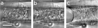
Experimental setup.— We refer to Thoumine and Ott (1997); Fernández et al. (2006); Fernández (2006) for details. A 3T3 fibroblast Todaro and Green (1963) adheres between two fibronectin-coated glass microplates. One of them is flexible: its deflection gives the perpendicular force acting on the cell. This plate is translated by a piezoelectric actuator controlled by a personal computer. The computer calculates force and cell length , and adjusts the piezo position via a proportional feedback loop to impose a given experimental protocol. Experiments are performed at 35∘C, in standard medium with Lysophosphatidic acid 50 M (Sigma). Cells are left to adhere for 30 min before measuring. All results described here are fully reproducible for fibroblasts which adhere sufficiently strongly to sustain pulling forces of 100 nN for several hours, which means about 30% of the cells in culture.
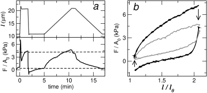
Loading and unloading at constant rate— We stretch the cell by 100% at a constant rate while measuring the force (Fig. 2a). The slope initially decreases, reaching a constant value at an elongation (Fig. 2b). Beyond 10% and up to 100% stretch, the relation is in most cases linear. After loading, the cell length is held constant for a few minutes; the force relaxes to a steady non-zero value which does not evolve faster than nN/s. An analogous response is observed upon unloading. The procedure is repeated with different rates between 3 nm/s and 10 m/s. The asymptotic slope and the equilibrium force are independent of the loading rate in the explored range.
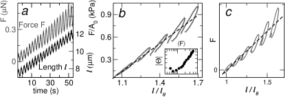
Small amplitude stiffening, large amplitude linearity— To explore small and large deformation amplitudes simultaneously we perform a loading ramp with superimposed harmonic oscillations, imposing . Fig. 3 shows a typical experiment. The response to small oscillations indeed stiffens with increasing stress. Yet, the averages over an oscillation period of the force and length are linearly related as inferred from the position of the loops. Therefore, we observe both responses simultaneously: stiffening at small amplitudes, as reported in Fernández et al. (2006), and linearity at large amplitudes.
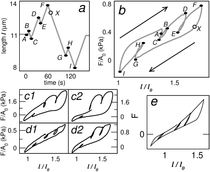
Small amplitude reversibility, large amplitude irreversibility— We study the amplitude dependence at a constant deformation rate . An essential feature of the protocol (Fig. 4a) are the turning points separated at various distances in order to study the reversibility of the response. Similar procedures can be found in plasticity textbooks Lubliner (1990). As Fig. 4b shows, the reversibility of the response upon a change of direction is determined by the distance to the previous turning point. Where turning points are separated by less than 10% stretch, the response is reversible (elastic). More than 10% stretch beyond a turning point, the response becomes irreversible (inelastic): the curve does not retrace its path upon direction reversal. In this inelastic regime the relation is approximately linear. Its nonzero slope leads to a translation of the elastic region by the inelastic deformation, a behavior known in plasticity as linear kinematic (or directional) hardening Prager and Geiringer (1934); Lubliner (1990); Nemat-Nasser (2004); Krempl (1996). Alternatively, the inelastic contraction under pulling tension between X and G in Fig. 4b is a strong Bauschinger effect (a decrease in yield stress upon unloading) Lubliner (1990); Kocks (1987).
Large amplitude stiffening after glutaraldehyde fixation.—
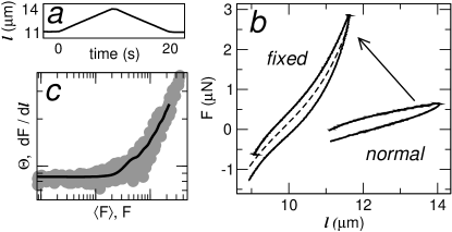
We add glutaraldehyde 0.1% in order to prevent slippage of cytoskeletal connections. Loading at constant rate (Fig. 5a) reveals a positive curvature (Fig. 5b). The numerical derivative of the relation obtained from fixed (hence dead) cells is the same as the differential master-relation obtained on living fibroblasts (Fig. 5c, from Ref. Fernández et al. (2006)). The relation after fixation closely resembles the exponential stress-stretch relations known from whole tissues (Fung, 1993; fab, ).
Rate dependence– In the inelastic regime the width of the hysteresis loops increases with stretch rate (Fig. 6a). To characterize this rate-dependence, we define the overstress as the extent of force relaxation after unloading (Fig. 6a). The overstress behaves as , approaching zero at a non-zero pulling speed of about 10 nm/s (Fig.6b). Below such rates the behavior becomes active and erratic and the overstress ill-defined.
Viscoplasticity– We now propose a minimal constitutive relation for fibroblasts under uniaxial extension. First we decompose the measurable cell length into inelastic rest length and elastic stretch ratio ,
| (1) |
The force is a function solely of the elastic strain,
| (2) |
where for concreteness we use exponential elasticity Fung (1993), according to Fig. 5b, c. As a flow rule relating the inelastic strain rate to the force , we propose an exponential function of the overstress , according to Fig. 6b:
| (3) |
with a vanishing dissipation as the flow rate approaches . The equilibrium force is the essential internal variable to describe kinematic hardening Prager and Geiringer (1934); Lubliner (1990); Krempl (1996). The drag force sets the scale where the overstress induces inelastic flow. To obtain an increased dissipation at large forces (e.g. the increase in the area of the loops in Fig. 3), we take the drag as proportional to the nonlinear modulus, . Finally, we have linear kinematic hardening:
| (4) |
This is an empiric description along the lines of modern viscoplasticity Lubliner (1990); Kocks (1987); Krempl (1996), without explicit history dependencies. As Figs. 3c and 4e show, it captures the essence of the phenomenology. At small amplitudes, , the deformation is essentially elastic: . At large amplitudes the overstress approaches the drag stress and the deformation becomes increasingly inelastic: . Nevertheless, this constitutive relation is not yet a full description. The details of the linear regime Fabry et al. (2001); Desprat et al. (2005) and fluidization at large flow rates Fernández et al. (2006) still have to be incorporated to it, whereas active contraction and inelastic deformation at rates below , as well as the force fluctuations seen in Fig. 2 may require a different approach.

Discussion— Our glutaraldehyde fixation experiments show that stress stiffening in fibroblasts Fernández et al. (2006) is due to the nonlinear elasticity of the cytoskeleton, unrelated to biological signalling or restructuring. In agreement, very similar stiffening is known from biopolymer networks Storm et al. (2005). To date the precise microscopic mechanism remains unclear; stretching Storm et al. (2005); Kuhl et al. (2005) and bending Fernández et al. (2006); Kabla and Mahadevan (2007) of single filaments as well as filament alignment Onck et al. (2005) have been proposed.
The energy to reach the linear inelastic regime in loading experiments (from Fig. 2) is . During stretch, first elastic elements must be loaded, until they dissipate the stored energy upon bond rupture. Taking typical orders of magnitude for bond energies Mogilner and Oster (1996) and dissipation Décavé et al. (2002) and a mesh size of 100 nm Bray (2001), over 10% of the actin cytoskeletal bonds must be ruptured to reach stationary flow. However, in order to observe stress stiffening ubiquitously during the inelastic deformation, the actin gel must be always above percolation threshold. For a gel with one bond relaxation time one expects a rate-independent overstress Gerbal et al. (1999); we observe an exponential rate-overstress dependence. In general, dissociation rates depend on the force per bond as , with a force scale 10 pN Bell (1978); Simson et al. (1999). In agreement, in fibroblasts the drag is about 100 nN, corresponding to 1–10 pN per filament. Interestingly, the inelastic stretch rate where the cell flows without hysteresis is of the order of , a typical rate for active processes such as crawling and contraction Pullarkat et al. (2007); Bray (2001). Thus, spontaneous bond dissociation may be what limits active phenomena to long timescales Thoumine and Ott (1997).
A living cell can neither be purely elastic, nor possess a yield stress within the physiological “working range”. Kinematic hardening viscoplasticity can be understood as a consequence of these conditions. Combined with a sharply rising rate-overstress dependence, it prevents cell breakage in our large deformation experiments: a cell portion under increased stress will readily flow and increase its equilibrium stress to reach a stable situation. Rather than break at a given spot, the cell prolongs homogeneously along its length. This homogeneous deformation may be behind the robust linearity of the kinematic hardening response, since integration of a constant magnitude along the cell length naturally gives a linear length-dependence. However, identification of the precise molecular mechanisms behind this unusual behavior in a soft system is a task for the future. At least, one can rule out a trivial interpretation in terms of a Hookean spring element (in form of intermediate filaments, for example) in parallel with a stiffening viscoelastic liquid: since the liquid cannot sustain an average stress, a non-mechanical coupling between the two elements is needed for the cell to stress-stiffen. Thus, in a mechanical interpretation hardening and stiffening must originate in one and the same mechanical element. If intermediate filaments Bray (2001) play a role, they must be interconnected with actin into a single network. This scenario reminds of composite alloys Kocks (1987) and granular materials Nemat-Nasser (2004), where kinematic hardening arises as the inelastic flow induces alterations of directional nature to the microstructure.
Summarizing, we have shown that cell mechanical properties in uniaxial stretching experiments can be thoroughly described by the superposition of two simple relations: exponential elasticity, and viscoplasticity with linear kinematic hardening. Given the cytoskeletal complexity, this is unexpected. A complete picture of passive cell rheology spanning from molecular details to a simple phenomenological description and straightforward theoretical concepts seems in close reach.
We thank P. A. Pullarkat for his invaluable advice, and K. Kroy for inspiring discussions and support. This work has been funded by the Universität Bayreuth.
References
- Bray (2001) D. Bray, Cell Movements : from molecules to motility (Garland Publishing, Inc., New York, 2001), 2nd ed.
- Thoumine and Ott (1997) O. Thoumine and A. Ott, J. Cell. Sci. 110, 2109 (1997).
- Pullarkat et al. (2007) P. A. Pullarkat, P. A. Fernández, and A. Ott, Phys. Rep. 449, 29 (2007).
- Fabry et al. (2001) B. Fabry, G. N. Maksym, J. P. Butler, M. Glogauer, D. Navajas, and J. J. Fredberg, Phys. Rev. Lett. 87, 148102 (2001).
- Wang et al. (1993) N. Wang, J. P. Butler, and D. E. Ingber, Science 260, 1124 (1993).
- Wang et al. (2002) N. Wang, I. M. Tolic-Nørrelykke, J. Chen, S. M. Mijailovich, J. P. Butler, J. J. Fredberg, and D. Stamenović, Am. J. Physiol. Cell Physiol. 282, C606 (2002).
- Fernández et al. (2006) P. Fernández, P. A. Pullarkat, and A. Ott, Biophys. J. 90, 3796 (2006).
- Wakatsuki et al. (2000) T. Wakatsuki, M. S. Kolodney, G. I. Zahalak, and E. L. Elson, Biophys. J. 79, 2353 (2000).
- Fernández et al. (2007) P. Fernández, L. Heymann, A. Ott, N. Aksel, and P. A. Pullarkat, New J. Phys. (2007).
- Yang and Saif (2005) S. Yang and T. Saif, Exp. Cell Res. 305, 42 (2005).
- Desprat et al. (2005) N. Desprat, A. Richert, J. Simeon, and A. Asnacios, Biophys. J. 88, 2224 (2005).
- Fernández (2006) P. Fernández, Ph.D. thesis, Universität Bayreuth (2006).
- Todaro and Green (1963) G. J. Todaro and H. Green, J. Cell. Biol. 17, 299 (1963).
- Lubliner (1990) J. Lubliner, Plasticity theory (Macmillan Publishing Company, New York, 1990), 1st ed.
- Prager and Geiringer (1934) W. Prager and H. Geiringer, Ergebnisse der exakten Naturwissenschaften 13 (1934).
- Nemat-Nasser (2004) S. Nemat-Nasser, Plasticity: A Treatise on Finite Deformation of Heterogeneous Inelastic Materials (Cambridge University Press, 2004).
- Krempl (1996) E. Krempl, in Unified constitutive laws of plastic deformation, edited by A. S. Krausz and K. Krausz (Academic Pres, San Diego, 1996), pp. 281–318.
- Kocks (1987) U. F. Kocks, in Unified constitutive equations for creep and plasticity, edited by A. Miller (Elsevier applied sciences, Essex, 1987), pp. 1–88.
- Fung (1993) Y. C. Fung, Biomechanics: Mechanical properties of living tissues (Springer Verlag, New York, 1993).
- (20) Exponential elasticity can also be revealed by microrheological techniques. B. Fabry, personal communication.
- Storm et al. (2005) C. Storm, J. J. Pastore, F. C. MacKintosh, T. C. Lubensky, and P. A. Janmey, Nature 435, 191 (2005).
- Kuhl et al. (2005) E. Kuhl, K. Garikipati, E. M. Arruda, and K. Grosh, J. Mech. Phys. Solids 53, 1552 (2005).
- Kabla and Mahadevan (2007) A. Kabla and L. Mahadevan, J. R. Soc. Interface 4, 99 (2007).
- Onck et al. (2005) P. R. Onck, T. Koeman, T. van Dillen, and E. van der Giessen, Phys. Rev. Lett. 95, 178102 (2005).
- Mogilner and Oster (1996) A. Mogilner and G. Oster, Biophys. J. 71, 3030 (1996).
- Décavé et al. (2002) E. Décavé, D. Garrivier, Y. Bréchet, F. Bruckert, and B. Fourcade, Phys. Rev. Lett. 89, 108101 (2002).
- Gerbal et al. (1999) F. Gerbal, V. Noireaux, C. Sykes, F. Jülicher, P. Chaikin, A. Ott, J. Prost, R. M. Golsteyn, E. Friederich, D. Louvard, et al., Pramana - J. Phys. 53, 155 (1999).
- Bell (1978) G. I. Bell, Science 200, 618 (1978).
- Simson et al. (1999) D. A. Simson, M. Strigl, M. Hohenadl, and R. Merkel, Phys. Rev. Lett. 83, 652 (1999).