Control of sub-excitable waves in neural networks by nonlocal coupling
Abstract
nonlinear dynamical systems, excitability, control, nonlocal and time delay coupling
keywords:
dahlem@physik.tu-berlin.de1,2 schneider@itp.physik.tu-berlin.de1 nastyap@imath.kiev.ua1,3 hiller@itp.tu-berlin.de1 schoell@physik.tu-berlin.de1 Institut für Theoretische PhysikTechnische Universität Berlin, Germany Klinik für Neurologie IIOtto-von-Guericke-Universität Magdeburg, Germany Institute of Mathematics National Academy of Sciences of Ukrainedahlem@physik.tu-berlin.de1,2
schneider@itp.physik.tu-berlin.de1
nastyap@imath.kiev.ua1,3
hiller@itp.tu-berlin.de1
schoell@physik.tu-berlin.de1
Institut für Theoretische PhysikTechnische Universität Berlin, Germany
Klinik für Neurologie IIOtto-von-Guericke-Universität Magdeburg, Germany
Institute of Mathematics National Academy of Sciences of Ukraine
1 Introduction
During migraine attacks, localized pathological excitation can spread through cortical tissue and invade large areas before it abates. This activity causes migraine aura, that is, neurological symptoms preceding the headache phase [1]. The underlying process is a phenomenon called cortical spreading depression (SD). It is assumed to be a reaction-diffusion process in the cortex, although reactions and diffusion processes that provide the mechanism of propagtion are still under debate [2, 3]. However, the generic dynamics of reaction-diffusion systems are largely independent of the interaction details and shared among various biological systems [4]. Therefore, to describe the spatio-temporal patterns of SD, the cortex can be approximated as a continuous excitable media supporting reaction diffusion waves [5, 6, 7].
Psychophysical studies on visual processing in migraine patients suggest that changes in their networks of cortical neurons lead to an interictal state of changed excitability, i. e., an anomalous cortical state in the interval between migraine attacks [8, 9]. This motivates efforts to understand how the spread of reaction diffusion waves is controlled by nonlocal network connectivity. To include this, we investigate in this work the hypothesis that the emergence of SD waves can be attributed to these network changes as well. Previously, we have investigated how to change parameters of an excitable medium so as to efficiently protect cortical tissue surrounding a stimulus against recruitment [10]. Our current results suggest that failures in synaptic transmission result in increased susceptibility of cortical tissue to SD. Such a modulation of excitability becomes of crucial importance when the cortical state is close to the bifurcation of the onset of wave propagation. The clinically relevant conclusion to be drawn from this is that therapy might target network connectivity that modulates cortical tissue excitability, even though a specific network connectivity is not required for the initiation or propagation of SD in a regime far from the bifurcation.
2 Neural network with diffusive and nonlocal coupling
A variety of neural network models for SD have been proposed, though there is not yet consent on the mechanism. Roughly speaking, two classes of models exist. One taking a bottom up approach based on biophysical laws including several ionic currents, ion pumps, membrane potentials, and osmotic forces [13, 14, 15, 16]. The other approach is top down trying to incorporate the system properties without detailing any first-level subsystems. Hodgkin proposed the first simplified reaction-diffusion approach to SD. The model was never published, but communicated to Grafstein [17, 18]. According to this references, Hodgkin suggested to consider a balance equation for potassium with a cubic source function and diffusion. The three roots of the cubic function being the resting state, a threshold, and a potassium ceiling level, respectively. Based on methods introduced by Huxley, solving the equation led to the approximate speed of SD. However, the diffusion coefficient used was four times higher than that of potassium in aqueous solution. This may either indicate the anomalous nature of potassium migration in the cortex [19], or the leading role of a faster transcelluar reaction-diffusion mechanism [20].
Another model [5] followed Grafstein’s potassium hypothesis with two extensions. We shortly summarize this work, because we follow a similar approach but with a different interpretation and aim. Firstly, the model included a second dynamic variable describing a refractory phase of SD. The source term in the balance equation of potassium is replaced by a quartic polynomial as the major nonlinearity of this activator-inhibitor system. Secondly, the reaction-diffusion model was connected to a neural network building together a hybrid model. The neural network was originally used to study cortical dynamics and sensory map reorganization. In the hybrid model it was used to explain visual field defects occurring during migraine with aura. We also use a reaction-diffusion system combined with a nonlocal interaction coming from a neural network. Our goal is to investigate which neural network connectivity can prevent SD, as suggested and studied using cellular automatons in [21].
We use the spatially extended FitzHugh-Nagumo (FHN) system [22, 23], which has a cubic nonlinearity, as a generic model of SD waves
| (1) | |||||
| (2) | |||||
The model approximates the cerebral cortex as a two-dimensional surface with the ability to support sustained SD wave propagation. As a generic model this system does not make an explicit distinction between the various species involved in SD. In effect it lumps together sodium inward currents and extracellular potassium concentration into a single activator variable and their combined kinetics into the cubic source term. Likewise, a single inhibitor variable is related to recovery processes, such as effective regulation of by - ion pumps and the glia-endothelial system [20, 14, 15]. Whether a transcellular or extracellular route is taken, is at this level not specified. The main reason we use the FHN mechanism as the reaction-diffusion part of the model is that it has been shown to successfully reproduce the two-dimensional spatio-temporal pattern of SD [7, 24]. Our study is essentially based on these pattern formation properties of SD waves and less on its detailed biophysical mechanism.
We extend the FHN system to encompass cortical lateral interactions, i. e., connections running parallel to the cortical surface. They are accounted for in the form of nonlocal coupling terms
| (3) |
The signal can either be the activator or inhibitor . A connection in the cortex can extend over several millimeters and it either mediates competitive or cooperative interactions. The parameter describes the connection length and the coupling strength of the interaction.
Lateral connections in the cortex can form clusters at regular intervals [25]. Their structure and how they might interact with SD waves will be further considered in Sec. 4 Until then, we consider only one nonlocal coupling term occurring either in the activator (1) or inhibitor (2) balance equation, with fixed values for and . This leads to four different coupling schemes: two of cross coupling (CC) activator and inhibitor, and two in which each dynamic variable is coupled via Eq. (3) into its own balance equation (NCC).
3 Suppression of waves by nonlocal interaction

The investigations were done in a one-dimensional spatial FHN system. We chose the parameter of the FHN system such that it is without the nonlocal coupling term in Eqs. (1-2) above but near to the excitability boundary of a one-dimensional system. This boundary was obtained by transforming the system into a co-moving frame and searching for homoclinic orbits. These orbits correspond to pulse solutions in the original coordinates. The excitability boundary () is defined by a saddle-node bifurcation at which the stable and unstable homoclinic orbit disappear [26]. The parameter values of , , and at which homoclinic orbits disappear constitute a boundary of codimension one. In other words, separates the parameter plane into a regime where local stimulations is transmitted without damping and a regime where such sustained 1D reaction-diffusion waves do not exist (Fig. 2).
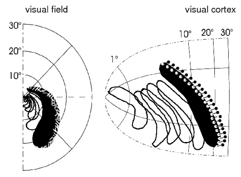
We chose a FHN system near because the transient nature of the observed symptomatic and electrophysiological events during migraine suggest such a regime [11, 27]. In the regime below but close to transient wave forms exist in a one-dimensional system [10]. Above transient wave forms exist in two-dimensional systems untill excitability reaches a boundary called . There, sustained wave segments, called “critical fingers”, propagate without reentering tissue (c.f. Fig. 1). The regime between and is therefore called sub-excitable. The regime in which this transition takes place is also well investigated in chemical model systems in experiment and theory, for a review see [28].
To investigate the influence of various nonlocal connectivity schemes on wave propagation in the regime of sub-excitability, we start by setting a super-threshold stimulation in the one-dimensional system, choosing a particular FHN system with parameter values , , , and . Once a stable one-dimensional wave profile is obtained, the nonlocal lateral network is switched on. Different networks for various parameter values and are classified by their effect on the wave. We distinguish two cases. Either the wave is suppressed. This indicates that the excitability boundary of the combined system is shifted to higher excitability values (upwards in Fig. 2) into a regime where without the nonlocal coupling pulse solutions would exit. Or the wave continues to spread, though its profile and speed might change. From a clinical point of view, the wave suppression is a desirable control goal for the network achieved within the solid black regions in the -planes in Fig. 3.
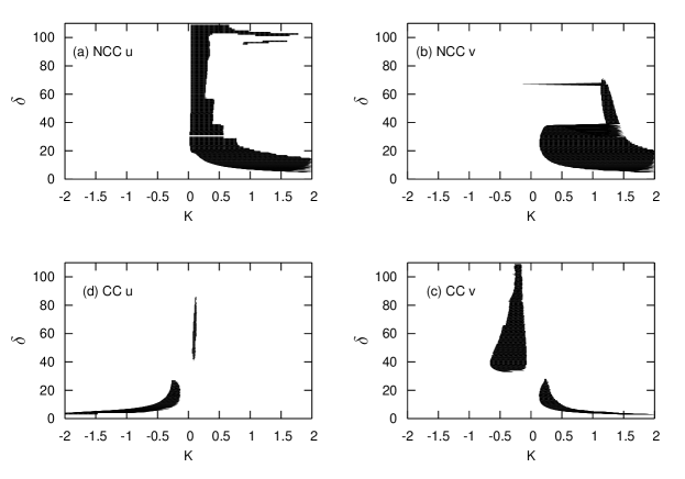
We find that wave propagation can be suppressed with a NCC (non-cross-coupled) setup only with positive coupling strength . When the NCC term appears in the activator balance equation, the desired control goal is achieved largely independent of the connection length (Fig. 3 a), as long as is in the range of the wave width, including its refractory tail. When the nonlocal coupling term appears in the inhibitor balance equation, a similar picture arises, though waves are suppressed for connection lengths ranging into the refractory tail of the wave () only for a narrow regime of . Suppression completely fails for (Fig. 3 b).
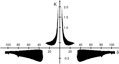
Cross coupling of inhibitor and activator achieves the desired control goal for both positive and negative coupling strengths , depending on the connection length (Fig. 3 c-d). The area in the parameter plane where this control goal is achieved resembles a Mexican-hat-type network connectivity. This is readily seen in Fig. 4. When the nonlocal term appears in the inhibitor balance equation (2) the regimes of successful control in the direction is much wider (Fig. 3 d) than the regime for cross coupling in the activator balance equation (1, Fig. 3 c).
4 On the nature of nonlocal coupling in migraine
In the previous section, we have shown which network failures lead to the emergence of reaction-diffusion waves. From this alone it is not deducible whether some of these network failures can also explain the anomalous cortical state in the interval between migraine attacks. It would be a plausible hypothesis, however, that the same network changes that cause the ictal migraine events, i. e., SD waves, lead to the anomalous interictal state. Changes causing the latter have been attributed to abnormal aspects in early visual processing in the cortex [9]. Various, seemingly contradictory explanations have been given, such as lack of both intra-cortical inhibition and excitation. They are also referred to as cortical hypo- or hyperexcitability (see [29] and references therein).
There is substantial work on the functional role of lateral connectivity for cortical processing, but little is known how the mechanism of SD is coupled to it. Evidence supporting a coupling comes from two independent sources. On the one hand, there is the structure of the hallucinatory aura patterns, in particular the typical zigzags (see Fig. 1). Such patterns were suggested to reflect the cortical network organization [30, 31]. The main idea is that the approaching wave initially affects cortical cells which possess the highest spontaneous activity and are clustered in patches. Within these patches the neuronal response properties remain relatively constant. Their feature distribution corresponds to the organization of the receptive field structure in the cortex. For example, the connection pattern in the visual cortex is unspecific in the immediate vicinity of each neuron, while long-range connections primarily run between so-called iso-orientation columns [25]. Cells in an orientation column have the same oriented receptive field structure, thus they are responsive to edges with the same orientation. These edges are literally seen during a migraine attack as the building blocks of the hallucinatory zigzag pattern. Therefore, it is reasonable to assume that the SD wave interacts with this neural network structure in form of a synchronization process that occurs at the front of the SD wave and extends over the typical spatial length scale of iso-orientation columns.
The other line of evidence comes from in vivo studies in animal research on SD. In [20] Herreras et al. showed that a synchronization of the firing pattern is possible up to the order of millimeters ahead of the SD wave. The peculiarity of this activity is that it is resistant to synaptic transmission blockade. This led to the hypothesis of direct neuron-to-neuron communication by previously closed gap junctions. They suggested that SD propagates through transcellular pathways using a reaction-diffusion mechanism. Computer simulations of Shapiro support this scheme [16]. A complete description of SD, however, must additionally include the full network connectivity of synaptic transmission when SD occures close to a bifurcation. Such a description of SD is beyond the scope of the present study. The time and space scales of these dynamics differ by several orders of magnitude such that a separate treatment is justified. Therefore we investigate the stability of the suggested neuron-to-neuron communication by gap junctions separately. We assume that the way this transcellular pathway interacts with synaptic transmission is in principle described in the previous section.
5 Time-delayed diffusive electrical coupling
In a previous study [32] we used the FHN system modeling two individual neurons with a diffusive coupling in the activator variable. We showed that two FHN-neurons, each oscillating under its own source of noise, can synchronize. The application of time-delayed feedback to only one of two subsystems was shown to change coherence and time scales globally. Time delayed feedback is also able to induce stochastic synchronization under certain conditions. This motivates the approach pursued here to examine a time-delayed coupling between two identical neurons. Since the time-delay can introduce rich dynamics we study the case without random fluctuations.

To distinguish this system of two neurons from the spatially extended FHN reaction diffusion system describing the cortical tissue in Eqs. (1-2), we use the variables and with a subscript 1 and 2 identifying the two neurons. The variables represent the membrane potential and the gating, respectively. The diffusive coupling occurs in the membrane potential. This is a discrete model of a gap junction-mediated electrical coupling, because ionic currents through gap-junctions give rise to strong electrical coupling of the neurons. Gating mechanisms of neuronal gap junctions have not been described as yet. Therefore, we do not consider any gating. But we include a time delay because if the spread in the transcellular pathway is diffusion limited, as the slow propagation speed of SD clearly suggests, the transmission time can be in the order of the excitation cycle.
Individual neurons have only one stable fixed point (for ). It is readily shown that when , the coupled system also has only one stable fixed point. With a non-vanishing delay time the phase space is infinite dimensional. Then, the fixed point is given by the four coordinates above as well as their respective history functions of length , which need to be constant. Along the lines of [33], it can be shown that this fixed point is stable. We find that for adequate parameter values , and , the system is multi-stable. It can avoid the stable fixed point and instead exhibit a mutual resonance phenomenon. In the 4D phase space section at time , this results in a stable firing oscillation of period between the two sub-systems (see Fig.5). Thus, for two FHN-Neurons in the excitable regime, a non-vanishing delay enables a synchronous operation of the two subsystems.
6 Discussion
We showed that certain control schemes of an inhibitor-activator type system shift the emergence of wave propagation towards higher values of excitability. The control we investigated is of the form of a nonlocal coupling given in Eq. (3). This nonlocal transaction was added to the reaction-diffusion mechansim either in the inhibitor or the activator balance equation. The sum of all individual cross coupling terms that achieve a clinically desirable control goal takes the shape of an upright or inverted Mexican hat, respectively. This supports our assumption that the nonlocal coupling results from intrinsic lateral cortical connections.
Dichotomic lateral interaction is an architecture widely used in models of topographic feature maps. The prototypical example of such maps is the orientation preference in primary visual cortex, which is activated by the SD wave. The link between SD and the neuronal network architecture is still missing. One possibility is that gap-junction-mediated oscillatory patterns trigger SD. If so, these oscillatory patterns are likely to be modulated by lateral synaptic connections, although their existence is in general resistant to synaptic transmission blockade [20]. However, when SD is close to the bifurcation of the onset of wave propagation, as suggested by the spatio-temporal patterns (e.g. in Fig. 1), therapy might target network connectivity as to prevent spread.
Although we are still far from modeling the full mechanism of migraine with aura, neural network models have become sophisticated enough to constrain and validate possible underlying cortical circuitry of involved subsystems. To understand the origin of the gap-junction-mediated oscillatory patterns better, we performed simulations in a system of two gap-junction-coupled neurons. We showed that a time-delay is sufficient to produce sustained oscillations in an otherwise merely excitable ensemble. Thus, opening gap junctions between neurons, which are closed in a healthy state, can explain a localized pathological synchrony in the cortex when there exits a time delay.
To summarize, in modeling migraine a major objective is to understanding cortical susceptibility to focal neurological symptoms in terms of neural circuitry [10, 21, 5]. This could open up to us new strategies for therapy using methods of controlling complex dynamics. Control of complex dynamics has evolved during the last decade as one of the central issues in applied nonlinear science [34]. Progress toward clinical implementation of nonlinear methods has been done so far in neurology in particular in Parkinson’s disease, a neurological diseases also characterized by pathological brain synchrony. There, techniques based on control of complex dynamics [35] are now tested in clinical studies and fundamentally novel therapy methods are being evolved [36]. It is hoped that this success can be expanded.
Acknowledgments
This work has been partially supported by the DFG within the framework of Sfb 555 and the Sachbeihilfe DA-602/1-1.
References
- [1] M. Lauritzen: Pathophysiology of the migraine aura. the spreading depression theory [see comments], Brain 117, 199 (1994).
- [2] A. J. Strong: Dr. Bernice Grafstein’s paper on the mechanism of spreading depression, J Neurophysiol 94, 5 (2005).
- [3] O. Herreras: Electrical prodromals of spreading depression void Grafstein’s potassium hypothesis, J Neurophysiol 94, 3656 (2005).
- [4] B. Hess: Periodic patterns in biology, Naturwissenschaften 87, 199 (2000).
- [5] J. A. Reggia and D. Montgomery: Modeling cortical spreading depression, Proc. Annu. Symp. Comput. Appl. Med. Care pp. 873–877 (1994).
- [6] M. A. Dahlem and S. C. Müller: Migraine aura dynamics after reverse retinotopic mapping of weak excitation waves in the primary visual cortex, Biol Cybern 88, 419 (2003).
- [7] M. A. Dahlem and S. C. Müller: Reaction-diffusion waves in neuronal tissue and the window of cortical excitability, Ann. Phys. 13, 442 (2004).
- [8] K. M. Welch, G. D’Andrea, N. Tepley, G. Barkley, and N. M. Ramadan: The concept of migraine as a state of central neuronal hyperexcitability, Neurol Clin 8, 817 (1990).
- [9] A. J. Shepherd: Increased visual after-effects following pattern adaptation in migraine: a lack of intracortical excitation?, Brain 124, 2310 (2001).
- [10] M. A. Dahlem, F. M. Schneider, and E. Schöll: Efficient control of transient wave forms to prevent spreading depolarizations, submitted to J. Theo. Biol. (2007).
- [11] K. Lashley: Patterns of cerebral integration inicated by scotomas of migraine, Arch. Neurol. Psychiatry 46, 331 (1941).
- [12] V. Hakim and A. Karma: Theory of spiral wave dynamics in weakly excitable media: asymptotic reduction to a kinematic model and applications, Phys. Rev. E 60, 5073 (1999).
- [13] H. C. Tuckwell and M. R. M.: A mathematical model for spreading cortical depression, Biophysical J. 23, 257 (1978).
- [14] H. Kager, W. J. Wadman, and G. G. Somjen: Simulated seizures and spreading depression in a neuron model incorporating interstitial space and ion concentrations, J. Neurophysiol. 84, 495 (2000).
- [15] G. G. Somjen: Mechanisms of spreading depression and hypoxic spreading depression-like depolarization, Physiol. Rev. 81, 1065 (2001).
- [16] B. E. Shapiro: Osmotic forces and gap junctions in spreading depression: a computational model, J. Comput. Neurosci. 10, 99 (2001).
- [17] B. Grafstein: Neural release of potassium during spreading depression., in Brain Function. Cortical Excitability ans Steady Potentials., edited by M. A. B. Brazier (University of California Press, Berkely, 1963), pp. 87–124.
- [18] J. Bureš, O. Burešova, and J. Křivǎnek: The mechanism and applications of Leão‘s Spreading Depression (Academia, New York, 1974).
- [19] C. Nicholson and J. M. Phillips: Ion diffusion modified by tortuosity and volume fraction in the extracellular microenvironment of the rat cerebellum, J Physiol 321, 225 (1981).
- [20] O. Herreras, C. Largo, J. M. Ibarz, G. G. Somjen, and R. Martin del Rio: Role of neuronal synchronizing mechanisms in the propagation of spreading depression in the in vivo hippocampus, J Neurosci 14, 7087 (1994).
- [21] L. H. A. Monteiro, D. C. Paiva, and J. R. C. Piqueira: Spreading depression in mainly locally connected cellular automaton, J. of Biol. Systems 14, 617 (2006).
- [22] R. FitzHugh: Impulses and physiological states in theoretical models of nerve membrane, Biophys. J. 1, 445 (1961).
- [23] J. Nagumo, S. Arimoto, and S. Yoshizawa.: An active pulse transmission line simulating nerve axon., Proc. IRE 50, 2061 (1962).
- [24] M. A. Dahlem and S. C. Müller: Self-induced splitting of spiral-shaped spreading depression waves in chicken retina, Exp. Brain Res. 115, 319 (1997).
- [25] C. D. Gilbert and T. N. Wiesel: Columnar specificity of intrinsic horizontal and corticocortical connections in cat visual cortex, J Neurosci 9, 2432 (1989).
- [26] Y. A. Kuznetsov: Elements of Applied Bifurcation Theory (Springer, New York, 1995).
- [27] N. Hadjikhani, M. Sanchez Del. Rio., O. Wu, D. Schwartz, D. Bakker, B. Fischl, K. K. Kwong, F. M. Cutrer, B. R. Rosen, R. B. Tootell, A. G. Sorensen, and M. A. Moskowitz: Mechanisms of migraine aura revealed by functional MRI in human visual cortex, Proc. Natl. Acad. Sci. USA 98, 4687 (2001).
- [28] A. S. Mikhailov and K. Showalter: Control of waves, patterns and turbulence in chemical systems, Phys. Rep. 425, 79 (2006).
- [29] M. A. Dahlem and E. P. Chronicle: A computational perspective on migraine aura, Prog. Neurobiol. 74, 351 (2004).
- [30] E. L. Schwartz: A quantitative model of the functional architecture of human striate cortex with application to visual illusion and cortical texture analysis, Biol Cybern 37, 63 (1980).
- [31] M. A. Dahlem, R. Engelmann, S. Löwel, and S. C. Müller: Does the migraine aura reflect cortical organization, Eur J Neurosci 12, 767 (2000).
- [32] B. Hauschildt, N. B. Janson, A. G. Balanov, and E. Schöll: Noise-induced cooperative dynamics and its control in coupled neuron models, Phys. Rev. E 74, 051906 (2006).
- [33] N. B. Janson, A. G. Balanov, and E. Schöll: Delayed feedback as a means of control of noise-induced motion, Phys. Rev. Lett. 93, 010601 (2004).
- [34] E. Schöll and H. G. Schuster (Editors): Handbook of Chaos Control (Wiley-VCH, Weinheim, 2007).
- [35] P. A. Tass, J. Klosterkotter, F. Schneider, D. Lenartz, A. Koulousakis, and V. Sturm: Obsessive-compulsive disorder: development of demand-controlled deep brain stimulation with methods from stochastic phase resetting, Neuropsychopharmacology 28 Suppl 1, 27 (2003).
- [36] C. Hauptmann and P. A. Tass: Therapeutic rewiring by means of desynchronizing brain stimulation, Biosystems 89, 173 (2007).