Dependence of the energies of fusion on the inter-membrane separation: optimal and constrained
Abstract
We calculate the characteristic energies of fusion between planar bilayers as a function of the distance between them, measured from the hydrophobic/hydrophilic interface of one of the two nearest, cis, leaves to the other. The two leaves of each bilayer are of equal composition; 0.6 volume fraction of a lamellar-forming amphiphile, such as dioleoylphosphatidylcholine, and 0.4 volume fraction of a hexagonal-forming amphiphile, such as dioleoylphosphatidylethanolamine. Self-consistent field theory is employed to solve the model. We find that the largest barrier to fusion is that to create the metastable stalk. This barrier is the smallest, about 14.6 , when the bilayers are at a distance about 20 percent greater than the thickness of a single leaf, a distance which would correspond to between two and three nanometers for typical bilayers. The very size of the protein machinery which brings the membranes together can prevent them from reaching this optimum separation. For even modestly larger separations, we find a linear rate of increase of the free energy with distance between bilayers for the metastable stalk itself and for the barrier to the creation of this stalk. We estimate these rates for biological membranes to be about 7.1 /nm and 16.7 /nm respectively. The major contribution to this rate comes from the increased packing energy associated with the hydrophobic tails. From this we estimate, for the case of hemagglutinin, a free energy of 38 for the metastable stalk itself, and a barrier to create it of 73 . Such a large barrier would require that more than a single hemagglutinin molecule be involved in the fusion process, as is observed.
1 Introduction
Although it is essential to a host of biological processes in which material enters, exits, or changes location within the cell, (e.g. viral entry, exocytosis, and intracellular trafficking) the process of membrane fusion is not well understood. Some basic concepts, however, are clear. The membranes to be fused must be put under tension, i.e. their free energy per unit area must be increased, so that the fused state with smaller area has a lower free energy than the unfused system. This tension is brought about by bringing the membranes to be fused in close proximity to one another, on the order of a few nanometers, thereby removing some water from the hydrophilic headgroups of the amphiphiles comprising the membrane and consequently raising the system free energy. This additional energy is supplied by fusion proteins. Even though the free energy of the system is reduced by fusion, the rearrangement of lipids required by the process can only occur if the system surmounts free energy barriers. The calculation of these barriers has been the subject of much attention (1, 2, 3, 4, 5).
From the above argument, it follows that the barrier to the fusion process must be a function of the tension. It also depends on the pathway to fusion that the system takes (6, 7), as well as several other factors. Among these are the average compositions of the different amphiphiles comprising the membrane and, in particular, their composition in the cis, or proximal, leaves (8, 9). We have examined each of these factors, and the upshot is, that for bilayers in which the relative fraction of hexagonal-forming and lamellar-forming amphiphiles in the cis leaves are similar to that in biological membranes, the largest barrier to fusion, in either the standard (1) or non-standard (10, 11, 12) pathways, is that to form the initial stalk. This is the initial local junction formed by the rearrangement of lipids in the two apposing cis leaves (1). Further, this barrier is not large; it was estimated (8) to be on the order of 13, with the absolute temperature and Boltzmann’s constant. That the rate-limiting barrier to fusion should be so small led us to conclude that fusion should occur rapidly once the two membranes were brought sufficiently close to initiate the process.
This conclusion highlights the question of what is “sufficiently close”, i.e. the issue of the dependence of the fusion barrier on the distance between the membranes to be fused. It is an interesting issue which speaks to the interplay between the lipids and a fusion protein. An example is provided by hemagglutinin, the fusion protein associated with the influenza virus (13). It is anchored in the viral membrane. A cluster of between three and six of them around the eventual site of fusion are required (14). A first conformational change of hemagglutinin is accompanied by removal of the receptor binding domains. A second conformational change exposes the hydrophobic fusion peptide which anchors in the target membrane. At this point the conformation of the several hemagglutinins, which are essentially normal to the membranes, keep the viral and target membranes at a distance of 13.5 nm from one another (15). A final conformational change brings the membranes much closer, on the order of 4 nm, with the hemagglutinin now parallel to the membranes and pointing away from the fusion site (16). This conformational change releases a great deal of energy, on the order of 60 per hemagglutinin (17), which presumably is expended in pulling the membranes to this distance and in bringing about the formation of the stalk. The question is why this distance is what it is. Is it because a smaller distance between membranes would cause fusion to be energetically less expensive, but the very size of the hemagglutinin prevents a closer approach, or is it that the machinery is such that it does bring the membranes to the optimal separation? Just what is the competition that sets the distance at which fusion occurs? Similar questions apply to the SNARE machinery which promotes fusion (18).
There has been little theoretical work on the distance dependence of the barrier to fusion (19, 20, 21). It was considered explicitly by Kozlovsky and Kozlov (19) using a phenomenological model. They found that the energy of an isolated stalk was practically independent of the distance between membranes, and approached a value of about 43 as the distance between membranes increased without limit. This result can be traced to a few assumptions. First the membranes are assumed to be tensionless. Hence, the additional membrane area needed to create a stalk between two membranes at a large distance costs no free energy by assumption. This assumption is presumably quite good when the distance between membranes is greater than that of the hydrophobic repulsion, on the order of a few nm (22). The second assumption is that the membranes can bend to take a shape which minimizes the curvature energy of the system. Given the constraints on the membrane separation placed by the presence of the fusion proteins, this is probably not the case. Finally, the phenomenological free energy employed does not capture the energy associated with packing the tails efficiently into the axially symmetric stalk structure, a structure very different from the planar bilayer, the membrane configuration of lowest free energy.
In order to clarify these issues, particularly that of the packing, we employ a microscopic model to study the dependence of the barriers to fusion on the distance, , between the hydrophobic/hydrophilic interfaces of the apposed leaves of planar membranes. The membranes are under either zero or a small tension. The membranes are composed of a mixture of two amphiphiles, one lamellar- and the other hexagonal-forming. The leaves are of equal composition, one that mimics the mix of these two classes of amphiphiles in the cis leaves of red blood cell membranes. This choice is made because previous work (19, 9) shows that the free energy of fusion intermediates is most sensitive to the composition of the cis leaves, and rather insensitive to that of the trans leaves. Only the standard fusion mechanism is considered. We do this because we have found very little difference in the barriers of the two different mechanisms when membranes with a mix of hexagonal and lamellar formers were considered (8). In addition, this restriction significantly simplifies the calculation.
We find, once again, that the largest barrier to fusion is that associated with the formation of the initial stalk. We also can understand the dependence of this barrier on the separation between membranes as follows. When the membranes are very close, the barrier to fusion increases with decreasing distance for two reasons. Not only does the repulsive, hydrophobic, interaction, essentially a depletion force, increase with decreasing separation, but also the energy required for the amphiphiles to rearrange into a stalk of such short extent becomes larger with smaller membrane separation. Due to this effect, the stalk is not a metastable structure. As a consequence fusion would have to proceed directly to a fusion pore without a stalk intermediate, an absence which would make the process much less likely. When the membranes are farther apart, the stalk becomes a stable intermediate, and the barrier to fusion decreases. As the distance between membranes increases still further, the barrier to fusion now increases rapidly with increasing distance due to the packing energy of the initial stalk connecting the membranes, an energy which scales with the length of the stalk. We find this rate of increase to be about 7 per nm. Consequently the lowest barrier to fusion occurs when the two membranes are at a distance large enough that membrane repulsion is not too great, and the stalk is metastable, but small enough that the stalk is relatively short and energetically inexpensive. In our system we find the optimum distance to be about twenty percent greater than the thickness of a single leaf of our bilayer, a distance which would correspond to between two and three nanometers for typical membranes. This is in reasonable agreement with the observed distance to which laboratory membranes must be brought in order to fuse (23). The lowest barrier to fusion corresponds to about for a biological membrane. To fuse membranes which are at a somewhat larger distance, as in the case when the very size of hemagglutinin prevents a closer approach, requires traversing a larger barrier. At a distance between headgroups of 4 nm applicable to the case of hemagglutinin, we estimate that the barrier is on the order of 73. It is not surprising, then, that more than a single fusion protein would be required.
2 The model
To investigate the effect of the distance between planar membranes on the free energy barrier to fuse them, we extend the application of self-consistent field theory to microscopic models of membranes initiated earlier (6, 7, 8). The basic assumption of this approach is that the self-assembly into bilayer vesicles and the processes which these vesicles can undergo, such as fusion, are common to systems of amphiphiles, of which lipids are but one example. Recent work on vesicles which consist of diblock copolymers serves to illustrate this point (24). It follows that these processes can be explored in whatever system of amphiphiles proves to be most convenient. For the application of self-consistent field theory, that system is one of block copolymers in a homopolymer solvent. While the processes that amphiphiles undergo are presumably universal, the energy scales of these processes are system-dependent, and thus it is necessary to be able to compare the energy scale in a biological bilayer with the energy scale in our system of block copolymers. This will be done below.
Here we consider a system of two bilayers each composed of two different amphiphiles that resemble dioleoylphosphatidylcholine, (DOPC), and dioleoylphosphatidylethanolamine, (DOPE), in their hydrophobic/hydrophilic ratios. The two leaves of each bilayer are of the same composition. The system is incompressible and occupies a volume . The two amphiphiles are each AB diblock copolymers. Type 1, a lamellar-former, consists of monomers and has a molecular volume . The fraction of hydrophilic monomers, arbitrarily chosen to be of type , is denoted and is assigned the value as such a diblock has a “spontaneous curvature” similar to that of DOPC (6). The amphiphile of type 2 consists of monomers and has a molecular volume of . The fraction of hydrophilic monomers, is chosen to be as this produces a spontaneous curvature similar to that of DOPE. We set such that hydrophobic tails of different types of amphiphiles have equal length. For our chosen and , . The solvent is an A homopolymer with volume .
We denote the local volume fraction of hydrophilic elements of amphiphile 1 to be , of amphiphile 2 to be , and of the solvent to be . The total local volume fraction of hydrophilic elements is denoted
| (1) |
Similarly the total local volume fraction of hydrophobic elements is
| (2) |
The amounts of each of the components are controlled by activities, , , and . Because of the incompressibility constraint, only two of the activities are independent. Cylindrical coordinates, , are employed.
Within the self-consistent field approximation, the free energy, , of the system containing a bilayer, or bilayers, each of area , is given by the minimum of the functional
| (3) | |||||
where , and are the configurational parts of the single chain partition functions of amphiphiles 1 and 2 and of solvent. They have the dimensions of volume, and are functions of the temperature, , which is inversely related to the Flory interaction , and functionals of the fields and . These fields, and the Lagrange multiplier , which enforces the local incompressibility condition, are determined by the self-consistent equations which result from minimizing the free energy functional. Insertion of these fields into the free energy functional, Eq. (3), yields the free energy within the self-consistent field approximation:
| (4) | |||||
The free energy of the system without the bilayer, i.e. a homogeneous solution, is denoted . The difference between these two free energies, in the thermodynamic limit of infinite volume, defines the excess free energy of the system with one, or more, membrane:
| (5) |
With the excess free energy known, the surface free energy per unit area, or equivalently, the surface tension, , is obtained from the excess free energy of a single, flat, bilayer
| (6) |
In order to calculate the free energy of stalk or hemifusion intermediates as a function of their radius, that radius must be fixed (6, 25) by a local Lagrange multiplier Similarly, to constrain the membranes to be separated by a specified distance, at some point , we must introduce an additional Lagrange multiplier, . The distance is chosen to be the distance between the hydrophilic/hydrophobic interfaces of the contacting, cis, leaflets as shown in Fig. 1. With these additional constraints, the free energy functional to be minimized now reads
It is clear that one cannot constrain the bilayers to be a distance apart at a position at which the stalk or hemifusion diaphragm come in contact. Consequently in the last integral in Eq. (2), the region of integration over is restricted to be greater than , where is the radius of the fusion intermediate, and is positive and at least as large as the hydrophilic thickness of the bilayer. The condition that the free energy functional of Eq. (2) be minimized yields a set of self-consistent equations that we solve in real space. A detailed description on the derivation of Eq. (3) and the real space solution algorithm can be found elsewhere (6, 26, 27).
Finally we need to compare the energies in a biological system with those in our homopolymer system. There are various choices for the energy of the biological system. One could choose a property of a single bilayer, such as the energy per unit area of a hydrophobic, hydrophilic interface. Alternatively a property of two interacting bilayers coud be chosen, such as the attractive energy per unit area between them. As the former is so well known, we shall employ it, but will show below that this gives essentially the same result had we chosen the latter. We consider the dimensionless quantity , where N/m is the oil, water interfacial tension, and m is a typical bilayer thickness. With Nm, this ratio is about 150 for a biological system. The analogous quantity in the polymer system is , where is the surface tension between coexisting solutions of hydrophobic and hydrophilic homopolymers, and is the thickness of our bilayers. We calculate , so that energy scales in a biological system are about a factor of 150/56.7=2.6 greater than in our polymer model.
3 Results and Discussion
We first consider some properties of a single bilayer composed of lamellar-forming amphiphiles, chosen to mimic DOPC, whose volume fraction is 0.6, and hexagonal-forming amphiphiles, chosen to mimic DOPE, whose volume fraction is 0.4. The leaves are of equal composition. As in our previous work, we have chosen the volume of amphiphile 1 to be , where is the radius of gyration of the polymer. The bilayer thickness, measured between the planes at which the volume fractions of the hydrophilic part of the amphiphiles and that of the solvent are equal, is . The hydrophobic thickness, measured between the planes at which the volume fractions of hydrophilic and hydrophobic parts of the amphiphiles are equal, is
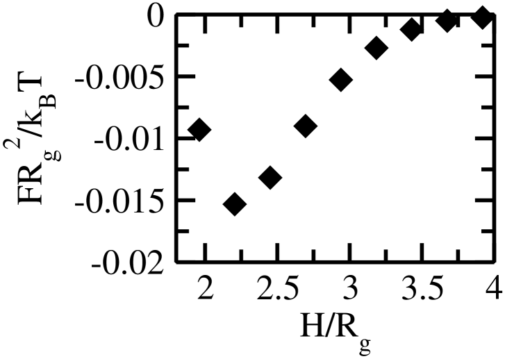
Two such bilayers have a weak attraction between them due to depletion forces induced by expulsion of some solvent when they are brought together. To see this, we calculate the excess free energy of a system of two flat bilayers a distance apart, and define the free energy per unit area
| (9) |
By definition, this quantity asymptotes to zero for large , and is negative when the bilayers attract one another. For the case of bilayers under zero tension, the dimensionless quantity is plotted in Fig. 2.
This energy of attraction per unit area can be compared with those measured between phospholipid bilayers provided we know the length scale given by , the radius of gyration of the polymers in our system. To obtain this we note that the thickness of our bilayers is approximately 4.3. If we take a typical bilayer thickness to be 4 nm, then nm. With this and J, our calculated value of the free energy per unit area at the equilibrium distance between membranes corresponds to 0.07 mJ/m2. This should be increased by the factor of 2.6 if the energy scale we obtained by comparison with the hydrophilic, hydrophobic repulsion, is correct. Thus we expect that the energies of attraction per unit area between two phospholipid bilayers should be approximately 0.18 mJ/m2. This agrees extremely well with the results presented by Marra and Israelachvili (28) in their Fig. 2. It shows that we could have obtained our energy scale equally well from the interaction energy of two bilayers.
The excess free energy of an intermediate, such as a stalk, is calculated as follows. We compute the excess free energy, , of the system of two bilayers which are connected by the intermediate, and which, far from it, are separated by a distance . The excess free energy of the intermediate is, then
| (10) |
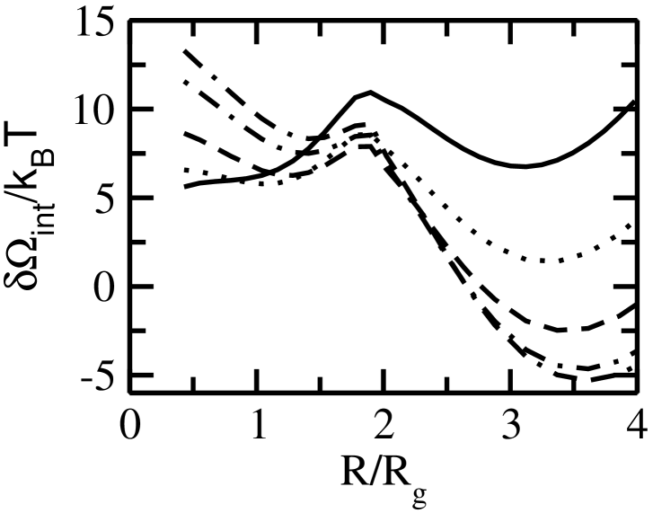
In Figure 3, we show the excess surface energy of the stalk as a function of its radius, , at different bilayer separations . Again, the tension of the bilayer is zero. Each leaf of the bilayers shown here have compositions, and , which are almost the same as the cis leaves of the asymmetric membranes we considered previously (8). We note that for stalk radii which are quite small, less than about 0.5 , we find no solution for a stalk-intermediate. This reflects the fact that the process by which the stalk initially forms cannot necessarily be thought of as one which produces a stalk of infinitesimal radius which then expands. At large radii, the stalk expands into a hemifusion diaphragm. We find that as the membrane separation increases, this hemifusion diaphragm becomes indistinguishable from a single bilayer membrane. Hence for all large the free energy increases linearly with with a slope directly related to a line tension, one which arises from the junction of the hemifusion diaphragm with the two bilayer membranes.
The most important result in Fig. 3 is that the increase of separation between fusing bilayers causes the energy of the metastable stalk to increase significantly. It follows that the barrier to the formation of this stalk also increases significantly with separation. As an estimate to this barrier, we take the energy of the stalk with the smallest radius for which we find a solution of our equations. This should be considered an upper bound, as there may be less expensive paths to the creation of the stalk. A second result of note is that there is no metastable stalk if the bilayers are too close to one another. This is because the energy associated with the rearrangement of amphiphiles needed to make the stalk is simply too large at small membrane separations. As the intermembrane distance increases, the stalk does become metastable with a radius on the order of 1.3. This is reasonable as the diameter of this stalk is about the same as the hydrophobic thickness of our bilayers, , so that amphiphiles that make up the stalk can take configurations somewhat similar to those of amphiphiles in the unperturbed bilayers.
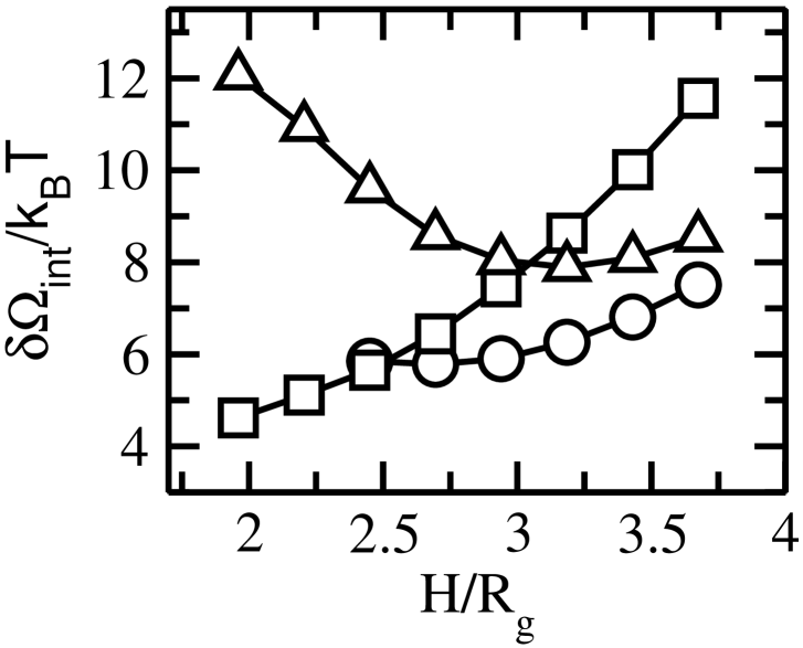
The importance of the stalk being metastable can be seen in Fig. 4, which summarizes the results of our calculation. We have plotted, as a function of separation, , the energy of the stalk with the smallest radius for which we find a solution, (squares), the energy of the metastable stalk (circles), and the barrier (triangles) which is associated with the expansion of the stalk into a hemifusion diaphragm before pore formation. For the smallest two interbilayer separations shown, and , there is no metastable stalk. Consequently one large activation energy of approximately (corresponding to 29 for a biological membrane) is required before a fusion pore can form. However, for separations for which there is a metastable stalk, , fusion can occur in two steps: formation of the initial stalk which relaxes to the metastable stalk, and expansion into a hemifusion diaphragm with formation of a fusion pore. An additional activation energy is required for this second step, and is given by the difference between the energy of the second barrier and that of the metastable stalk. A third point of interest concerns the range from , at which the stalk first becomes metastable, to at which the barrier to make the hemifusion diaphragm (triangles in Fig. 4) is no longer larger than the barrier to make the initial stalk, (squares in Fig. 4). Within this range, the additional energy needed by the metastable stalk to surmount the second barrier and go forward to the hemifusion diaphragm is larger than that required for the process to reverse itself by means of the disappearance of the stalk. In other words, in this range successful fusion is a less likely outcome of stalk formation than the simple disappearance of the stalk. The probabilities of these outcomes are not reversed until exceeds 3.25. But at this larger separation, the barrier to form the initial stalk is also larger. Thus we expect that most of the time a metastable stalk actually forms, it does not lead to successful fusion.
A fourth point we wish to make is the following: once a metastable stalk becomes possible, the additional activation energy needed to pass to the hemifusion diaphragm is always less than the barrier to create the initial stalk. Hence this barrier to create the initial stalk, whose magnitude is shown by the squares in Fig. 4, becomes the largest barrier to fusion. Its magnitude is the smallest when the stalk first becomes metastable, which occurs when the bilayers are at a distance which exceeds by about 20% a distance equal to half the hydrophobic thickness of our bilayers. This small membrane separation, again defined between the hydrophilic/hydrophobic interfaces of the apposed cis leaflets can be compared with the results of Weinreb and Lentz (23) who found optimum fusion at a distance between hydrophobic/hydrophilic interfaces that was comparable to half the hydrophobic thickness of their bilayers. The value of this smallest barrier for stalk formation is, from Fig. 4, about 5.6 for the copolymer membranes, which corresponds to about 14.6 for a biological membrane.
We note from Fig. 4 that the free energies of the metastable stalk and of the barrier to its creation become linear functions of even for values of which are not too large. The rate of increase of the free energy of the metastable stalk with intermembrane distance, (circles in Fig. 4), is 2.5 . We can convert this rate of change of free energy with distance to practical units as follows. We increase the energy by a factor of 2.6 to account for the difference between our amphiphilic bilayers and those composed of lipids and utilize the length scale nm obtained earlier. From these we find that the above rate of increase of the metastable stalk free energy with thickness becomes
| (11) |
As we have set the tension of the bilayers to zero, this increase in stalk free energy does not arise simply from the additional surface area of a longer stalk. We have repeated our calculations taking a surface tension equivalent to 2.68 mN/m, a value in the range of tensions which can cause rupture (29), and found that the rate of change of metastable stalk energy with membrane separation increased from the value of 7.1 kBT/nm only to 9.4 kBT/nm. Therefore we conclude that the increased area associated with a stalk of greater length is not the major contribution to the stalk free energy. Rather it is plausible that the dominant contribution to the length dependence of the metastable stalk free energy comes from the packing of the hydrophobic tails. That is, although the stalk has a diameter comparable to the hydrophobic thickness of the bilayer, the axially symmetric configuration is very different from the planar bilayer. If the density of headgroups in the stalk is comparable to that in the bilayer, then the tails become crowded near the center of the stalk. Conversely, if the tail density at the center is comparable to that of the interior of the bilayer, then the density of headgroups must be considerably less than that of the bilayer causing a significant energy penalty of contact between solvent and tails. This conjecture is strengthened by the observation, from Fig. 4, that the rate of increase with distance, , of the barrier to stalk formation is greater than that for the metastable stalk itself. This is reasonable as the intermediate that we consider, and which corresponds to the barrier, is a stalk of diameter smaller than that of the metastable stalk, and also smaller than the thickness of an unperturbed bilayer. Hence the hydrophobic tails are packed quite densely. From Fig. 4, (squares), this slope is which for a biological membrane translates to
| (12) |
These results permit us to discuss the interesting case which arises when the apposing membranes cannot be brought to the optimum, small, distance which the amphiphiles would like simply because of the very size of the protein machinery which brings the membranes together. This is the case with hemagglutinin whose approximate 4nm width (30) keeps the head groups of apposing membranes this distance apart. If we assume a headgroup of 1 nm (28), then the minimum distance between hydrophilic/hydrophobic interfaces is on the order of 6nm. The free energy of the metastable stalk and the barrier to its creation when the apposing bilayers are constrained to be at such a distance can be estimated from Fig. 4 and the linear behavior at large distances given above. We find the metastable stalk to have an excess free energy of 38 . The barrier to be overcome to create this metastable stalk is about 73 . It is understandable that more than a single hemagglutinin molecule is required to bring about the amphiphile reorganization needed to produce a stalk linking membranes at such a distance, one imposed by the very machinery of fusion itself.
4 Acknowledgments
This work was supported by the National Science Foundation under Grant No. DMR-0503752.

5 Figure Captions
-
Figure 1 Apposed bilayers separated by distance . Circles represent hydrophobic head groups and curved lines hydrophobic tails. The separation is measured between the hydrophilic/hydrophobic interfaces of the contacting leaflets.
-
Figure 2 Free energy per unit area of apposed bilayers, of eq (9), in units of as a function of separation distance, , between bilayers composed of 60% lamellar formers and 40% hexagonal formers and under zero tension.
-
Figure 3 Excess surface energy of stalk-like fusion intermediates as a function of stalk radius, for (solid), (dotted), (dashed), (dot-dashed), and (dot double-dashed) for systems composed of 60% DOPC-like and 40% DOPE-like diblocks under zero tension.
-
Figure 4 Various energies related to fusion in the standard mechanism as a function of separation for bilayers shown in Fig. 3. Squares represent the initial barrier to create a stalk, circles the metastable stalk energy, and triangles the second barrier as the stalk expands to a hemifusion diaphragm. For the lowest two values of separation ( and ), metastable stalks do not exist.
References
- Kozlov and Markin (1983) M. M. Kozlov and V. S. Markin, Biofizika 28, 255 (1983).
- Siegel (1993) D. P. Siegel, Biophys. J. 65, 2124 (1993).
- Kuzmin et al. (2001) P. I. Kuzmin, J. Zimmerberg, Y. A. Chizmadzhev, and F. S. Cohen, Proc. Natl. Acad. Sci. U.S.A. 98, 7235 (2001).
- Lentz et al. (2000) B. R. Lentz, V. Malinin, M. E. Haque, and K. Evans, Curr. Opinion in Struct. Biol. 10, 607 (2000).
- Chernomordik and Kozlov (2003) L. V. Chernomordik and M. M. Kozlov, Annu. Rev. Biochem 72, 175 (2003).
- Katsov et al. (2004) K. Katsov, M. Müller, and M. Schick, Biophys. J. 87, 3277 (2004).
- Katsov et al. (2006) K. Katsov, M. Müller, and M. Schick, Biophys. J. 90, 915 (2006).
- Lee and Schick (2007) J.-Y. Lee and M. Schick, Biophys. J. xx, xx (2007).
- Kasson and Pande (2007) P. Kasson and V. S. Pande (2007).
- Noguchi and Takasu (2001) H. Noguchi and M. Takasu, J. Chem. Phys. 115, 9547 (2001).
- Müller et al. (2002) M. Müller, K. Katsov, and M. Schick, J. Chem. Phys. 116, 2342 (2002).
- Müller et al. (2003) M. Müller, K. Katsov, and M. Schick, Biophys. J. 85, 1611 (2003).
- Eckert and Kim (2001) D. Eckert and P. Kim, Annu. Rev. Biochem. 70, 777 (2001).
- Blumenthal et al. (1996) R. Blumenthal, D. Sarkar, S. Durell, D. Howard, and S. Morris, J. Cell Biol. 135, 63 (1996).
- Wiley and Skehel (1987) D. Wiley and J. Skehel, Annu. Rev. Biochem. 56, 365 (1987).
- Bentz et al. (1993) J. Bentz, H. Ellens, and D. Alford, in Viral fusion mechanisms (1993).
- Kozlov and Chernomordik (1998) M. Kozlov and L. Chernomordik, Biophys. J. 75, 1384 (1998).
- Söllner et al. (1993) T. Söllner, S. W. Whiteheart, M. Brunner, H. Erdjument-Bromage, S. Geromanos, P. Tempst, and J. E. Rothman, Nature 362, 318 (1993).
- Kozlovsky and Kozlov (2002) Y. Kozlovsky and M. M. Kozlov, Biophys. J. 82, 882 (2002).
- Knecht and Grubmüller (2003) V. Knecht and H. Grubmüller, Biophys. J. 84, 1527 (2003).
- Kasson et al. (2006) P. Kasson, N. Kelley, N. Singhal, M. Vrlic, A. T. Brunger, and V. S. Pande, PNAS 103, 11916 (2006).
- Israelachvili and Pashley (1982) J. Israelachvili and R. Pashley, Nature 300, 341 (1982).
- Weinreb and Lentz (2007) G. Weinreb and B. R. Lentz, Biophys. J. 92, 4012 (2007).
- Discher et al. (1999) B. D. Discher, Y.-Y. Won, D. S. Ege, J. C.-M. Lee, F. S. Bates, D. E. Discher, and D. A. Hammer, Science 284, 1143 (1999).
- Matsen (1999) M. W. Matsen, J. Chem. Phys 110, 4658 (1999).
- Fredrickson (2006) G. Fredrickson, The Equilibrium Theory of Inhomogeneous Polymers (Oxford University Press, Incorporated, Oxford, 2006).
- Müller et al. (2006) M. Müller, K. Katsov, and M. Schick, Physics Reports 434, 113 (2006).
- Marra and Israelachvili (1985) J. Marra and J. Israelachvili, Biochemistry 24, 4608 (1985).
- Evans et al. (2003) E. Evans, V. Heinrich, F. Ludwig, and W. Rawicz, Biophys J. 85, 2342 (2003).
- Wilson et al. (1981) I. Wilson, J. Skehel, and D. Wiley, Nature 289, 366 (1981).