Polaron tunneling dynamics in the DNA molecule
Abstract
The formation of polaron and its migration in a DNA chain are studied within a semiclassical Peyrard-Bishop-Holstein polaron model. Comparing the energetics of the polaron system found from the quantum chemical and semiclassical calculations, we extract the charge-phonon coupling constant for poly DNA sequences. The coupling constant is found to be larger for the G-C than for the A-T pairs. With this coupling constant we study tunneling in the DNA molecule. The rates and the nature of tunneling have strong dependence on the DNA sequence. By changing the trap positions in the molecular bridge the tunneling rate can be varied up to seven orders of magnitude.
The discovery of conductance in DNA has attracted many researchers to investigate the transport properties review ; ratner ; berlin ; yoo ; kasumov ; kawai ; porath ; conw ; jortner of DNA. For the (G-C)(A-T)N(G-C)3 DNA sequences the mechanism of charge transfer is more or less clear review ; ratner ; berlin ; jortner and is described by the competiton between tunneling and hopping transfer. But for the poly and mixed DNA sequences the experimental data observed by different groups are often contradictory. In some experiments a high conductivity was obtained yoo ; kasumov , while in others the conductivity was rather low kawai ; porath . In the works devoted to simulation of charge transfer in the DNA molecule within the tight-binding Hamiltonian macia ; wang or the system of kinetic equations jortner ; berlin , no explanation of this phenomena was found. Within these models the charge transfer integral between the nearest base pairs and the energy gap between the states were the key parameters. At the same time the models did not take into account consistently the effect of geometry fluctuations or phonons on the charge transfer processes in the DNA molecule. This is despite the fact that already in 1956 Marcus pointed out mark1 that geometry fluctuations can activate and strongly affect an electronic transition between two states. Therefore, the polaron model Bishop1 ; conw ; Bishop2 ; yoo , which takes into account all these parameters should be invoked for an adequate investigation of the charge transfer in DNA.
In this paper, we demonstrate the possibility to design an artificial DNA molecule with semiconductor or insulating behaviors simply by placing a trap at the correct points. The study is based on the analysis of charge transfer from the donor to the acceptor through a molecular bridge composed of the potential barriers (A-T pairs) and the charge traps (G-C pairs). We show that in the mixed DNA molecules the transfer rate of the charge strongly depends on the sequences, i.e., on the positions of the traps between the donor and the acceptor. This position determines the relation between the rate of charge trapping and the rate of charge escape from the trap. Depending on this ratio the tunneling between the donor and the acceptor can be described either as a sequential tunneling or coherent tunneling through a trap. This difference strongly affects the final rate of charge transfer. We show that the time of polaron tunneling can be changed by times by changing the position of the trap. The slower transfer occurs when the polaron trapping rate is much slower than the escape rate. On the other hand, the fastest polaron transfer occurs when the trapping and the escape rates are equal. In this case the tunneling has the coherent nature and the polaron only partially occupies the trap.
We focus here on the DNA structures where the guanine and the adenine are stacked in one DNA strand. In this case, there is only one degree of freedom for the charge transfer – the longitudinal one-dimensional charge tunneling through a single strand. The polaron tunneling is described by the system of equations within the Peyrard-Bishop-Holstein (PBH) model Bishop1 ; Bishop2 , where the charge motion is treated quantum-mechanically while the polaron tunneling classically. The Schrödinger equation describing the dynamics of the charge within the DNA chain with sites is determined by the Hamiltonian , which includes the tight-binding and the charge-lattice interaction terms. The Schrödinger equation has the form
| (1) |
where is the probability amplitude for the charge to be on the -th base pair, is the transfer integral between the nearest base pairs, is the charge-vibrational coupling constant for the -th site, is the on-site energy, determines the stretching at the -th site, i.e. the displacement of the atomic structure. The motion of the stretching displacement is described by the Newton equation as Bishop2
| (2) | |||||
where is the base pair mass, is the friction parameter ( ps-1 Bishop1 ), is the vibronic potential of the deformation energy of the hydrogen bonds in the base pair taking into acount the repulsive interactions of the phosphates and is the nearest-neighbor potential of interactions of the stacked base pairs Bishop2 . The parameters for the deformation potential and the interaction potential are taken from Ref. Bishop2 .
| (eV) 111Simulation results are from Ref. berash | (eV/Å) | |
|---|---|---|
| (G-C)N=1 | 0.360 | 1.60 |
| (G-C)N=2 | 0.310 | 1.15 |
| (G-C)N=3 | 0.265 | 0.90 |
| (G-C)N=4 | 0.225 | 0.60 |
| (A-T)N=1 | 0.185 | 1.05 |
| (A-T)N=2 | 0.165 | 0.53 |
| (A-T)N=3 | 0.140 | 0.40 |
| (A-T)N=4 | 0.125 | 0.30 |
The probability amplitude and the position of the stretching displacement in time are evaluated from the self-consistent solution of the time-dependent Schrödinger and Newton equations. Here the Schrödinger equation (1) is presented as
| (3) |
where the Hamiltonian is a function of the stretching displacements . The three-point difference scheme for the first order time derivation of the stretching parameter has been used in Eq. (2). For the stacionary solution of Eqs. (1-2), the initial occupation probability is assumed to be close to unity on the donor site. This solution is taken as the initial state (=0) for studying the polaron dynamics.
At first we analyze the equilibrium stationary polaronic states within a finite region of the DNA chain. The charge-vibration coupling constant is the main parameter regulating the stretching of the polaron to the nearest sites Bishop2 and the magnitude of . The value of depends on the geometries of the DNA sites participating in the formation of the polaron. The shift of the state energy due to the polaron occupation in the absence of the DNA-solvent interactions can be described by the inner-sphere reorganization energy ( in Ref. berash )
| (4) |
where is number of sites occupied by the polaron. Recently, the exponential decrease of the inner-sphere reorganization energy with the elongation of the DNA chain was found within the quantum chemical calculations for the (G-C)N and the (A-T)N chains berash . The geometry relaxation was found to have a maximum at the center of the polaron, which agrees with the results obtained within the PBH model Bishop2 .
The reorganization energy found in Ref. berash are shown in Table I for the (G-C)N and the (A-T)N chains. From these data we can estimate the coupling constants for different systems. The corresponding results for are shown in Table I. We have found that the coupling constant is smaller for the A-T base pair than for the G-C pair. We also determine the tendency in the dependence of the coupling constant on the size of the complexes; the coupling constant decreses with increasing sizes of the (G-C)N and (A-T)N chains.
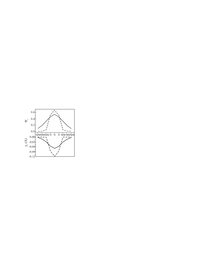
With the values of coupling constants derived for different base pairs we now study the properties of the polaronic state in the poly and mixed DNA chains. In the poly(dG)-poly(dC) and the poly(dA)-poly(dT) DNA molecules, according to our calculations the polaron occupies mostly 7–9 sites. In the mixed DNA chain, the polaron stretching is limited by the difference between the coupling constants , and on-site energies , for A-T and G-C pairs (see Table 1). The results for GGGGGGGGG and AAAGGGAAA chains are shown in Fig. 1. The polaron in the AAAGGGAAA structure is mostly localized within the GGG due to high potential barriers between guanines and adenines (1.08 eV) Saito .
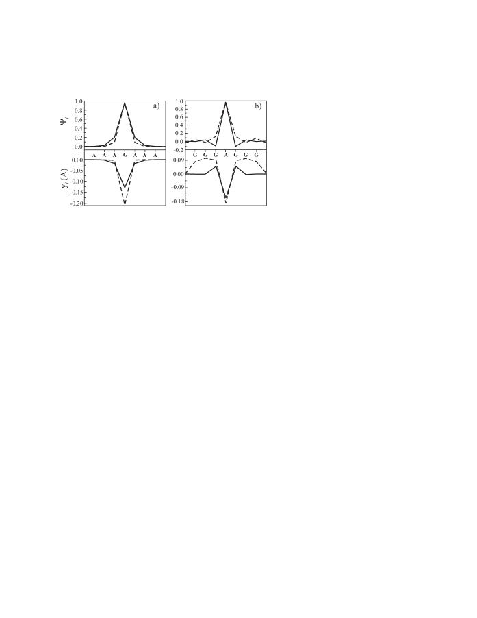
The value of the coupling constant also determines the polaron stretching, but its effect is strong only in the structure with low potential barriers. An example of such structures is the AAAAGAAAA chain where the energy gap between the A-T and the G-C is only 0.4 eV. The results for the AAAAGAAAA structure are shown in Fig. 2 (a) for two cases: (i) when the value of coupling constant is the same over the whole chain and is equal to =0.6 eV/Å Saito , and (ii) when the coupling constant is different for G-C and A-T base pairs. Clearly, the introduction of different coupling constants for A-T and G-C pairs provides stronger localization of the polaron within the G-C trap.
The effect of the coupling constant on the polaron localization in the GGGGAGGGG chain is illustrated in Fig. 2 (b). Actually, for this chain the polaron vibration mode is outside of the lattice band of the A-T site Bishop2 . When the coupling constant =0.6 eV/Å is the same over the whole DNA chain, the vibration mode is only marginally delocalized. The energy of this state is eV, while the potential barrier between (G-C)N and A-T site is 0.87 eV. The polaron in this case is almost localized at the A-T site. When we introduce the dependence of the coupling constant on the site type [Fig. 2 (b) (dashed line)], the energy of the state becomes eV and the polaron becomes delocalized over three nearest G-C sites from each side of the A-T pair.
To study polaron tunneling between the DNA traps, we first compare the energies of the polaronic states in different types of traps. Localization of the polaron in the (G-C)N traps shifts the on-site energy to a lower value. This is the energy of the polaron which is the eigenvalue of the Hamiltonian corresponding to Eq. (1). This energy can also be estimated from the site energy and the electronic energy Bishop2
| (5) |
For the energy difference between the polaronic states in different traps we have found the values eV for G-C and (G-C)2 traps and eV for G-C and (G-C)3 traps. Inclusion of inner-sphere reorganization energy into the charge transfer model has brought down these values from eV and eV, respectively Saito . A direct comparison with the experimental results in the solvent lewisa would require evalution of the solvent reorganization energy berash1 , which is beyond the scope of this work. However, for the results that follow, in particular for the polaron migration dynamics, the solvent contribution perhaps is not the dominant one.
The low energy gap between the states of the (G-C)N traps results in the competition between two processes in the mixed DNA Schuster2 : (i) the trapping of the polaron within the trap and (ii) the tunneling of the polaron between the (G-C)N traps. To study the problem of polaron tunneling between the DNA traps we have performed numerical simulation of the polaron dynamics in a mixed DNA chain. Here the first G-C trap is a donor with localized charge on it in the initial state and the (G-C)3 trap is an acceptor. Since the system is initially in the nonequlibrium state the polaron will tunnel from the donor to the acceptor. For the system without any additional traps, i.e. a DNA chain with only the donor and the acceptor [(G)(A)n(G)3], we have found an exponential dependence of the tunneling rate on the tunneling distance. Let be the tunneling time for the structure (G)(A)n(G)3. We then have for the normalized rate, for , for , and for ( ns). These data are in a good agreement with the tunneling rates in the experimental results giese .
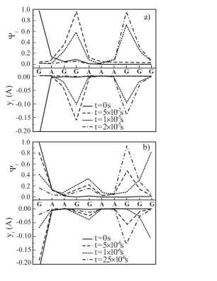
The new features in the polaron tunneling process is observed for the mixed DNA structure with additional trap between the donor and the acceptor. Here we study the system with the (G-C)2 trap in the (A-T)6 molecular bridge. The dispersion of the site energies of the G-C base pairs within the (G-C)3 where the guanine at the end site has higher site energy than guanines located close to the sequence center berash , has been taken into account. In the case of the (G-C)2 trap located close to the donor site (G-C) the polaron is stretched over the donor and the trap. As a result the polaron quickly tunnels to the (G-C)2 trap [Fig. 3(a)]. The polaron occupation process takes some time and finally the polaron tunnels to the acceptor. In this case the tunneling from the donor to the acceptor states has the sequential nature and the tunneling processes from the donor to the trap and from the trap to the acceptor are uncorrelated.
When the (G-C)2 trap is placed exactly in the middle of the (A-T)6 bridge, a significant change in the polaron tunneling dynamics is observed [Fig. 3 (b)]. In this case, the rate of charge tunneling from the donor to the trap is almost equal to the rate of tunneling from the trap to the acceptor. Therefore, the polaron is only partially localized on the trap and the final polaron tunneling from the donor to the acceptor is a coherent process. The curve for sec in Fig. 3 (b) shows the occurence of the resonance effect due to the coincidence of the trap site energy with the site energy of the last guanine within the (G-C)3 acceptor. The rate of charge tunneling in Fig. 3 is in good agreement with the experimental results giese2 , where the transfer from a donor to an acceptor in similar systems was estimated to be sec.
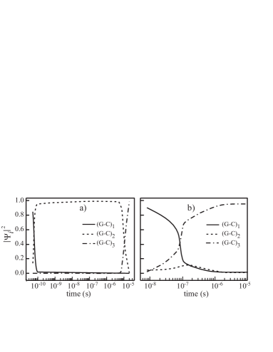
In Fig. 4 the dependence of the occupation probability of different traps within the DNA chain is shown for (a) G(A)1GG(A)3GGG and (b) G(A)2GG(A)2GGG structures. We again see a completely different nature of tunneling for different positions of the trap. When the trap is close to the donor, the charge transfer process is the sequential incoherent tunneling, i.e., when the polaron spends a long time within the trap. But if the trap is moved closer to the center of the tunneling bridge then the tunneling becomes coherent. However, when the position of the (G-C)2 trap is closer to the acceptor as in the G(A)3GG(A)1GGG structure, the polaron is not localized on the trap but tunnels directly from the donor to the acceptor. Since the trap does not participate in the charge transfer, the width of the potential barrier for the polaron covers the whole molecular bridge (A)3GG(A)1 and coherent tunneling from the donor to the acceptor occurs in the range of sec. This is times slower than the time for coherent tunneling through the trap [see in Fig.3(b) and Fig.4(b)]. Therefore, the transfer mechanism for the G(A)3GG(A)1GGG sequence is similar to that for the G(A)6GGG structure.
In conclusion, from the results of the ab initio quantum mechanical calculations, we obtained the charge-vibration coupling constants in the Peyrard-Bishop-Holstein model for polarons formed in the (G-C)N and the (A-T)N DNA molecules. We have found that the coupling constants are larger for the (G-C)N complex than for the (A-T)N. From the calculated values of the coupling constants we have studied the energetics and the structure of the polaron in different DNA sequences. In the poly-DNA molecule, the polaron occupies nine DNA base pairs, while in the mixed DNA the size of the polaron is strongly affected by the potential gap between the A-T and the G-C sites. In addition to the properties of the stationary polaronic state, we have also studied the dynamics of the polaron tunneling from the donor to the acceptor. We have found a very strong dependence of the tunneling rates on the structure of the tunneling bridge. The position of additional traps within the bridge strongly affects the nature of the tunneling process and the rates. By changing the position we can change the tunneling rate upto seven orders of magnitude. To have the fastest tunneling rate we need to have coherent tunneling, i.e. tunneling to each of the traps should be almost equal to the escape rate from the trap. Our calculations are restricted to low temperatures. Therefore, the dynamics of the polaron in our study is completely due to quantum mechanical processes.
This work has been supported by the Canada Research Chair Program and a Canadian Foundation for Innovation (CFI) grant.
References
- (1) Electronic mail: tapash@physics.umanitoba.ca
- (2) Long-range charge transfer in DNA, edited by, G.B. Schuster (Springer-Verlag, Heidelberg, New York 2004).
- (3) M.A. Ratner, and J. Jortner, Molecular Electronics (Blackwell, Oxford, 1997).
- (4) J. Jortner, M. Bixon, T. Langenbacher, and M.E. Michel-Beyerle, Proc. Natl. Acad. Sci. (USA) 95, 12759, (1998).
- (5) Y.A. Berlin, A.L. Burin, and M.A. Ranter, J. Phys. Chem. A 104, 443 (2000).
- (6) E. Conwell, Top. Curr. Chem. 237, 73 (2004).
- (7) K.-H. Yoo, et al., Phys. Rev. Lett. 87 198102 (2001).
- (8) A.Y. Kasumov, et al. Science 291 280 (2001).
- (9) M. Taniguchi, and T. Kawai, Physica E 33, 1 (2006).
- (10) D. Porath, A. Bezryadin, S. deVries, C. Dekker, Nature 403, 635 (2000).
- (11) X.F. Wang and T. Chakraborty, Phys. Rev. Lett. 97, 106602 (2006)
- (12) S. Roche, E. Macia, Mod. Phys. Lett. B. 18 847 (2004).
- (13) R.A. Marcus, J. Chem. Phys. 24, 966 (1956).
- (14) P. Maniadis, et al. Phys. Rev. E 72, 021912 (2005).
- (15) P. Maniadis, et al. Phys. Rev. B. 68, 174304 (2003).
- (16) J. Berashevich, and T. Chakraborty, cond-mat/0703764.
- (17) H. Sugiyama and I. Saito, J. Am. Chem. Soc. 118, 7063 (1996).
- (18) F.D. Lewis, et al. J. Am. Chem. Soc. 122 12037 (2000).
- (19) J. Berashevich, and T. Chakraborty, J. Chem. Phys. 126, 035104 (2007).
- (20) J. Joseph, G.B. Shuster, J. Am. Chem. Soc. 128, 6070 (2006).
- (21) F.D. Lewis, et al. Angew. Chem. Int. Ed. 45, 7982 (2006).
- (22) F.D. Lewis, et al. J. Am. Chem. Soc. 125 4850 (2003).