What are the benefits of bound (protonation) states for the electron-transfer kinetics?
Abstract
We describe a model of electron transfer reactions affected by local binding to the donor or acceptor sites of a particle in equilibrium with the solution. The statistics of fluctuations of the donor-acceptor energy gap caused by binding/unbinding events are non-Gaussian, and the resulting free energy surfaces of electron transfer are non-parabolic. The band-width of the charge-transfer optical transition is predicted to pass through a maximum as a function of the concentration of binding particles in the solution. The model is used to rationalize recent observations of pH-dependence of electron transfer rates involving changes in the protonation state of the donor-acceptor complex.
I Introduction
Many redox reaction in chemistry and biology involve bound states which are weaker than common chemical bonds, but stronger than intermolecular interactions in bulk molecular liquids. A prominent example of such association is binding of water molecules to solutes via hydrogen bonds. Since the strength of such bonds typically varies between different electronic states of the solute, water association can be recorded by optical solvatochromism.Reichardt:94 ; DMjpcb:97 ; SolvPolarity:04 ; Agmon:05 Another example is the one of proton equilibria involved in biological electron transfer (ET) reactions responsible for photosynthesis and respiration.McEvoy:06 In Photosystem II, the primary donor P is oxidized by tyrosine which changes its pKa-value from 10 to upon oxidation. As a result, it cannot hold onto its phenolic proton in aqueous solution releasing it in what is argued to be a concerted electron-proton transfer mechanism.Cukier:04 Another related example is photoacidity when photoexcited intramolecular charge transfer lowers pKa for the release of a proton to the solvent.Agmon:05 In view of the wide spread of such reactions, often referred to as proton-coupled ET,Cukier:96 ; Soudackov:00 ; Hammes-Schiffer:01 ; Cukier:04 in biological systems in general and enzymetic reactions in particular,Ferguson-Miller:96 ; Kirby:97 one wonders if their occurrence is just a coincidence caused by ubiquity of acid-base equilibria in proteins, or, alternatively, protonation/deprotonation transitions are used by nature to fine-tune the ET energetics. This question, also used for the paper title, is the subject of this study.
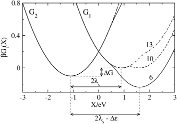
It has been argued that coupling of ET to the change in protonation alters the reaction free energy, often turning uphill reactions along the ET coordinate into downhill reactions along a combined electron-proton reaction path.Mayer:04 In order to make this argument more qualitative, at least one more parameter, the reorganization energy, needs to be specified.Marcus:93 ; Borgis:96 The picture of crossing parabolas used to calculate barriers of charge-transfer reactionsMarcus:65 involves in fact three components: the reaction free energy representing the vertical shift of the minima, the curvature given in terms of the reorganization energy , and the horizontal shift of the parabolas’ minima responsible for the Stokes shift, (Figure 1).

The reaction coordinate , used to monitor the progress of a charge-transfer reaction, is the instantaneous energy gap between the donor and acceptor electronic energy levels.King:90 The vertical axis in Figure 1 refers to free energies as functions of energy . They cross at zero energy gap allowing electronic tunneling ( refer to reactants and products, respectively). In fact, the Marcus-Hush description of ETMarcus:89 imposes one more additional constraint requiring the Stokes shift to be identically equal to ,
| (1) |
where . Therefore, only two parameters, and , are required to determine the activation barrier. The strong link between the Stokes shift and the reorganization energy is a consequence of the Gaussian statistics of . Once the statistics become non-Gaussian, the free energy surfaces are non-parabolic and eq 1 does not hold.DMacc:07 Here, we propose a simple model for a charge-transfer reactions triggering the change in the state of binding of some particle dissolved in the solvent. In this development, we are less concerned with the details of a particular binding mechanisms, but more interested in addressing a more general question of whether the involvement of bound states can potentially provide a new mechanism of tuning the energetics of ET not incorporated in the picture of crossing parabolas. We find that the statistics of fluctuations of the donor-acceptor energy gap, induced by local binding/unbinding events, are non-Gaussian thus violating the link between the Stokes shift and reorganization energy given by eq 1. The main consequence of this result is more flexibility, compared to the standard picture, in tuning the energetics of ET.
II Model
We will consider a somewhat simplified situation in which the bound state is fully thermalized on the time-scale of ET i.e. on the time required for the system to climb the top of the potential barrier from the equilibrium bottom of the free energy surface ( is the ET rate constant). This assumption implies that populations of bound and unbound states follow Boltzmann statistics along the ET reaction coordinate, a situation similar to the treatment of intramolecular vibrations in Sumi-Marcus model.Sumi:86 This condition can be achieved when the rate of release of the binding particle B, , is much higher that the rate of ET
| (2) |
When this requirement is satisfied, one can use statistical mechanics to construct a one-dimensional free-energy surface for the ET reaction in which the exchange between localized and delocalized states of the binding particle produce fluctuations of the donor-acceptor energy gap, just as any other solvent mode interacting with the transferred electron.Stuchebrukhov:03

The release of one particle from the bound localized state requires the Gibbs energy balancing the binding energy with an entropy gain from moving the particle into the bulk. The binding energy is generally different in the initial, D–A (), and final, D+–A- (), configurations of the donor-acceptor complex (Fig. 2). The difference in free energies needed to release particle B to the solution is equal to the difference in binding energies, . Therefore, this quantity can be estimated from the difference of the equilibrium dissociation constants pK:
| (3) |
The requirement of non-adiabaticity of ET, which we implicitly assume here, imposes an additional constraint on the rate of bound particle release. The change of the donor and acceptor energy levels caused by the motion of the bound particle should not break the Landau-Zener non-adiabaticity condition close to donor-acceptor energy resonance:
| (4) |
Here, is the donor-acceptor electronic coupling. Assuming the rate of binding energy change as and of the order of 1 cm-1, eq 4 requires the rate of release of B to be faster than 1 ns-1. This threshold rate is comparable to the rate of proton release from bound protonation states of proteins,Gutman:97 while the time of water exchange between the protein surface and the bulk is faster, in the range of tens of picoseconds.Halle:04
Once particle B is released from its bound state, it becomes a part of the bulk, which is the aqueous solution or a solvent mixture for water binding or the ionic atmosphere for ionic (proton) association. The interaction of the bulk with electronic states of the donor and acceptor is relatively well understood. For water binding, fluctuations of the dipolar polarization produce the thermal noise. For electrolytes, the Debye-Hückel electric field both shifts the donor-acceptor energy gap and creates its fluctuations thus producing a corresponding reorganization energy.German:92 This ionic reorganization energy scales quadratically with the inverse Debye-Hückel length and linearly with a solute dimension . Since it is also inversely proportional to the static dielectric constant of the solvent , the effect of fluctuations of the ionic atmosphere in polar solvents is commonly small relative to the reorganization energy of polarization fluctuations. We will therefore assume that once particle B is released from its bound state, it does not interact any more with the transferred electron and becomes a part of the many-particle electrostatic potential.
This approximation allows us to apply the tight-binding approximation and to use the following expression for the Hamiltonian of the donor-acceptor system in a polar solvent
| (5) |
Here, incorporates the vacuum energy and the free energy of solvation by the electronic degrees of freedom of the solvent and denotes the vacuum electric field of the donor-acceptor complex in two ET states () and two binding states (u,b). Also, and in eq 5 describe the population operators for the bound (b) and unbound (u) states of particle B with the binding energy . One can expect only a small change in the electric field of the donor-acceptor complex for weak binding of a neutral molecule, . However, the dependence of the electric field on the binding state needs to be incorporated for equilibria of charged particles.
In application to protonation/deprotonation equilibria, the Hamiltonian considered here (eq 5) is different from the ones used by CukierCukier:96 and Soudackov and Hammes-Schiffer.Soudackov:00 In their case, the proton was considered to move between two localized states within the donor-acceptor complex, while in the problem considered here the proton is released to the bulk and loses any connection to electronic transitions within the donor-acceptor pair except for the influence of the Debye-Hückel field it becomes a part of. The different physics of the problem considered here demands the different Hamiltonian.
The electric field interacts with the (nuclear) dipolar polarization of the solvent , and the asterisk in eq 5 implies both the tensor contraction and the volume integration. The statistics of are Gaussian with the response function such that the solvent reorganization energy is
| (6) |
Although in eq 6 are formally the reorganization energies of the dipolar polarization field of the solvent, we will attach a more general meaning to them as the reorganization energies of the nuclear degrees of freedom of the bulk. The corresponding formal definition is easy to obtain from eq 5 by replacing the Gaussian polarization noise with a sum of statistically independent Gaussian fields.
The free energies of ET are obtained from the Hamiltonian in eq 5 by projecting all possible nuclear motions on the reaction coordinate :
| (7) |
Here, implies functional integration over the field and “Tr” refers to the sum over bound, , and unbound, , states of particle B with statistical weights :
| (8) |
The ratio of the statistical weights gives the entropy of releasing particle B to the bulk, .
Taking the integral and trace in eq 7 results in the following equation for the free energy surfaces of ET
| (9) |
In eq 9, is the free energy of the donor-acceptor complex in the unbound state. The solvation free energy entering is thus of electrostatic origin and does not include the free energy of binding particle B. Correspondingly, the vertical energy gap in the second summand is given as for the forward transition and as for the backward transition . Binding of particle B is expressed by the term under the logarithm where function is the ratio of Boltzmann factors for activation through bound and unbound states:
| (10) |
Here, are the vertical ET gaps in the bound state of particle B which are given as () and ().
In writing eq 9 we have also represented the Boltzmann factor for the release of particle B in terms of pK and pB as follows
| (11) |
In this equation is the free energy of releasing the bound particle from the molecular fragment in the donor-acceptor complex (e.g. tyrosine) and is the change in the solvation energy of the entire donor-acceptor complex caused by unbinding event. The equilibrium constants thus reflects the equilibrium of the entire complex and can be modified compared to the equilibrium constants of the molecular fragment alone.
The free energies can be used to calculate the first and second moments of the reaction coordinate , e.g.
| (12) |
where
| (13) |
One gets for the average
| (14) |
and for the variance
| (15) |
| (16) |
is the equilibrium population of the bound site.
Equations 7–16 provide a general description of ET when binding affects both the statistics of the energy gap fluctuations and the electronic density responsible for the electric field. In order to reduce the number of independent parameters, we first neglect the second effect assuming that the electric field does not change with binding, i.e. , , and . This situation most closely corresponds to binding of a neutral molecule (water) while the case of a charged particle (protonation) is postponed to the next section.
The free energy surfaces of ET can then be written as follows:
| (17) |
where refers to the free energy of the donor acceptor complex with particle B released to the solution.
Figure 3 illustrates the change of the free energy surfaces with pB according to eq 17 (pK values of tyrosine). For the reaction , the system needs to climb the activation barrier from the bottom of the free energy well to the crossing point at . The bottom of the potential well rises with increasing pB at pBpK thus resulting in a smaller activation barrier. The reaction rate depends on pB. The barrier height stops changing once pB reaches pK, and the reaction rate is insensitive to pB at pBpK. Notice that the rate of the reverse transition remains unchanged in the whole range of pB values. This invariance makes the effect of pB on the ET rate distinct from the effect of the driving force which alters the activation barriers for both the forward and backward reactions.
When binding does not affect the electric field of the donor-acceptor complex the equations for the first and second cumulants (eqs 14 and 15) simplify to
| (18) |
and
| (19) |
Several important observations follow from eqs 18 and 19. First, the binding/unbinding fluctuations break the connection (eq 1) between the reorganization energy from the free energy curvature
| (20) |
and the Stokes shift
| (21) |
This effect is the consequence of the local character of the unbinding events contrasting with the quasi-macroscopic nature of the bulk fluctuations resulting in eq 1. Second, the reorganization energy in eqs 19 and 20 arising from binding fluctuations depends on the state of the donor-acceptor complex, i.e. . This condition implies that the free energy surfaces must be non-parabolicDMacc:07 to comply with the energy conservation,King:90 . The effect of thermalized bound states on ET cannot therefore be accounted for within the Marcus-Hush formalism. We also note that the variance of the reaction coordinate in eq 16 does not follow the fluctuation-dissipation theoremLandau5 requiring and instead has a more complex temperature dependence arising from the equilibrium dissociation constant entering the equilibrium population in eqs 15 and 19.
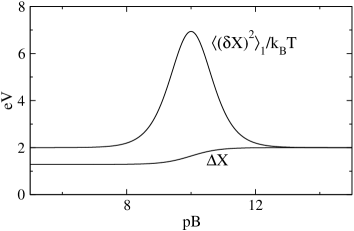
The dependence of both the energy gap variance and the Stokes shift on pB are shown in Figure 4. The equality of and , expected from eqs 1, is seen when both the initial and final ET states are in the same binding state. They are shifted by the binding energy when the the binding states are different in the two ET states (left corner in Figure 4). The most interesting region is when pBpKa and the term proportional to in eqs 15 and 19 can potentially dominate the energy gap variance. Our model thus makes a simple, experimentally testable prediction that the band-width of a charge-transfer optical transition (absorption for binding to the donor) is expected to pass through a maximum as a function of pB.
III Protonation affecting electron transfer
Measurements of oxidation rates of primary pair P by tyrosine in Photosystem II have produced intriguing results.Ahlbrink:98 The reaction rate increases with increasing pH until it levels off at pH approximately equal to pKa of phenolic proton. Similar results were reported by Hammarström and co-workers for intramolecular ET from tyrosine to Ru(III) covalently connected in a donor-acceptor complex.Sjodin:00 ; Lomoth:06 In order to explain the observed pH-dependence, they used the Marcus equation for the activation free energy in which a pH-dependent redox potential was substituted. The pH-dependence of the rate arises in their analysis from the dependence of the tyrosine reduction potential on the concentration of protons in the solution.
This practice,Sjodin:00 ; Carra:03 which had not been anticipated in the original Marcus formulation,Marcus:65 was criticized by KrishtalikKrishtalik:03 and, more recently, by Savéant and co-workers.Costenin:07 Krishtalik argued that the Gibbs free energy of ET reactions cannot possibly depend on the ideal mixing entropy of protons in the bulk, which is the origin of the pH-dependent term in the Nernst equation for the redox potential.Bockris:70 We can only add to this, absolutely correct, argument that using a pH-dependent in the Marcus equation clearly violates the Franck-Condon principle. The vertical energy gap does not involve entropy change since the nuclei do not move on the time-scale of electronic transitions. When the entropic pH term appears in and is not compensated by a corresponding term in , the unphysical entropy of protons mixing appears in the vertical transition energy. The present model allows us to account for the pH-dependence of the ET rate without introducing unphysical approximations.

For protonation affecting ET, BH and pBpH. This problem is, however, potentially more complex than binding/unbinding of neutral molecules. The process of deprotonation proceeds by formation of a geminate pair (on the time-scale of picoseconds for photoexcited statesAgmon:05 ) followed by a slower diffusion of the released proton in the Coulomb potential of the deprotonated negative charge.Agmon:05 ; Perez:07 This slow process may imply that the unbound proton will not be able to sample all possible states available to a particle in an ideal solution on the time-scale of ET, . A full account of such effectsDMacc:07 presently appears hard to achieve. We will therefore introduce an empirical non-ergodicity multiplier to account for the effects of insufficient sampling. The term pKpH in eq 9 is replaced with , where is a non-ergodicity coefficient between zero and unity. With this correction, the model qualitatively reproduces the dependence of the ET rate on pH observed by Sjödin et al.Sjodin:00 for the oxidation of tyrosine by Ru(III).
Sjödin et al.Sjodin:00 have monitored the recovery of Ru(II) from Ru(III) produced by flash photolysis and accelerated by electron transfer from tyrosine covalently linked to the ruthenium complex. Their observed rates, monitoring the arrival of electrons, mathematically correspond to projecting the complex, potentially multi-coordinate,Agmon:83 ; Sumi:86 ; Soudackov:00 dynamics of the system onto one single ET reaction coordinate, which is exactly the scenario considered here. Since proton is a charged particle, we need the full formulation given by eq 9 to analyze the experimental rates. The reaction Gibbs energies and reorganization energies of ET will potentially be different for the bound and unbound ET pathways and indeed the reaction free energies are eV and eV for the unbound and bound proton states, respectively.Carra:03 The unknown parameters are the rate preexponent, reorganization energies , and the non-ergodicity multiplier . This latter parameter is expected to be small because of the slow rate of proton dissociation from tyrosineSjodin:00 and thus a lower extent of modulation of the donor-acceptor gap by unbinding events than it would be possible for full thermalization.

In order to see if the model can qualitatively account for the experimental observations, we have used the rate preexponent, reorganization energies , and as free parameters to fit the data from ref Sjodin:00, . The result is shown in Figure 5. The fitting curve ( eV, eV, and ) of vs pH rises linearly with the slope when pH is below pK and levels off after reaching this value. The reorganization energies here include reorganization of classical intramolecular vibrations in addition to solvent reorganization. The parameters extracted from the fit of the rates result in a dramatic violation of eq 1 as is shown in Figure 6.
Fitting the experimental jump in the rate at requires a higher value of the rate preexponent as is shown by the fragment of the curve obtained by using the same parameters as in the low-pH fit, but varying the preexponent. The increase of the rate at has been addressed by Carra et al.Carra:03 The low-pH portion of the curve corresponds to electronic transition accompanied by simultaneous proton release (proton-coupled ETHammes-Schiffer:01 ). The nonadiabatic matrix element in the rate preexponent then includes small Franck-Condon overlap between proton bound and free states.Cukier:96 This overlap disappears in the flat portion of the plot at when tyrosine is deprotonated in both ET states. Only electronic coupling enters the rate preexponent in this regime resulting in a higher rate. A more quantitative analysis would require extensive calculations. Since the model presented here does not aim at a detailed quantitative analysis of proton-coupled ET, we limit our discussion to qualitative observations only.
IV Conclusions
Traditional theories of ET in polar liquids emphasize activation of electronic transitions by long-range, quasi-macroscopic solvent fluctuations.Marcus:65 Local binding to the donor and acceptor sites provides an additional modulation of the donor-acceptor energy gap. This study addressed the question of whether this additional thermal noise can be accounted for within the standard framework of Gaussian fluctuations of the energy gap, that is by adjusting the magnitudes of the driving force and reorganization energy.
We have found that the statistics of binding events are non-Gaussian, and the resultant free energy surfaces cannot be reduced to crossing parabolas. The model predicts a regime of a significant dependence of the activation barrier on the concentration of the binding particles in the solution. When the concentration pB is close to the binding equilibrium constant pKa, the variance of the energy gap passes through a maximum which can be observed by spectroscopy of charge-transfer bands. Despite these new features, the main effect of binding is in shifting the free energy gap of ET, as was suggested previously,Mayer:04 leaving the reorganization energy mostly unaffected and within the realm of standard models. Finally, the model helps to rationalize some recent observations of the dependence of rates of intramolecular ET, involving deprotonation of reactants, on the pH of the solution.
Acknowledgements.
This work was supported by the NSF (CHE-0616646).References
- (1) Reichardt, C. Chem. Rev. 1994, 94, 2319-2358.
- (2) Matyushov, D. V.; Schmid, R.; Ladanyi, B. M. J. Phys. Chem. B 1997, 101, 1035.
- (3) Katritzky, A. R.; Fara, D. C.; Yang, H.; Tämm, K. Chem. Rev. 2004, 104, 175.
- (4) Agmon, N. J. Phys. Chem. A 2005, 109, 13.
- (5) McEvoy, J. P.; Brudvig, G. W. Chem. Rev. 2006, 106, 4455.
- (6) Cukier, R. I. Biochim. Biophys. Acta 2004, 1655, 37-44.
- (7) Cukier, R. J. Phys. Chem. 1996, 100, 15428.
- (8) Soudackov, A.; Hammes-Schiffer, S. J. Chem. Phys. 2000, 113, 2385.
- (9) Hammes-Schiffer, S. Acc. Chem. Res. 2001, 34, 273.
- (10) Ferguson-Miller, S.; Babcock, G. Chem. Rev. 1996, 96, 2889.
- (11) Kirby, A. Acc. Chem. Res. 1997, 30, 290-296.
- (12) Mayer, J. M. Annu. Rev. Phys. Chem. 2004, 55, 363.
- (13) Marcus, R. A. Rev. Mod. Phys. 1993, 65, 599.
- (14) Borgis, D.; Hynes, J. J. Phys. Chem. 1996, 100, 1118.
- (15) Marcus, R. A. J. Chem. Phys. 1965, 43, 679.
- (16) King, G.; Warshel, A. J. Chem. Phys. 1990, 93, 8682.
- (17) Marcus, R. A. J. Phys. Chem. 1989, 93, 3078.
- (18) Matyushov, D. V. Acc. Chem. Res. 2007, 40, 294.
- (19) Sumi, H.; Marcus, R. A. J. Chem. Phys. 1986, 84, 4894.
- (20) Stuchebrukhov, A. A. J. Theor. Comp. Chem. 2003, 2, 91.
- (21) Gutman, M.; Nachliel, E. Annu. Rev. Phys. Chem. 1997, 48, 329.
- (22) Halle, B. Phil. Trans. R. Soc. Lond. 2004, 359, 1207.
- (23) German, E. D.; Kuznetsov, A. M. Electrokhimiya 1992, 28, 294.
- (24) Landau, L. D.; Lifshits, E. M. Statistical Physics; Pergamon Press: New York, 1980.
- (25) Ahlbrink, R.; Haumann, M.; Cherepanov, D.; Bögershausen, O.; Mulkidjanian, A.; Junge, W. Biochemistry 1998, 37, 1131.
- (26) Sjödin, M.; Styring, S.; Åkermark, B.; Sun, L.; Hammarström, L. J. Am. Chem. Soc. 2000, 122, 3932.
- (27) Lomoth, R.; Magnuson, A.; Sjödin, M.; Huang, P.; Styring, S.; Hammarström, L. Photosynth. Res. 2006, 87, 25.
- (28) Carra, C.; Iordanova, N.; Hammes-Schiffer, S. J. Am. Chem. Soc. 2003, 125, 10429.
- (29) Krishtalik, L. I. Biochim. Biophys. Acta 2003, 1604, 13.
- (30) Costenin, C.; Robert, M.; Savéant, J.-M. J. Am. Chem. Soc. 2007, 129, 5870.
- (31) Bockris, J. O. Modern Electrochemistry; McDonald: London, 1970.
- (32) Pérez-Lustres, J. L.; Rodriguez-Prieto, F.; Mosquera, M.; Senyushkina, T. A.; Ernsting, N. P.; Kovalenko, S. A. J. Am. Chem. Soc. 2007, 129, 19.
- (33) Agmon, N.; Hopfield, J. J. J. Chem. Phys. 1983, 78, 6947.