Title: Identification of Characteristic Protein Folding Channels in a
Coarse-Grained Hydrophobic-Polar Peptide Model
Authors: Stefan Schnabel, Michael Bachmann, and Wolfhard Janke
Author affiliation: Institut für Theoretische Physik and Centre for Theoretical Sciences (NTZ),
Universität Leipzig, Augustusplatz 10/11, D-04109 Leipzig, Germany
Corresponding author: Prof. Dr. Wolfhard Janke
Address:
Institut für Theoretische Physik,
Universität Leipzig,
Augustusplatz 10/11, D-04109 Leipzig, Germany
Phone:
+49 341 9732725
Fax:
+49 341 9732548
E-mail:
Wolfhard.Janke@itp.uni-leipzig.de
Abstract:
Folding channels and free-energy landscapes of hydrophobic-polar heteropolymers are discussed on the basis of a minimalistic off-lattice coarse-grained model. We investigate how rearrangements of hydrophobic and polar monomers in a heteropolymer sequence lead to completely different folding behaviors. Studying three exemplified sequences with the same content of hydrophobic and polar residues, we can reproduce within this simple model two-state folding, folding through intermediates, as well as metastability.
Text:
The understanding of protein folding is one of the major challenges of modern interdisciplinary science. Proteins are linear chains of amino acids connected by peptide bonds and typically consist of many hundreds of these acid residues. Except proline and glycine, all amino acids occurring in natural proteins possess a backbone with identical atomic composition, while they differ strongly in their flexible side chains connected with the carbon atom in the backbone. Secondary structures, such as helices, sheets, and turns, are mainly formed and stabilized by hydrogen bonds between backbone atoms, while side chains widely influence the three-dimensional conformation, i.e., the tertiary structure, of the protein. Roughly, side chains can be characterized as polar or hydrophobic, depending on their chemical structure. Through attractive polarisation effects with the aqueous environment, polar residues tend to form hydrogen bonds with surrounding water molecules, whereas hydrophobic side chains disturb the surrounding polar “network” and effectively attract each other. For this reason, hydrophobic monomers rather form a dense hydrophobic core in the interior of the protein, which is surrounded by a shell of polar residues. It is widely believed that the hydrophobic effect is the main driving force in the folding process of proteins, which in many cases happens spontaneously following the generation of the genetically coded amino acid sequence in the ribosome.
Computer simulations of protein folding are difficult, mainly for two reasons. Firstly, the folding process is so slow (microseconds to seconds) that molecular dynamics simulations of the whole folding trajectory are currently still impossible employing realistic models. Secondly, one reason for the slow folding dynamics is the assumed funnel-like free-energy landscape [1, 2, 3] with “hidden” barriers. This means that in many cases probably no single order parameter or reaction coordinate exists that is suitable to describe the folding channel(s) in the free-energy landscape. The problem is partly due to the enormous differences in the flexibilities of the degrees of freedom. While covalent bond lengths and bond angles between covalent bonds are “rigid” (and fixed in several all-atom protein models), dihedral torsional angles are much more flexible, although the flexibility is frequently hindered by strong torsional barriers. In consequence, studies of folding kinetics and thermodynamics revealing the conformational transitions accompanying the folding process are therefore also difficult using Monte Carlo methods. In particular, investigating effects of mutation and permutation of a wild-type amino acid sequence on folding channels is very time consuming when employing realistic protein models. For just this purpose, simplified lattice and off-lattice coarse-grained models were designed, some of which only incorporate two types of amino acids: hydrophobic and polar residues [4, 5]. This will not allow the study of secondary structures, but it should be possible to focus on qualitative aspects that help to systemize the understanding of tertiary heteropolymer folding, as, for example, hydrophobic-core formation [6, 7, 8, 9, 10, 11, 12].
Another widely used class of models in studies of folding cooperativity are Gō models, where the native conformation enters as input and the energy function is defined as the similarity of a conformation with the native fold [13, 14, 15, 16, 17, 18, 19]. This and similar simple structure-stabilizing models have been employed to gain a better insight into the folding kinetics of an important class of proteins – the so-called two-state folders [20, 21, 22, 23, 24, 25, 26, 27]. It should be stressed, however, that the latter models are knowledge-based, i.e., that they are gauged for the sequence considered. This is not the case in the above-mentioned coarse-grained model used in the present work. A big advantage of these more physical models is that a variety of folding behaviors can be studied, because the kinetics is not guided towards a certain structure. This enables the comparison of different, related sequences and has particular implications for non-two-state folding and metastability, the latter primarily concerning designed synthetic peptides or mutated biopolymers.
The main focus of this paper is on investigations of folding channels in dependence of a suitable “order” or system parameter that allows for studies of cooperative, global changes [28, 29] of the macrostates dominating the ensemble. To this end, we employ the simplest coarse-grained off-lattice hydrophobic-polar heteropolymer model, the so-called AB model [5], and perform comparative studies of changes in folding channels by permuting AB sequences.
Model and Definitions
For the present study, we employ the AB model [5] in its original form, with the exception that the heteropolymer conformations can extend into three dimensions. In this model, only hydrophobic () and hydrophilic or polar () monomers are distinguished. The reason is that these two classes of amino acids are mainly responsible for the tertiary fold and the folding process is governed by the hydrophobic forces and usually ends up in a conformation with a compact hydrophobic core surrounded by a polar shell.
In the following, we denote the spatial position of the th monomer in a heteropolymer consisting of residues by , , and the vector connecting nonadjacent monomers and by . For covalent bond vectors, we set . The bending angle between monomers , , and is () and symbolizes the type of the monomer. In the AB model [5], the energy of a conformation is given by
| (1) |
where the first term is the bending energy and the sum runs over the bending angles of successive bond vectors. The second term partially competes with the bending barrier by a potential of Lennard-Jones type. It depends on the distance between monomers being non-adjacent along the chain and accounts for the influence of the AB sequence on the energy. The long-range behavior is attractive for pairs of like monomers and repulsive for pairs of monomers:
| (2) |
Exploring this model by means of multicanonical Monte Carlo simulations [30, 31] and a spherical update mechanism described in Ref. [12], we study in the following the folding properties of the three sequences listed in Table 1. This is a subset of deliberately designed sequences given in Ref. [32]. All sequences have the same content of hydrophobic (14 each) and polar (6 each) residues – only the order of the different types of monomers is exchanged. For each sequence, 10 independent simulations (including estimation of the multicanonical weight factors) were performed and statistics of conformations in each of the 10 production runs per sequence was accumulated. A detailed analysis of thermodynamic and structural properties was recently performed in a separate multicanonical Monte Carlo study [12, 33].
Results and Discussion
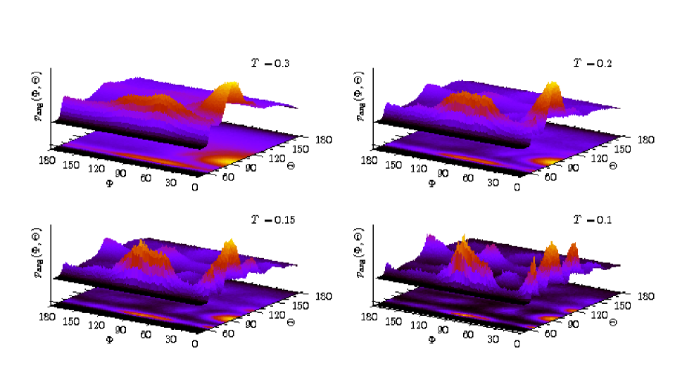
The folding process of proteins is necessarily accompanied by cooperative conformational changes. Although not phase transitions in the strict sense, it should be expected that one or a few parameters can be defined that enable the description of the structural ordering process. The number of degrees of freedom in most all-atom models is given by the dihedral torsional backbone and side-chain angles. In coarse-grained Cα models as the AB model used in this study, the original dihedral angles are replaced by a set of virtual torsional and bond angles. In fact, the number of degrees of freedom is not necessarily reduced in simplified off-lattice models. Therefore, the complexity of the space of degrees of freedom is comparable with more realistic models, and it is also a challenge to identify a suitable order parameter for the folding in such minimalistic heteropolymer models. On the other hand, the computational simplicity of these models allows for a more systematic and efficient analysis of the heteropolymer folding process. In Fig. 1, we show the probability distributions of all successive pairs of virtual bond angles and torsion angles [34] for the exemplified AB sequence S3 at several temperatures. This plot can be considered as the AB analogue of the Ramachandran map for real proteins. Although this representation is not appropriate to describe the folding process, which will be rather complicated for this example as described later on, a few interesting features can already be read off from this figure. At the temperature , we observe two domains in this landscape, i.e., a structural pre-ordering has already taken place. The distribution is noticeably peaked for bond angles around 90∘ and torsion angles close to 0∘, i.e., almost perfectly planar cis conformations are favored in the ensemble as well as segments with bond angles between 60∘ and 70∘ for a broad distribution of torsion angles mainly between 40∘ and 100∘. The reason for the large width of the torsion-angle distribution in this region is that the temperature is still to high for fine-structuring within the conformations. Explicit torsional barriers might stabilize these segments even at this temperature but are disregarded in the model. Decreasing the temperature down to , we see that the landscape of this accumulated distribution of the degrees of freedom becomes very complex, and the peaks are much sharper. In fact, close to , we observe a conformational transition towards the formation of the ground states. Actually, the complexity of this landscape can be understood better when considering the folding channels in the following, where we will see that this heteropolymer exhibits metastability and therefore rather glassy behavior. A remarkable aspect is the formation of the peaks in the bond-angle distribution at low temperatures close to 60∘, 90∘, and 120∘, as these angles are typical base angles in face-centered cubic crystals. As this concerns only segments of the conformations, the conformational transition is actually not a crystallization. Concluding, distributions of degrees of freedom are not quite useful to describe the folding process. For this reason it is necessary to define a suitable effective system parameter [35, 36]. A useful choice will be discussed in the following.
In analogy to studies of the specific folding behavior in all-atom protein models [37, 38], we use here a generalized variant of the overlap order parameter as introduced in Ref. [12]. The idea is to define a simple and computationally low-cost measure for the similarity of two conformations, where the differences of the angular degrees of freedom are calculated [39]. In order to consider this parameter as kind of order parameter, it is useful to compare conformations of the actual ensemble with a suitable reference conformation , which is preferably chosen to be the global-energy minimum conformation. We define the overlap parameter as follows:
| (3) |
With and being the respective numbers of bond angles and torsional angles , the angular deviation between the conformations is calculated according to
| (4) |
where
Here we have taken into account that the AB model is invariant under the reflection symmetry . Thus, it is not useful to distinguish between reflection-symmetric conformations and therefore only the larger overlap is considered. Since and , the overlap is unity, if all angles of the conformations and coincide, else . It should be noted that the average overlap of a random conformation with the corresponding reference state is for the sequences considered close to . As a rule of thumb, it can be concluded that values indicate weak or no significant similarity of a given structure with the reference conformation.
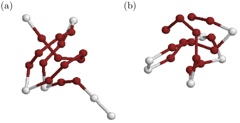
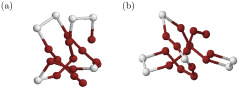
The global energy minimum conformations for the three sequences, which will be used as reference states in Eq. (3), are shown in Figs. 2(a), (b), and 3(a), respectively. The conformation rendered in Fig. 3(b) has a similar energy compared with the one in Fig. 3(a), but possesses a different geometry. This means that sequence S3 exhibits a kind of metastable behavior at low temperatures. The values of the lowest energies associated with the conformations in Figs. 2 and 3 are listed in Table 1. These minimum energies were identified within the multicanonical simulations and are in perfect agreement with previous results [12] from energy-landscape paving (ELP) optimizations [40].
For the qualitative discussion of the folding behavior it is useful to consider the histogram of energy and angular overlap obtained from the multicanonical simulations,
| (5) |
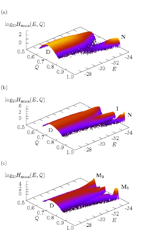
where the sum runs over all Monte Carlo sweeps . In Figs. 4(a)–(c), the multicanonical histograms are plotted for the three sequences listed in Table 1. Ideally, multicanonical sampling yields a constant energy distribution
| (6) |
An exemplified plot of the actual multicanonical distribution and the density of states for the sequence S1 is shown in Fig. 5, where is the multicanonical weight factor. In consequence, the distribution can suitably be used to identify the folding channels, independently of temperature. This is more difficult with temperature-dependent canonical distributions , which can, of course, be obtained from by a simple reweighting procedure, . Nonetheless, it should be noted that, since there is a unique one-to-one correspondence between the average energy and temperature , regions of changes in the monotonic behavior of can also be assigned a temperature, where a conformational transition occurs.
Interpreting the ridges of the probability distributions in Fig. 4 as folding channels, it can clearly be seen that the heteropolymers exhibit noticeable differences in the folding behavior towards the native conformations (N). Considering natural proteins it would not be surprising that different sequences of amino acids cause in many cases not only different native folds but also vary in their folding behavior. Here we are considering, however, a highly minimalistic heteropolymer model and hitherto it was not clear whether it would be possible to separate characteristic folding channels in this simple model, but as Fig. 4 demonstrates, in fact, it is. For sequence S1, we identify in Fig. 4(a) a typical two-state characteristics. Approaching from high energies (or high temperatures), the conformations in the ensemble D have an angular overlap with the lowest-energy reference state shown in Fig. 2(a), which means that there is no significant similarity with the reference structure, i.e., the ensemble D consists mainly of unfolded peptides. For energies a second branch opens. This channel (N) leads to the native conformation (for which and ). The constant-energy distribution, where the main and native-fold channels D and N coexist, exhibits two peaks noticeably separated by a well. Therefore, the conformational transition between the channels looks first-order-like, which is typical for two-state folding. The main channel D contains the ensemble of unfolded conformations, whereas the native-fold channel N represents the folded states.
The two-state behavior is confirmed by analysing the temperature dependence of the minima in the free-energy landscape. The free energy as a function of the “order” parameter at fixed temperature can be suitably defined as:
| (7) |
In this expression,
| (8) |
is related to the probability of finding a conformation with a given value of in the canonical ensemble at temperature . The formal integration runs over all possible conformations . In Fig. 6(a), the free-energy landscape at various temperatures is shown for sequence S1. At comparatively high temperatures (), only the unfolded states () in the main folding channel D dominate. Decreasing the temperature, the second (native-fold) channel N begins to form (), but the global free-energy minimum is still associated with the main channel. Near , both free-energy minima have approximately the same value, the folding transition occurs. The discontinuous character of this conformational transition is manifest by the existence of the free-energy barrier between the two macrostates. For even smaller temperatures, the native-fold-like conformations () dominate and fold smoothly towards the reference conformation, which is the lowest-energy conformation found in the simulation.
A significantly different folding behavior is noticed for the heteropolymer with sequence S2. The corresponding multicanonical histogram is shown in Fig. 4(b) and represents a folding event through an intermediate macrostate. The main channel D bifurcates and a side channel I branches off continuously. This branching is followed by the formation of a third channel N, which ends in the native fold. The unique -fold is plotted in Fig. 2(b). The characteristics of folding-through-intermediates is also confirmed by the free-energy landscapes as shown for this sequence in Fig. 6(b) at different temperatures. Approaching from high energies, the ensemble of denatured conformations D () is dominant. Close to the transition temperature , the intermediary phase I is reached. The overlap of these intermediary conformations with the native fold is about . Decreasing the temperature further below the native-folding threshold close to , the hydrophobic-core formation is finished and stable native-fold-like conformations with dominate (N).
The most extreme behavior of the three exemplified sequences is exhibited by the heteropolymer S3. The main channel D does not decay in favor of a native-fold channel. In fact, we observe both, the formation of two separate native-fold channels M1 and M2. Channel M1 advances towards the fold as shown in Fig. 3(a) and M2 ends up in a completely different conformation with approximately the same energy, which is shown in Fig. 3(b). The spatial structures of these two conformations are noticeably different and their mutual overlap is correspondingly very small, . It should also be noted that the lowest-energy conformations in the main channel D have only slightly larger energies than the two native folds. Thus, the folding of this heteropolymer is accompanied by a very complex folding characteristics. In fact, this multiple-peak distribution near minimum energies is a strong indication for metastability. A native fold in the natural sense does not exist, the conformation is only a reference state but the folding towards this structure is not distinguished as it is in the folding characteristics of sequences S1 and S2. This explains also, why the bond- and torsion-angle distribution in Fig. 1 possesses so many spikes: it represents rather the ensemble of amorphous conformations than a distinct footprint of a distinguished native fold. The amorphous folding behavior is also seen in the free-energy landscapes in Fig. 6(c). Above the folding transitions () the typical sequence-independent denatured conformations with dominate (D). Then, in the annealing process, several channels are formed and coexist. The two most prominent channels (to which the lowest-energy conformations belong that we found in the simulations) eventually lead for to ensembles of macrostates with (M1), which are similar to the reference conformation shown in Fig. 3(a), and conformations with (M2). The lowest-energy conformation found in this regime is shown in Fig. 3(b) and is structurally different but energetically degenerate compared with the reference conformation.
Summary
The purpose of this paper is to show that even with a very simple coarse-grained model, the AB model [5], complex sequence-dependent folding behavior of hydrophobic-polar heteropolymers can be investigated. This is very advantageous, since folding studies of all-atom protein models with full force fields are very time consuming and a systematic, comparative study of folding channels for a set of mutants would presently be still not feasible with realistic efforts. Although a one-to-one correspondence with real proteins cannot be claimed employing such a minimalistic model, it is a useful tool for understanding general mechanisms of tertiary heteropolymer folding. For this purpose, we have performed multicanonical simulations for three exemplified hydrophobic-polar sequences. It should be noted that these sequences are a subset of 20-mers studied within a different context [32] and, therefore, were not designed for the present study. Nonetheless, we found surprisingly complex folding behaviors, which are, in fact, qualitatively comparable to known characteristics of bioproteins and synthetic peptides. Beside the typical two-state folding behavior of sequence S1, we also observed folding through an intermediate macrostate (sequence S2), as well as folding into metastable conformations (sequence S3).
Since the study of phase transitions in complex disordered systems such as, e.g., diluted ferromagnets, spin glasses, or structural glasses is successfully performed by means of simple models, we expect that also the understanding of conformational transitions of heteropolymers (which are intrinsically disordered by the nonhomogeneous sequence of hydrophobic and hydrophilic monomers [41, 42, 43]) can be advanced by studies of minimalistic models.
This work is partially supported by the DFG (German Science Foundation) under Grant contract No. JA 483/24-1. Some simulations were performed on the supercomputer JUMP of the John von Neumann Institute for Computing (NIC), Forschungszentrum Jülich under grant No. hlz11.
References
- [1] Onuchic, J. N., Luthey-Schulten, Z., & Wolynes, P. G. (1997) Annu. Rev. Phys. Chem. 48, 545.
- [2] Clementi, C., Maritan, A., & Banavar, J. R. (1998) Phys. Rev. Lett. 81, 3287.
- [3] Onuchic, J. N. & Wolynes, P. G. (2004) Curr. Opin. Struct. Biol. 14, 70.
- [4] Lau, K. F. & Dill, K. A. (1989) Macromolecules 22, 3986.
- [5] Stillinger, F. H., Head-Gordon, T., & Hirshfeld, C. L. (1993) Phys. Rev. E 48, 1469; Stillinger, F. H. & Head-Gordon, T. (1995) Phys. Rev. E 52, 2872.
- [6] Sorenson, J. M. & Head-Gordon, T. (1999) Proteins 37, 582.
- [7] Bachmann, M. & Janke, W. (2003) Phys. Rev. Lett. 91, 208105.
- [8] Bachmann, M. & Janke, W. (2004) J. Chem. Phys. 120, 6779.
- [9] Bachmann, M. & Janke, W. (2005) Comp. Phys. Comm. 169, 111.
- [10] Hsu, H.-P., Mehra, V., Nadler, W., & Grassberger, P. (2003) Phys. Rev. E 68, 021113.
- [11] Liang, F. (2004) J. Chem. Phys. 120, 6756.
- [12] Bachmann, M., Arkın, H., & Janke, W. (2005) Phys. Rev. E 71, 031906.
- [13] Clementi, C., Nymeyer, H., & Onuchic, J. N. (2000) J. Mol. Biol. 298, 937.
- [14] Li, L. & Shakhnovich, E. I. (2001) Proc. Natl. Acad. Sci. USA 98, 13014.
- [15] Koga, N. & Takada, S. (2001) J. Mol. Biol. 313, 171.
- [16] Kaya, H. & Chan, H. S. (2003), J. Mol. Biol. 326, 911.
- [17] Kaya, H. & Chan, H. S. (2003), Phys. Rev. Lett. 90, 258104.
- [18] Schonbrun, J. & Dill, K. A. (2003) Proc. Natl. Acad. Sci. USA 100, 12678.
- [19] Head-Gordon, T. & Brown, S. (2003) Curr. Opin. Struct. Biol. 13, 160.
- [20] Jackson, S. E. & Fersht, A. R. (1991) Biochemistry 30, 10428.
- [21] Mayor, U., Johnson, C. M., Daggett, V., & Fersht, A. R. (2000) Proc. Natl. Acad. Sci. USA 97, 13518.
- [22] Ozkan, S. B., Bahar, I., & Dill, K. A. (2001) Nature Struct. Biol. 8, 765.
- [23] Karanicolas, J. & Brooks III, C. L. (2002) Protein Science 11, 2351.
- [24] Fersht, A. R. & Daggett, V. (2002) Cell 108, 1.
- [25] Kaya, H. & Chan, H. S. (2003) Proteins 52, 510.
- [26] Weikl, T. R. & Dill, K. A. (2003) J. Mol. Biol. 332, 953.
- [27] Weikl, T. R., Palassini, M., & Dill, K. A. (2004) Protein Science 13, 822.
- [28] Dill, K. A. & Chan, H. S. (1993) Proc. Natl. Acad. Sci. USA 90, 1942.
- [29] Chan, H. S., Shimizu, S., & Kaya, H. (2004) Methods Enzymol. 380, 350.
- [30] Berg, B. A. & Neuhaus, T. (1991) Phys. Lett. B 267, 249; (1992) Phys. Rev. Lett. 68, 9.
- [31] Janke, W. (1998) Physica A 254, 164; Berg, B. A. (2000) Fields Inst. Comm. 26, 1.
- [32] Irbäck, A., Peterson, C., Potthast, F., & Sommelius, O. (1997) J. Chem. Phys. 107, 273.
- [33] In the comparative multicanonical study of Ref. [12], the sequence S1 was denoted 20.1, S2 had the label 20.4, and S3 was 20.3.
- [34] For a polymer with monomers, there are bond and torsion angles. In order to form successive pairs of these angles, we have left out the last bond angle (counted from the first monomer in the sequence) in Fig. 1.
- [35] Du, R., Pande, V. S., Grosberg, A. Yu., Tanaka, T., & Shakhnovich, E. S. (1998) J. Chem. Phys. 108, 334.
- [36] Pande, V. S. & Rokhsar, D. S. (1999) Proc. Natl. Acad. Sci. USA 96, 1273.
- [37] Hansmann, U. H. E., Masuya, M., & Okamoto, Y. (1997) Proc. Natl. Acad. Sci. USA 94, 10652.
- [38] Berg, B. A., Noguchi, H., & Okamoto, Y. (2003) Phys. Rev. E 68, 036126.
- [39] Another, frequently used similarity measure is the root mean square deviation (rmsd) of two geometries. Since the rmsd calculation requires an optimization of the relative positions of the main axes, this procedure is computationally more demanding than the calculation of the overlap parameter used in this study.
- [40] Hansmann, U. H. E. & Wille, L. T. (2002) Phys. Rev. Lett. 88, 068105.
- [41] Wolynes, P. G. (1992) in Directions in Condensed Matter Physics, Vol. 6: Spin Glasses and Biology, ed. Stein, D. L. (World Scientific, Singapore), p. 225.
- [42] Pande, V. S., Grosberg, A. Yu., Joerg, C., & Tanaka, T. (1996) Phys. Rev. Lett. 76, 3987.
- [43] Pitard, E. & Shakhnovich, E. I. (2001) Phys. Rev. E 63, 041501.
List of figure captions
Fig. 1: (Color online) Bond and torsion angle distributions of sequence S3 at different temperatures. The distributions of the torsion angles are reflection-symmetric and therefore only the positive intervals are shown.
Fig. 2: (Color online) Lowest-energy reference conformations for sequences (a) S1 and (b) S2, both residing in the respective native-fold channels (N).
Fig. 3: (Color online) Lowest-energy conformations for sequence S3, considered as (a) reference conformation (M1) and (b) alternative metastable conformation (M2), whose angular overlap with is .
Fig. 4: (Color online) Multicanonical histograms of energy and angular overlap parameter for the three sequences (a) S1, (b) S2, and (c) S3. The different branches of these distributions are channels the heteropolymer can follow in the folding process towards the native state. The native folds are located in the right corner for and . Folding channels are labeled as D (denatured states), N (native folds), I (intermediates), and M (metastable states).
Fig. 5: Multicanonical energy histogram and density of states for sequence S1.
Fig. 6: Free energy as a function of the overlap parameter at four different temperatures for sequences (a) S1, (b) S2, and (c) S3. Note that the free energies are only determined up to a constant which was used to shift the curves for better discrimination. Labels of the free-energy minima refer to the folding channels in Fig. 4.


| label | sequence | global energy minimum | |
|---|---|---|---|
| S1 | |||
| S2 | |||
| S3 | , |