]Corresponding author: myqiang@nju.edu.cn
Structure and organization in inclusion-containing bilayer membranes
Abstract
Based on a considerable amount of experimental evidence for generic properties of lateral organization of lipid membranes containing inclusions, we first present a general model system of bilayer membranes embedded by nanosized inclusions, and account well for a series of unexpected behaviors in related experimental findings. (1) The appearance and disappearance of lipid/inclusion-rich rafts are observed with increasing the inclusion content. (2) The chain arrays of inclusions may form at high concentrations. (3) Location of inclusions changes with increasing the inclusion content, and may undergo a layering transition from one-layer located in the center of the bilayer to two-layer structure arranged in opposing leaflets of a bilayer. (4) The membrane fluidity is enhanced by the presence of inclusions. Our theoretical predictions address the complex interactions between membranes and inclusions, suggesting a unifying mechanism which reflects the competition between the conformational entropy of lipids favoring the formation of lipid-rich rafts and the steric repulsion of inclusions leading to the uniform dispersion. The present study advances our understanding of membrane organization by unifying these experimental evidences of real biomembranes with inclusions which can be different, but with the hydrophobic and rigid properties.
pacs:
87.16.Dg, 87.14.Cc, 87.68.+z, 64.75.+gRecently, there is growing evidence that due to the presence of inclusions within membranes, the distribution of lipids is inhomogeneous, where lateral segregation could induce the formation of lipid/inclusion-rich raft domains. For instance, cholesterol which is one of the most important regulators of lipid organization, prefers to have conformationally ordered lipid chains next to it due to its hydrophobically smooth and stiff steroid ring structureourit1 ; ne20 , and promotes the formation of lipid rafts(see, for example, reviewsourit1 ; ourit2 ; renew4 and recent research worksmmm4 ). On the other hand, it was reported ne9 that hydrophobic drugs such as taxol(paclitaxel)ne8 ; ne9 and dipyridamole(DIP)ne6 ) may assist the formation of lipid/drug-enriched raft domains, and increase with increasing the drug content, but disappear at high concentrations, which has also been observed in cholesterol-lipid systems renew5 . Particularly, the perturbed lipids may lead to chaining of cholesterols renew5 ; roger or drugs ne5 ; ne9 inside the bilayer. Furthermore, the introducing of taxol drug into the lipid layer will perturb the hydrocarbon chain conformation, which may increase membrane fluidityne5 ; ne8 ; ne9 . Most recently, some foreign inclusions such as silver nanoparticles were reported to have similar effects on the membrane fluiditya4 .
Despite the common properties of membrane organization due to distinct inclusions and despite its important significance in cellular functions such as signal transduction and membrane traffickingourit2 , the influence of the inclusions on such a change in lateral organization has not yet been considered in computational and theoretical investigations, and further insight into the mechanisms behind general evidence from lipid-inclusion complexes remains poorschick5 . Previous theoretical works were concerned with the hydrophobic mismatch interaction between inclusionsbsmit ; brr ; bsd and the possible formation of lipid raftsourit1 ; schick5 . However, a detailed structural change with varying the inclusion content has not been systematically elucidated. In this letter, we examine a simple model that not only allows us to present a unifying description of these phenomena with varying inclusion concentrations, but also sheds light on physical mechanism behind membrane organization due to distinct inclusions such as intrinsic membrane protein, cholesterol, hydrophobic drug, or other bionanoparticles.
Consider a lipid bilayer membrane containing inclusions of radius R in an aqueous environment(Fig. 1). The volume of system is , where and are lateral membrane lengths under periodical boundary conditions, and is the size of system along the membrane normal direction. The bilayer membrane is composed of one type of lipids with two hydrophobic tails. The number of lipids is given by , where is the number density of lipids in one leaflet. The headgroup of lipids has the volume , and two equal-length tails composed of segments each are assumed as flexible Gaussian chainsgh . The segment has the volume and the length . Thanks to the smooth and rigid property of bioinclusions compared with the flexible lipidsourit1 ; renew4 ; bsmit , we assume inclusions as
hard-sphere nanoparticles. The concentrations of the head and tails of lipids are and , and the inclusions . The solvent molecules have the volume and the concentration a5 . Recently, the self-consistent field theory (SCFT) has been proven to be powerful in calculating equilibrium morphologies in polymeric systemsadd3 ; m3 ; em3 , while nanoparticles can be treated by density-functional theory (DFT) add1 to account for steric packing effects of particles. Interestingly, Thompson et al. add3 developed a hybrid SCFT/DFT approach to study mixtures of diblock copolymer and nanoparticles. On the other hand, the SCFT method is extended to successfully study the phase behavior of pure lipid systemsgh ; leer3 ; schick5 . Here, we will extend the hybrid SCFT/DFT approachadd3 to calculate the structural organization of the bilayer membrane in the presence of inclusions. The resulting free energy for the present system dfg is given by
| (1) | |||||
where , and . , , , , , and are the Flory interaction parameters between tail-head, tail-solvent, tail-inclusion, head-solvent, head-inclusion, and solvent-inclusion, respectively. , , , and are the local volume fractions of lipid tail, head group, inclusion, and solvent, and , , , and are corresponding self-consistent fields. ensures the incompressibility of the system, and stands for the inclusion center distribution. , , and are respective partition functions for lipid, solvent, and inclusionsadd3 . The last term in Eq. (1) is the nonideal steric interaction term add1 with the weighted inclusion density add3 . The fields and densities are then determined by minimizing the free energy in Eq.(1), and the resulting self-consistent equations can be solved numericallyadd3 . To reasonably describe the interactions of hydrophobic inclusions dispersed in bilayer lipids with hydrophilic heads and hydrophobic tails, we choose , , , , , and . The other parameters are fixed to be , , , , and . Here, the chosen was large enough, ensuring the solvent concentration at and .
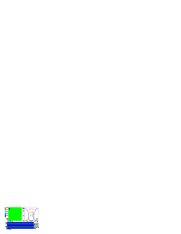
We first examine the case of a bilayer membrane in an aqueous environment in the absence of inclusions. Figure 2a shows the lateral distribution of lipid tails in one leaflet, which is uniform. The distribution is symmetrical with respect to the opposing leaflet. Figure 2b provides the average distribution of headgroups in the x-z cross-sections, reflecting that the shape of membrane surfaces is smooth in the absence of inclusions, since the lipid length is matched. In this case, the membrane is in a gel phase for the limited lateral mobilityrenew5 ; ne5 , where lipid tails tightly pack and hardly move in the pure membrane. Figure 2c shows the laterally averaged density profiles of , , and across a bilayer, which displays the basic structure of the bilayer and is favorably comparable with mesoscopic simulation and other coarse-grained models describing the bilayer compositionleer3 ; xe1 . This validates the used SCFT which can reasonably explore conformational properties of lipids, in contrast to phenomenological models that ignore much of the internal structure of the bilayerschick5 . Particularly, the approach will become powerful in exploring the lateral inhomogeneity of membrane with inclusions, in contrast to other field theory or simulation schemesschick5 , where the only one-dimensional density profiles along the membrane normal is shown in the cost of including molecular details of lipids.
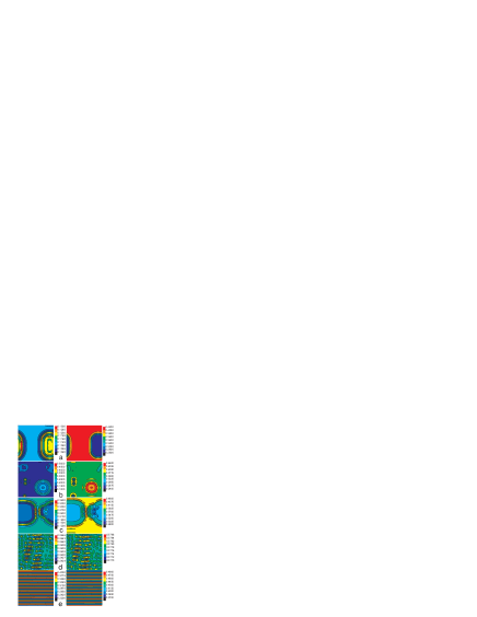

Figure 3 shows the in-plane density distributions of inclusions in the left column and the lipid tails in the right column with increasing the inclusion concentration . When is low, the translational entropy of inclusions has a significant contribution to the free energy of the system. Any compositional fluctuation of inclusions may lead to the lateral inhomogeneity of lipid composition, in which lipids are depleted in the inclusion-rich regions for ensuring the large translational entropy of inclusions(Fig. 3a). Astonishingly, as is increased, the lipid/inclusion-rich rafts appear(Fig. 3b). In this case, the entropic contribution of lipids becomes significant, and tail chains closing to the rigid surfaces of inclusions can get extra conformational flexibilityourit1 ; brr ; renew005 ; a4 , which enriches the lipids surrounding inclusions. Therefore, inclusions are accumulated in certain membrane regions, which leads to the formation of lipid-rich rafts. The raft size may be enlarged by increasing (Fig. 3c). Interestingly, as is added to a certain range(), lipid-rich rafts disappear, but instead the uniform distribution of both inclusion and lipid tail occurs (Fig. 3d). This unexpected behavior provides a strong support for the experimental findings in drug-membrane ne9 and cholesterol-membranerenew5 complexes. This is due to the strong steric repulsion from large numbers of inclusions, which leads to the uniform dispersion of inclusions. Further increase of leads to the deformation of lipids which produces the effective attraction between inclusions. The deformed conformational entropy can partially be released by chaining of inclusions under the lipid-mediated attraction, shown in Fig. 3e. Such a regularly modulated inclusion-rich stripe structure has been reported in the drug-membranene5 ; ne9 and cholesterol-membrane complexesrenew5 ; roger where the lipids form ribbons between the aligned cholesterol domains.
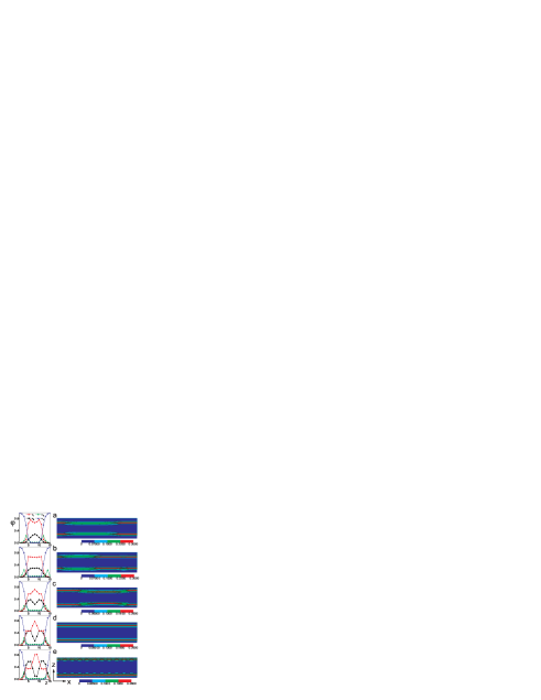
Figure 4a shows the translational entropy of inclusions. For low , the large increase of with indicates that plays an important role at first, which can account well for the formation of the weak inhomogeneous distribution of lipids(Fig. 3a) to ensure large translational entropy of inclusions. For large , decreases with the appearance of the chaining structure. Figure 4b shows the translational entropy of lipids. The curve goes up monotonously, meaning that the addition of inclusions increases the lateral membrane fluidity, which is a characteristic of gel-liquid transitionrenew5 ; ne5 . The reason is that lipid tails are stretched along the bilayer normal, which increases translational degrees of freedom and thus enhances the lateral mobility of lipidsourit1 . Figure 4c shows the conformational entropy of lipids. Beginning from a low conformational entropy in the gel phase of the pure membrane, lipids get more conformations with the perturbation of added inclusions where the lipid-enriched rafts are formed. However, there is a small decrease in the range with the disappearance of lipid rafts, where all lipids are strongly stretched with the same length. Figure 4d shows the steric repulsion energy of inclusions, which increases with increasing . Therefore, it is the steric repulsion between inclusions that suppresses lipid rafts. For high , the repulsion of inclusions is stronger, but at the same time, the deformed lipid-mediated attraction between inclusions becomes also stronger. The only way that the conformational entropy of deformed lipids is released(Fig. 4c), is to drive inclusions to assemble anisotropically along one direction. As a result, the deformed lipids provide an additional lateral anisotropic interaction between inclusions, which stabilizes the parallel chain arrays of inclusions.
Finally, Fig. 5 shows laterally averaged density profiles of different components across the bilayer in the left column and average density distributions of lipid heads in x-z cross-sections in the right column. For low (Fig. 5a), the inclusions assemble in the bilayer midplane for the membrane stability, which is displayed by one peak of density profiles () of inclusions. With increasing , one peak density profile may be saturated (Fig. 5b), and further increase of inclusions leads to the occurrence of two peaks of (Fig.5c-e), implying the relocation of inclusions where the two-layer distribution of inclusions is arranged in opposing leaflets of a bilayerrenew005 . Previous experimentne5 on drug-membrane complexes has also shown that the location of drugs in the bilayer depends on drug content. The two-layer distribution of inclusions ensures that the ends of lipid tails can still remain in the membrane midplane favoring the conformation of lipids. On the other hand, in Fig. 5a-c, the irregular density distribution of headgroups originates from the lipid chain-length mismatch, while in Fig. 5d, the membrane surfaces become flat with a matched length of strongly stretched lipids where lipid-rich domains disappear. By comparison of the distances between two peaks of head profiles () in the left side of Fig. 5, we also find that the thickness of membrane continuously increases with the addition of inclusions.
This work was supported by the National Natural Science Foundation of China, Nos. 10334020, 20674037, and 10574061.
References
- (1) O. G. Mouritsen, Life - as a matter of fat (Springer-Verlag, Berlin, 2005).
- (2) L. Miao et al., Biophys. J. 82, 1429(2002).
- (3) F. Maxfield and I. Tabas, Nature 438, 612(2005); K. Simons and W. Vaz, Annu. Rev. Biophys. Biomol. Struct. 33, 269(2004); H. McConnell and M. Vrljic, ibid 32, 469(2003); M. Edidin, ibid 32, 257(2003); D. Brown and E. London, Annu. Rev. Cell Dev. Biol. 14, 111(1998).
- (4) H. Ohvo-Rekila, et al., Prog. Lipid Res. 41, 66(2002).
- (5) K. Simons and E. Ikonen, Science 290, 1721(2000); T. Baumgart, S. Hess, and W. Webb, Nature, 425, 821(2003); S. Komura, et al. Europhys. Lett. 67, 011910(2003); H. McConnell and A. Radhakrishnan, Proc. Natl. Acad. Sci. USA 103, 1184(2006); R. Elliott, I. Szleifer, and M. Schick, Phys. Rev. Lett. 96, 098101(2006); S. Veatch and S. Keller, ibid 94, 148101(2005).
- (6) S. Feng, K. Gong, and J. Chew, Langmuir 18, 4061(2002).
- (7) C. Bernsdorff, R. Reszka, and R. J. Winter, J. Biomed. Mater. Res. 46, 141(1999).
- (8) P. Nassar, L. Almeida, and M. Tabak, Biochim. Biophys. Acta 1328, 140(1997); Langmuir 14, 6811(1998).
- (9) J. C. Lawrence et al., Biophys. J. 84, 1827(2003).
- (10) J. Rogers, A. G. Lee, and D. D. Wilton, Biochim. Biophys. Acta 552, 23(1979).
- (11) S. V. Balasubramanian and R. M. Straubinger, Biochemistry 33, 8941(1994).
- (12) S. H. Park et al., Colloids Surf. B: Biointerfaces 44, 117(2005).
- (13) M. Müller, K. Katsov, and M. Schick, Phys. Rep., 434, 113(2006), and references therein. This review has justified the recent applications of coarse-grained SCFT with Gaussian chain model to bilayer membrane.
- (14) M. Venturoli, et al. Phys. Rep. 437, 1(2006).
- (15) M. Sperotto, S. May, and A. Baumgaertner, Chemistry and Physics of Lipids 141, 2(2006); N. Dan, P. Pincus, and S. Safran Langmuir 9, 2768(1993).
- (16) R. Bruinsma and P. Pincus, Curr. Opin. Sol. St. Mater. Sci. 1, 401(1996); S. May, Langmuir 18, 6356(2002).
- (17) X. J. Li and M. Schick, Biophys. J. 78, 34(2000) compares the SCFT results of the lipid system with the experimental results, and a good agreement is found between them, while the real lipid is not completely flexible.
- (18) R. Elliott et al., J. Chem. Phys. 122, 044904-1(2005).
- (19) R. B. Thompson et al., Science 292, 2469(2001); Macromolecules 35, 1560(2002).
- (20) M. W. Matsen and M. Schick, Phys. Rev. Lett. 72, 2660(1994); F. Drolet and G. H. Fredrickson, ibid 83, 4317(1999); C. Ren and Y. Ma, J. Am. Chem. Soc. 128, 2733(2006).
- (21) F. Schmid, J. Phys.:Condens. Matter 10, 8105(1998); G. H. Fredrickson, The equilibrium theory of inhomogeneous polymers (Oxford University Press, Oxford, 2006); M. W. Matsen, in Soft Matter, eds. G. Gompper and M. Schick (Wiley-VCH, Weinheim, 2006), Volume 1.
- (22) P. Tarazona, Mol. Phys. 52, 81(1984); N. F. Carnahan and K. E. Starling, J. Chem. Phys. 51, 635(1969).
- (23) R. A. Kik, F. A. M. Leermakers, and J. M. Kleijn, Phys. Chem. Chem. Phys., 7, 19969(2005).
- (24) see Ref.em3 for technical details of the SCFT approach.
- (25) A. L. Frischknecht and L. J. D. Frink, Phys. Rev. E 72, 041924-1(2005); J. C. Shillcock and R. Lipowsky, J. Chem. Phys. 117, 5048(2002).
- (26) M. B. Sankaram and T. E. Thompson, Biochemistry, 29, 10676(1990).