Modulation of the Hydrophobic Effect through Confinement: A New Model for Chaperonin Action
Abstract
Despite the spontaneity of some in vitro protein folding reactions, native folding in vivo often requires the participation of barrel-shaped multimeric complexes known as chaperonins. Although it has long been known that chaperonin substrates fold upon sequestration inside the chaperonin barrel, the precise mechanism by which confinement within this space facilitates folding remains unknown. In order to address this question, we focused on the solvent of the folding reaction and investigated the effect of confinement within a chaperonin-like cavity on the configurational free energy of water. Using a simple theory for the thermodynamics of density and hydrogen bond order fluctuations in liquid water, we calculated solvent free energies for cavities corresponding to the different conformational states in the ATP-driven catalytic cycle of the prokaryotic chaperonin GroEL. Our findings suggest that one function of chaperonins may be to trap unfolded proteins and subsequently expose them to a micro-environment in which the hydrophobic effect, a crucial thermodynamic driving force for folding, is enhanced.
I Introduction
Efficient production of natively folded protein is an absolute requirement for cell survival. Although the amino acid sequence of a small polypeptide generally suffices to specify the chain’s native structure under conditions favorable to spontaneous folding in vitro, the protein folding problem in vivo is more challenging in several respects. To begin with, the cell must somehow protect the delicate homeostasis of its proteome against various environmental stresses that wreak havoc with the thermodynamics and kinetics of folding. Moreover, folding reactions that take place in the highly crowded intracellular milieu run the risk of never proceeding to completion because of the tendency of partially unfolded polypeptides to aggregate non-specifically and irreversibly at high concentrations. And even under more dilute conditions, a large protein that circuitously explores many non-native kinetic traps along the way to its native state may fold too slowly to be of any use in a biological context.
Proteins known as molecular chaperones enable the cell to overcome these obstacles Frydman2001chaprev ; Hartl2002chaprev . Chaperones are characterized by their ability to bind the exposed hydrophobic moieties of non-native polypeptides and guide these wayward chains back onto productive folding pathways. Perhaps the most intriguing type of chaperone is the chaperonin, a barrel-shaped multimeric complex that engulfs and releases its substrates in an ATP-dependent manner Horwich1998grorev ; Hartl2002chaprev ; Horwich2003grorev . Chaperonins sub-divide into groups I (eubacterial) and II (archaebacterial/eukaryotic), and the group I tetradecamer Hsp60/GroEL from E. coli is the most extensively studied model of chaperonin action. The open conformation of GroEL uses its largely hydrophobic inner surface to snare an unfolded substrate. Subsequent binding of ATP and the GroES co-factor induces a conformational change in GroEL, after which the substrate may attempt to fold inside a closed complex whose interior cavity surface displays many hydrophilic residues. Once ATP hydrolysis takes place, GroES dissociates from the complex and the barrel re-opens, releasing the substrate into the surroundings and completing the reaction cycle.
For some of its substrates, GroEL mainly provides an infinite-dilution environment in which a protein may fold without risk of aggregation. For others, however, it has been shown that confinement within the chaperonin barrel is accompanied by a marked increase in the rate of folding even under conditions that preclude aggregation Brinker2001twosubstr . The origins of this chaperonin foldase activity are still largely unclear, but recent experiments have begun to shed some light on the question Tang2006charge . Tang et. al. assayed the activity of a wide range of GroEL mutants and found that the acceleration of folding achieved by a chaperonin mutant correlated with the net charge and hydrophilicity of its interior cavity surface. This intriguing result suggests that the hydrophilic walls of the cavity somehow reshape the folding energy landscape of a confined protein to favor progression to the native state Horwich2003grorev . At present, the mechanism for this reshaping remains mysterious, and an explanation for how it might come about would undoubtedly do much to improve our understanding of chaperonin action.
This study aims to provide such an explanation by considering the role of water in folding under confinement. Folding reactions take place in aqueous solvent, and it is the strong tendency of water molecules to hydrogen bond with each other that generates the “hydrophobic effect” Chandler2005phobrev , a thermodynamic force that helps drive proteins to fold by causing non-polar amino acid residues to reduce their solvent-exposed surface area Spolar1989phob . Hydrogen bond quality is sensitive to the relative orientations of molecules, and numerous studies have documented substantial changes in the behavior of water once the liquid is confined in a small enough space that the organization of the hydrogen bond network becomes significantly constrained Raviv2001film ; Singh2006superphob ; Byl2006nanotube . It is therefore reasonable to hypothesize that the high degree of confinement experienced by water that participates in folding reactions inside GroEL may substantially alter the solvent free energy, and therefore the folding landscape, relative to what it would be in the cytosol.
Here, we investigate the capacity of a nanopore to modulate the free energy of water confined within it by undergoing shifts in surface chemistry that mimic the conformational changes in GroEL’s catalytic cycle. Using a phenomenological theory for the free energy of fluctuations in density and hydrogen bond order in bulk water, we compute the effect of confinement in hydrophilic and hydrophobic cavities on the “folding landscape” of a simple model protein. Our findings suggest that GroEL may be designed to preferentially bind unfolded substrates and allow them to fold in a micro-environment in which the hydrophobic effect is enhanced above cytosolic levels.
II Model
In order to examine the possible role of GroEL in remodeling the solvent of the folding reaction, we sought to develop an analytical framework for describing the thermodynamics of confined water. Microscopic theories of molecular liquids are notoriously difficult, especially for a molecule with water’s rare tendency to form extensive, tetrahedrally-coordinated networks of hydrogen bonds. For this reason, we chose to avoid microscopics in favor a phenomenological approach. Following past work Tanaka1998LG that attempted to explain the anomalous bulk properties of water using a Landau-like statistical field theory, we posited that the effective free energy of a fluctuation in the configuration of bulk water at a given temperature and chemical potential depends entirely on two order-parameters: the local density of the fluid and the local quality of the hydrogen bond network . Defining and to be the respective deviations of these quantities from their average values in the bulk, we can write the free energy of water for small configurational fluctuations in a volume as
| (1) |
The coefficients multiplying each term in the integral should, in principle, be functions of and , although we would expect the coupling between the density and hydrogen bond order to roughly equal the energy of a hydrogen bond. The derivative terms simply ensure that both order parameters not vary too rapidly over space, since they are only well-defined quantities on length scales larger than a water molecule. The squared terms ensure that larger deviations from bulk values of the order parameter carry higher free energetic costs, while the cross-term proportional to dictates that the formation of more hydrogen bonds lowers the energy of the system.
It is important to note here that because we did not include cubic and quartic terms in our free energy functional, we would be unable to use it to capture the trade-off between fluid density and hydrogen bond quality that leads to the anomalous decrease in the density of water as it cools to its freezing point. While such effects may, in fact, be important to adequately describing confinement effects in water, our initial analysis assumes for the sake of simplicity that the deviations from bulk behavior involved in the phenomenon in question are small enough that the quadratic functional written above will suffice.
Thus far, we have presented a simple theory of order fluctuations in bulk water. Before we can apply this theory to the study of confined water, however, we must augment it by introducing an additional contribution to the free energy functional that comes from the interaction between the liquid and the surface that encloses it. Thus, let our system consist of water confined within a shell of thickness , where is comparable to the size of a single water molecule. If is perfectly non-polar, then it cannot participate in hydrogen bonds and we must require that the contribution to the free energy due to hydrogen bonding that would be given by in the bulk must be zero in the shell. We may further introduce some potential that accounts for any additional effects from the surface chemistry of . Thus, we obtain a new term in our functional:
| (2) |
Here, is a parameter that reflects the degree of hydrophobicity of the surface, with corresponding to a completely non-polar surface.
To find the equilibrium density and hydrogen bond order, we minimize the total free energy with respect to the order parameter fields by using the calculus of variations. The equilibrium fields and will obey a system of Helmholtz equations that may be de-coupled by a change of basis. In the cases of spherically and planar symmetric systems, it is straightforward from there to obtain closed-form solutions whose integration constants must be set by plugging back into the functional and minimizing . For arbitrary cavity geometries and surface chemistries, however, it is necessary to seek a numerical solution. We therefore restrict our attention here to the analytically tractable cases, which are somewhat easier to deal with and nevertheless suffice as tools for examing the issues of interest in this study.
III Results
We modeled GroEL as a spherical pore of radius and the chaperonin substrate protein as a smaller sphere of radius . The chaperonin barrel was assumed to have two different states that reflected the open and closed conformations identified in structural studies of GroEL Braig2002groxtal . In the open state, the inner surface of the chaperonin was treated as a largely non-polar surface, meaning and . In the closed state, the chaperonin cavity was treated as being highly hydrophilic because of its richness in charged and polar residues that participate in hydrogen bonds with water molecules. Thus, we set and where accounts for the net attraction of favorably-oriented molecular electric dipole moments to the net charge of the cavity surface. The protein was also assumed to have two states: native and unfolded. The native conformation was assumed to have a hydrophilic surface chemistry described by , and the unfolded state was assumed to be partially hydrophobic, with and .
The solutions to the resulting Helmholtz equations were linear combinations of terms of the form , with the coefficients determined through minimization of the total free energy , and the correlation length scales set by the parameters of the theory. In order to plot these solutions, it was necessary to choose specific values for the parameters in the free energy functional. Since the questions we sought to answer were qualitative in nature, the precise parameter set used for the study was somewhat arbitrary. Indeed, we were unable to observe any qualitative change in the results from varying the coefficients so long as it was assured that bulk density and hydrogen bond order remained a free energy minimum. The constraints we ultimately made sure to satisfy were that the shell width and the characterstic correlation length scales for the order parameters were comparable to the size of a few water molecules and that the protein and chaperonin cavity were sized correspondingly.
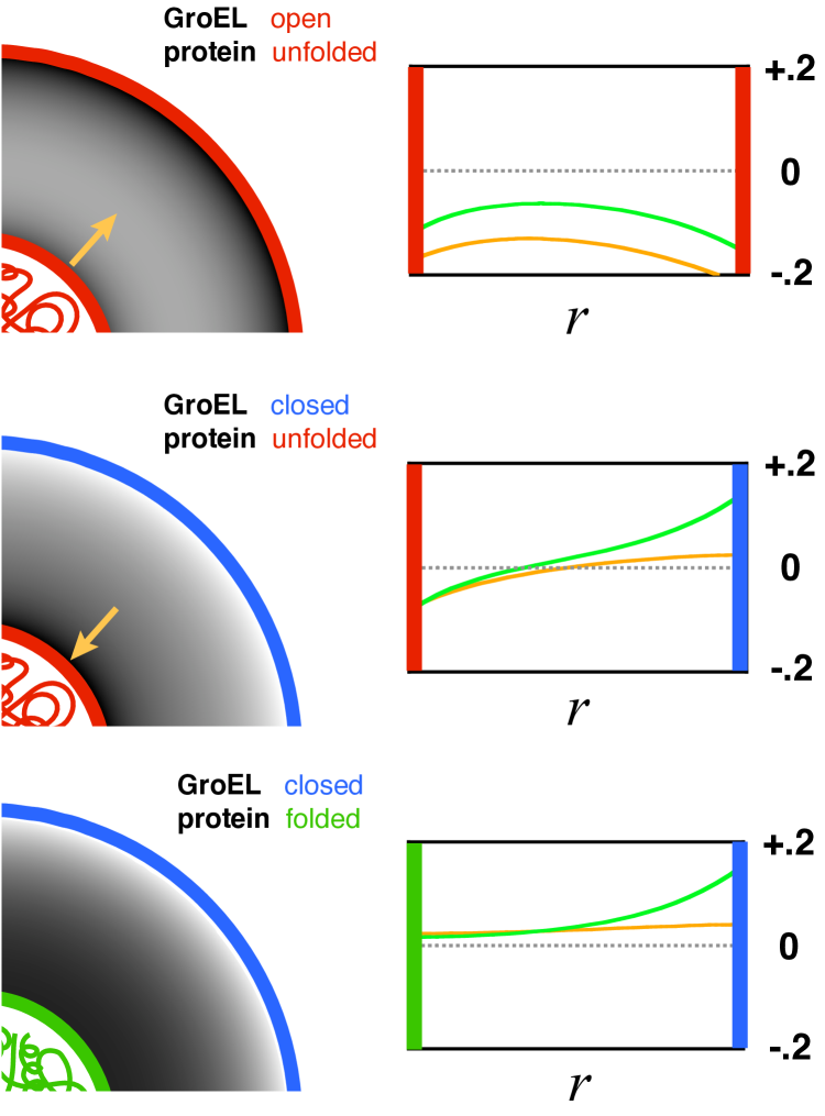
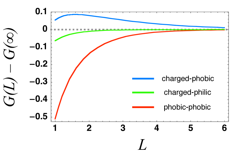
Figure 1 displays the equilibrium liquid density and hydrogen bond order profiles for the different states of GroEL and its substrate. For the case of a non-native substrate inside an open GroEL complex, both surfaces confining the solvent are hydrophobic (Fig. 1, top). As a result of the loss of hydrogen bonding at these surfaces, water withdraws from them and from the cavity as a whole, driving the average density below bulk levels. Moreover, it is apparent from a calculation of the free energy of solvent trapped between two planar surfaces of the same type that the force between the non-native substrate and the chaperonin wall is attractive (Fig. 2, red curve), and would favor binding. Once the chaperonin forms a closed complex and 89890undergoes its conformational change, however, the projection of charged and polar side-chains into the barrel’s interior causes the solvent order and resulting thermodynamic forces to change. With the inner surface of the barrel now highly hydrophilic, the solvent density is elevated near the chaperonin wall and depressed surrounding the unfolded protein (Fig. 1, middle). The result is a repulsive force between the surfaces that pushes the substrate towards the center of the cavity (Fig. 2, blue curve). Upon folding, the substrate surface becomes more hydrophilic, allowing the density of the nearby solvent to relax to bulk levels (Fig. 1, bottom) with the result that the force between chaperonin and substrate becomes very weak (Fig. 2, green curve).
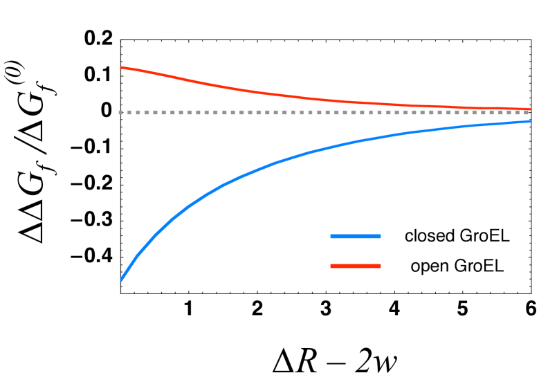
With equilibrium density and bond order profiles in hand, we were able to compute the solvent free energy changes for folding inside the different conformers of GroEL (Fig. 3). Within the framework of the model considered here, folding without the involvement of a chaperonin simply consists of a gradual reduction in hydrophobicity of the surface of a sphere of radius surrounded by aqueous solvent. In contrast, chaperonin-assisted folding divides into several stages. First, the hydrophobic interior of the barrel in its open conformation decreases its free energy by binding to hydrophobic moeities exposed by a partially unfolded substrate. Once the non-native protein is inside the cavity, the solvent free energy change associated with folding is made less negative by the surrounding hydrophobic walls, and the chaperonin therefore provides a thermodynamic drive towards unfolding that increases as the radius of confinement becomes smaller (Fig. 3, red curve). This effect comes about because the hydrophobic cavity walls deplete the amount of water in the cavity, making the cost of being unable to form hydrogen bonds at the protein surface lower than in bulk solution. After GroEL binds to GroES and ATP and undergoes its conformational change, however, the reverse is true: the solvent portion of the free energy of folding inside the cavity becomes more negative than that of folding in bulk solution, with the magnitude of the effect again increasing as the confinement becomes more severe (Fig. 3, blue curve). This dramatic shift takes place because the charged inner surface of the chaperonin has elevated the density of the water in the cavity, thereby raising the number of molecules who have difficulty finding a hydrogen bonding partner when hydrophobic substrate surface area is presented to the solvent. Thus, our model predicts that the conformational change in GroEL brings about a remodeling of the confined solvent that enhances the hydrophobic effect above its strength in bulk solvent and helps to drive the folding reaction to completion.
IV Discussion
Past attempts to explain the foldase activity of chaperonins have pointed to a diverse list of possible causes. While some researchers have focused on evidence that GroEL may assist in the unfolding of kinetically trapped substrates Betancourt1999groel ; Cheung2003nanopore , others see a role for the barrel-shaped cavity in accelerating folding by reducing the conformational entropy of a protein’s unfolded state Takagi2003confine , while still others propose that interactions between the protein and side-chains on the cavity wall might help to smooth out some of the pitfalls in the protein’s free energy landscape Baumketner2003chap ; Jewett2004chap . In this work, we examined a novel dimension of the chaperonin puzzle by studying the thermodynamics of the aqueous solvent in which the folding reaction takes place. By developing a simplified theory of order fluctuations in confined water, we were able compute the free energy of water in cavities that mimicked the salient features of a protein folding inside GroEL. Our findings underlined the potential importance of solvent effects in a complete description of how GroEL promotes folding.
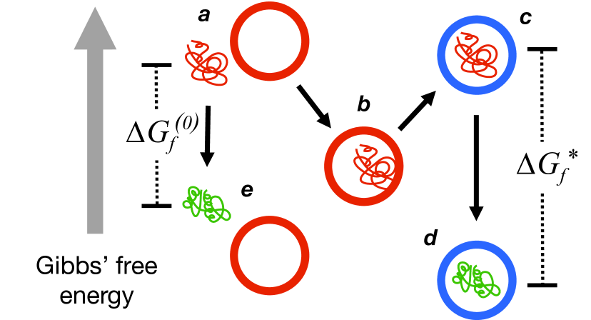
Figure 4 summarizes the model of chaperonin action that emerges from our study. A non-native protein that exposes hydrophobic groups to the bulk solvent will be engulfed by an open GroEL complex because of hydrophobic forces mediated by the solvent. At this point, the substrate becomes bound to the wall of the chaperonin. Once GroES and ATP bind, however, the solvent travels uphill in free energy as a conformational change in GroEL presents charged residues to the barrel’s interior. As a result, the substrate is forced away from the walls into the center of the cavity, a prediction that is consistent with experimentally measured increases in substrate fluorescence anisotropy following closing of the complex Tang2006charge . The subsequent folding reaction takes place inside a charged, hydrophilic chamber, and is therefore accompanied by a more drastic decrease in the free energy of the surrounding solvent. In other words, the interior of GroEL may provide a micro-environment in which the hydrophobic effect is substantially stronger than in bulk solution.
This finding leads us to propose a novel mechanism for the acceleration of folding by chaperonins. The hydrophobic effect is known to be a crucial contributor to the thermodynamic stability of natively folded proteins. However, several studies have also documented the importance of hydrophobic forces to folding kinetics Viguera2002phobstab ; Northey2002phobstab ; Calloni2003phobstab , both in terms of bringing about nucleation through hydrophobic collapse Northey2002phobstab and through stabilization of the folding transition state Viguera2002phobstab . It is therefore plausible that the stronger hydrophobic effects experienced by substrates inside closed GroEL complexes would hasten progression towards the native state. Thus, through its interaction with the confined solvent of the folding reaction, the chaperonin could reshape the free energy landscape in order to promote more rapid folding.
This hydrophobic enhancement model of chaperonin action provides us with a ready means to explain the puzzling relationship between foldase activity and cavity hydrophilicity reported in recent experiments. Our model assumes that charged groups on the interior of GroEL draw water into the barrel chamber, and that this elevation of solvent density leads directly to an enhancement of hydrophobic effects. The model therefore predicts that replacement of charged amino acid residues with neutral ones inside the GroEL complex should significantly reduce the chaperonin’s foldase activity, just as was observed in the recent mutational studies of Tang and co-workers Tang2006charge . Interestingly, the same study also reported a negative correlation between foldase activity and the volume of the GroEL folding chamber. Although this result was interpreted to be due to the reduction in conformational entropy of the unfolded protein that confinement causes, it is worth noting that since hydrophobic enhancement rises with the degree of confinement (Fig. 3), our model provides an alternative explanation for the outcome of those experiments as well.
Additional evidence pointing to charged surface residues as an important determinant of GroEL activity comes from the work of Wang et. al. Wang2002optgro , in which GroEL-GroES was optimized through directed evolution to fold green fluorescent protein (GFP) more rapidly. GFP is a small protein with an exceptionally well-buried hydrophobic core that would be likely to form more rapidly in response to enhancement of hydrophobic effects. The authors found that a key mutation for accelerating the folding of GFP was the replacement of an aromatic tyrosine on the interior surface of GroES with either arginine or histidine, both charged residues. These results are consistent with our proposal that charged cavity residues accelerate folding by strengthening the hydrophobic effect.
The existing evidence consistent with the mechanism of chaperonin foldase activity proposed here is substantial, but too indirect to be compelling by itself. Fortunately, our model’s most endearing quality is that it makes clear predictions that can easily be tested in silico and in the laboratory. At the most basic level, we would expect an all atom simulation of water and hydrophobic solutes inside a sufficiently hydrophilic cavity to show that hydrophobic forces between the solutes are enhanced by the confinement. We would also anticipate that a full-scale simulation of the closed GroE complex would show that water density inside the cavity was elevated (a prediction already confirmed by unpublished results), and that the degree of the corresponding elevation in simulations of previously characterized charged-to-neutral mutants would correlate roughly with their measured foldase activity. Finally, and most conclusively, we would predict that experiments that used Förster resonance energy transfer (FRET) or other techniques to measure the strength of hydrophobic forces between solutes inside GroEL would demonstrate that closure of the barrel through the binding of GroES and ATP is necessary and sufficient to enhance the hydrophobic effect inside the cavity.
The authors thank E. Miller, D. Kaganovich, D. Lucent for helpful comments J. England thanks the Fannie and John Hertz Foundation for support. This work was also supported by NIH NIGMS (R01 GM062868) and the NIH Nanomedicine Center for Protein Folding Machinery (PN1EY016525).
References
- (1) Frydman, J. (2001) Ann. Rev. Biochem. 70, 603–647.
- (2) Hartl, F. U & Hayer-Hartl, M. (2002) Science 295, 1852–1858.
- (3) Sigler, P. B, Xu, Z, Rye, H. S, Burston, S. G, Fention, W, & Horwich, A. L. (1998) Ann. Rev. Biochem. 67, 581–608.
- (4) Fenton, W. A & Horwich, A. L. (2003) Quart. Rev. Biophys. 36, 229–256.
- (5) Brinker, A, Guenther, P, Kerner, M. J, Naylor, D. J, Hartl, F. U, & Hayer-Hartl, M. (2001) Cell 295, 1852–1858.
- (6) Tang, Y, Chang, H, Roeben, A, Wischnewski, D, Wischnewski, N, Kerner, M. J, Hartl, F. U, & Hayer-Hartl, M. (2006) Cell 125, 903–914.
- (7) Chandler, D. (2005) Nature 437, 640–647.
- (8) Spolar, R, Ha, J, & Record, M. (1989) Proc. Natl. Acad. Sci. 86, 8382–8385.
- (9) Raviv, U, Laurat, P, & Klein, J. (2001) Nature 413, 51–54.
- (10) Singh, S, Houston, J, Swol, F. v, & Brinker, C. J. (2006) Nature 442, 526.
- (11) Byl, O, Liu, J, Wang, Y, Yim, W, Johnson, J. K, & Yates, J. T. (2006) J. Am. Chem. Soc. 101021, A–H.
- (12) Tanaka, H. (1998) Phys. Rev. Lett. 80, 5750–5753.
- (13) Braig, K, Otwinowski, Z, Hegde, R, Boisvert, D, Joachmiak, A, Horwich, A. L, & Sigler, P. B. (2002) Nature 371, 578–586.
- (14) Cheung, M & Thirumalai, D. (2003) J. Mol. Biol. 357, 632–643.
- (15) Betancourt, M & Thirumalai, D. (1999) J. Mol. Biol. 287, 627–644.
- (16) Takagi, F, Koga, N, & Takada, S. (2003) Proc. Natl. Acad. Sci. 100, 11367–11372.
- (17) Baumketner, A, Jewett, A, & Shea, J. E. (2003) J. Mol Biol. 332, 701–713.
- (18) Jewett, A. I, Baumketner, A, & Shea, J. E. (2004) Proc. Natl. Acad. Sci. 101, 13192–13197.
- (19) Viguera, A, Vega, C, & Serrano, L. (2002) Proc. Natl. Acad. Sci. 99, 5349–5354.
- (20) Northey, J. G. B, Di Nardo, A. A, & Davidson, A. R. (2002) Nat. Struc. Biol. 9, 126–130.
- (21) Calloni, G, Taddei, N, Plaxco, K, Ramponi, G, Stefani, M, & Chiti, F. (2003) J. Mol. Biol. 330, 577–591.
- (22) Wang, J. D, Herman, C, Tipton, K. A, Gross, C. A, & Weissman, J. S. (2002) Cell 111, 1027–1039.