Inhibition of DNA ejection from bacteriophage by Mg+2 counterions
Abstract
The problem of inhibiting viral DNA ejection from bacteriophages by multivalent counterions, specifically Mg+2 counterions, is studied. Experimentally, it is known that MgSO4 salt has a strong and non-monotonic effect on the amount of DNA ejected. There exists an optimal concentration at which the minimum amount of DNA is ejected from the virus. At lower or higher concentrations, more DNA is ejected from the capsid. We propose that this phenomenon is the result of DNA overcharging by Mg+2 multivalent counterions. As Mg+2 concentration increases from zero, the net charge of DNA changes from negative to positive. The optimal inhibition corresponds to the Mg+2 concentration where DNA is neutral. At lower/higher concentrations, DNA genome is charged. It prefers to be in solution to lower its electrostatic self-energy, which consequently leads to an increase in DNA ejection. By fitting our theory to available experimental data, the strength of DNADNA short range attraction energies, mediated by Mg+2, is found to be 0.004 per nucleotide base. This and other fitted parameters agree well with known values from other experiments and computer simulations. The parameters are also in aggreement qualitatively with values for tri- and tetra-valent counterions.
pacs:
81.16.Dn, 87.16.A-, 87.19.rmI Introduction
Most bacteriophages, or viruses that infect bacteria, are composed of a DNA genome coiling inside a rigid, protective capsid. It is well-known that the persistence length, , of DNA is about 50 nm, comparable to or larger than the inner diameter of the viral capsid. The genome of a typical bacteriophage is about 10 microns or 200 persistence lengths. Thus the DNA molecule is considerably bent and strongly confined inside the viral capsid resulting in a substantially pressurized capsid with internal pressure as high as 50 atm [Smith et al., 2001; Evilevitch et al., 2003; Castelnovo et al., 2003; Petrov, Lim-Hing, and Harvey, 2007]. It has been suggested that this pressure is the main driving force for the ejection of the viral genome into the host cell when the capsid tail binds to the receptor in the cell membrane, and subsequently opens the capsid. This idea is supported by various experiments both in vivo and in vitro [Letellier et al., 2004; Evilevitch et al., 2003; Black, 1989; Murialdo, 1991; Castelnovo et al., 2003; Purohit et al., 2005; Evilevitch et al., 2004, 2008]. The in vitro experiments additionally revealed possibilities of controlling the ejection of DNA from bacteriophages. One example is the addition of PEG (polyethyleneglycol), a large molecule incapable of penetrating the viral capsid. A finite PEG concentration in solution produces an apparent osmotic pressure on the capsid. This in turn leads to a reduction or even complete inhibition of the ejection of DNA.
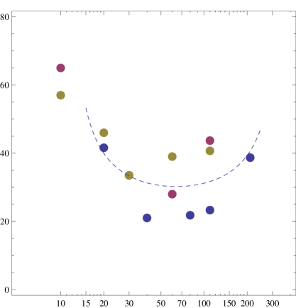
Since DNA is a strongly charged molecule in aqueous solution, the screening condition of the solution also affects the ejection process. At a given external osmotic pressure, by varying the salinity of solution, one can also vary the amount of DNA ejected. Interestingly, it has been shown that monovalent counterions such as NaCl have a negligible effect on the DNA ejection process [Evilevitch et al., 2003]. In contrast, multivalent counterions such as Mg+2, CoHex+3 (Co-hexamine), Spd+3 (spermidine) or Spm+4 (spermine) exert strong effect. In this paper, we focus on the role of Mg+2 divalent counterion on DNA ejection. In Fig. 1, the percentage of ejected DNA from bacteriophage (at 3.5 atm external osmotic pressure) from the experiment of Ref. Evilevitch et al., 2008; Fang, 2009 are plotted as a function of MgSO4 concentration (solid circles). The three colors correspond to three different sets of data. Evidently, the effect of multivalent counterions on the DNA ejection is non-monotonic. There is an optimal Mg+2 concentration where the minimum amount of DNA genome is ejected from the phages.
The general problem of understanding DNA condensation and interaction in the presence of multivalent counterions is rather complex, as evident by the large literature dedicated to this subject. This is especially true in the case of divalent counterions because many physical factors involved are energetically comparable to each other. Most studies related to DNA screening in the presence of divalent counterions have focused on ion specific effects. For example, in Ref. Evilevitch et al., 2008, hydration effects were proposed to explain the data of DNA ejection in the presence of MgCl2 salt where the minimum has not yet been observed for salt concentration upto 100 mM. In this paper, we focus on understanding the non-specific electrostatic interactions involved in the inhibition of DNA ejection by divalent counterions. We show that some aspects of the DNA ejection experiments can be explained within this framework. Specifically, we propose that the non-monotonic behavior observed in Fig. 1 has similar physical origin to that of the phenomenon of the reentrant condensation of macroions in the presence of multivalent counterions. It is the result of Mg+2 ions inducing an effective attraction between DNA segments inside the capsid, and the so-called overcharging of DNA by multivalent counterions in free solution.
Specifically, the electrostatics of Mg+2 modulated DNA ejection from bacteriophages is following. Due to strong electrostatic interaction between DNA and Mg+2 counterions, the counterions condense on the DNA molecule. As a result, a DNA molecule behaves electrostatically as a charged polymer with the effective net charge, per unit length, equal to the sum of the “bare” DNA charges, Å, and the charges of condensed counterions. There are strong correlations between the condensed counterions at the DNA surface which cannot be described using the standard Poisson-Boltzmann mean-field theory. Strongly correlated counterion theories, various experiments and simulations [Shklovskii, 1999; Grosberg, Nguyen, and Shklovskii, 2002; Moreira and Netz, 2002; Besteman, Eijk, and Lemay, 2007; Kanduč, Naji, and Podgornik, 2010] have showed that when these strong correlations are taken into account, is not only smaller than in magnitude but can even have opposite sign: this is known as the charge inversion phenomenon. The degree of counterion condensation, and correspoly the value of , depends logarithmically on the concentration of multivalent counterions, . As increases from zero, becomes less negative, neutral and eventually positive. We propose that the multivalent counterion concentration, , where DNA’s net charge is neutral corresponds to the optimal inhibition due to Mginduced DNA-DNA attraction inside the capsid. At counterion concentration lower or higher than , is either negative or positive. As a charged molecule at these concentrations, DNA prefers to be in solution to lower its electrostatic self-energy (due to the geometry involved, the capacitance of DNA molecule is higher in free solution than in the bundle inside the capsid). Accordingly, this leads to a higher percentage of ejected viral genome.
The fact that Mg+2 counterions can have such strong influence on DNA ejection is highly non-trivial. It is well-known that Mg+2 ions do not condense or only condense partially free DNA molecules in aqueous solution [Rau and Parsegian, 1992, Hud and Downing, 2001]. Yet, they exert strong effects on DNA ejection from bacteriophages. We argue that this is due to the entropic confinement of the viral capsid. Unlike free DNA molecules in solution, DNA packaged inside capsid are strongly bent and the thermal fluctuations of DNA molecule is strongly suppressed. It is due to this unique setup of the bacteriophage where DNA is pre-packaged by a motor protein during virus assembly that Mg+2 ions can induce attractions between DNA. It should be mentioned that Mg+2 counterions have been shown experimentally to condense DNA in another confined system: the DNA condensation in two dimension [Koltover, Wagner, and Safinya, 2000]. Recent computer simulations [Lee, Le, and Nguyen, 2010, Lyubartsev and Nordenskiöld, 1995] also show that if the lateral motion of DNA is restricted, divalent counterions can induced DNA condensation. The strength of DNADNA attraction energy mediated by divalent counterions is comparable to the results presented in this paper. These facts strongly support our proposed argument.
The dashed line in Fig. 1 is a fit of our theoretical result to the experimental data for MgSO4. The optimal Mg+2 concentration is shown to be mM. The Mgmediated attraction between DNA double helices is found to be /base ( is the Boltzmann constant and is the temperature of the system). As discussed later in Sec. IV, these values agree well with various known parameters of other DNA systems.
The organization of the paper as follows; In Sec. II, a brief review of the phenomenon of overcharging DNA by multivalent counterions is presented. In Sec. III, the semi-empirically theory is fit to the experimental data of DNA ejection from bacteriophages. In Sec. IV, the obtained fitting parameters is discussed in the context of various other experimental and simulation studies of DNA condensation by divalent counterions. Finally, we conclude our paper in Sec. V.
II Overcharging of DNA by multivalent counterions
In this section, let us briefly visit the phenomenal of overcharging of DNA by multivalent counterions to introduce various physical parameters involved in our theory. Standard linearized mean field theories of electrolyte solution states that in solutions with mobile ions, the Coulomb potential of a point charge, , is screened exponentially beyond a Debye-Hückel (DH) screening radius, :
| (1) |
The DH screening radius depends on the concentrations of mobile ions in solution and is given by:
| (2) |
where and are the concentration and the valence of mobile ions of species , is the charge of a proton, and is the dielectric constant of water.
Because DNA is a strongly charged molecule in solution, linear approximation breaks down near the DNA surface because the potential energy, , would be greater than in this region. It has been shown that, within the general non-linear meanfield Poisson-Boltzmann theory, the counterions would condense on the DNA surface to reduce its surface potential to be about . This so-called Manning counterion condensation effect leads to an “effective” DNA linear charge density:
| (3) |
In these mean field theories, the charge of a DNA remains negative at all ranges of ionic strength of the solution. The situation is completely different when DNA is screened by multivalent counterions such as Mg2+, Spd3+ or Spm4+. These counterions also condense on DNA surface due to theirs strong attraction to DNA negative surface charges. However, unlike their monovalent counterparts, the electrostatic interactions among condensed counterions are very strong due to their high valency. These interactions are even stronger than and mean field approximation is no longer valid in this case. Counterintuitive phenomena emerge when DNA molecules are screened by multivalent counterions. For example, beyond a threshold counterion concentration, the multivalent counterions can even over-condense on a DNA molecule making its net charge positive. Furthermore, near the threshold concentration, DNA molecules are neutral and they can attract each other causing condensation of DNA into macroscopic bundles (the so-called like-charged attraction phenomenon).
To understand how multivalent counterions overcharge DNA molecules, let us write down the balance of the electro-chemical potentials of a counterion at the DNA surface and in the bulk solution.
| (4) |
Here is the molecular volume of the counterion, is the counterion valency. is the electrostatic surface potential at the dressed DNA. Approximating the dressed DNA as a uniformly charged cylinder with linear charged density and radius , can be written as:
| (5) |
where and are Bessel functions (this expression is twice the value given in Ref. Winterhalter and Helfrich, 1988 because we assume that the screening ion atmosphere does not penetrate the DNA cylinder). In Eq. (4), is the local concentration of the counterion at the DNA surface:
| (6) |
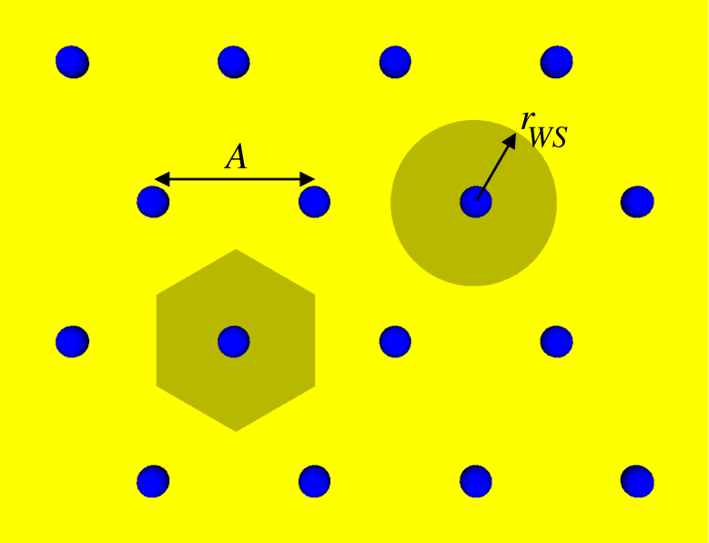
where is the bare surface charge density of a DNA molecule and the Gouy-Chapman length is the distance at which the potential energy of a counterion due to the DNA bare surface charge is one thermal energy . The term in Eq. (4) is due to the correlation energies of the counterions at the DNA surface. It is this term which is neglected in mean-field theories. Several approximate, complementary theories, such as strongly correlated liquid [Shklovskii, 1999; Perel and Shklovskii, 1999; Grosberg, Nguyen, and Shklovskii, 2002], strong coupling [Moreira and Netz, 2002, Kanduč, Naji, and Podgornik, 2010] or counterion release [Bruinsma, 1998, Gelbart et al., 2000] have been proposed to calculate this term. Although with varying degree of analytical complexity, they have similar physical origins. In this paper, we followed the theory presented in Ref. Grosberg, Nguyen, and Shklovskii, 2002. In this theory, the strongly interacting counterions in the condensed layer are assumed to form a two-dimensional strongly correlated liquid on the surface of the DNA (see Fig. 2). In the limit of very strong correlation, the liquid form a two-dimensional Wigner crystal (with lattice constant ) and is proportional to the interaction energy of the counterion with background charges of its Wigner-Seitz cell. Exact calculation of this limit gives [Grosberg, Nguyen, and Shklovskii, 2002]:
| (7) |
Here is the radius of a disc with the same area as that of a Wigner-Seitz cell of the Wigner crystal (see. Fig. 2). It is easy to show that for multivalent counterions, the so-called Coulomb coupling (or plasma) parameter, , is greater than one. Therefore, , and thus cannot be neglected in the balance of chemical potential, Eq. (4).
Knowing , one can easily solve Eq. (4) to obtain the net charge of a DNA for a given counterion concentration:
| (8) |
where the concentration is given by:
| (9) |
Eq. (8) clearly shows that for counterion concentrations higher than , the DNA net charge is positive, indicating the overcondensation of the counterions on DNA. In other words, DNA is overcharged by multivalent counterions at these concentrations. Notice Eq. (7) shows that, for multivalent counterions , is strongly negative for multivalent counterions, . Therefore, is exponentially smaller than and a realistic concentration obtainable in experiments.
Besides the overcharging phenomenon, DNA molecules screened by multivalent counterions also experience the counterintuitive like-charge attraction effect. This short range attraction between DNA molecules can also be explained within the framework of the strong correlated liquid theory. Indeed, in the area where DNA molecules touch each other, each counterion charge is compensated by the ”bare” background charge of two DNA molecules instead of one (see Fig. 3).
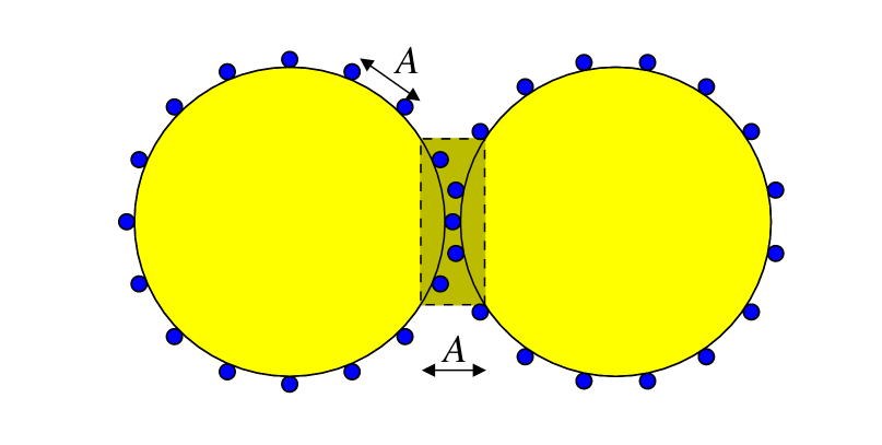
Due to this doubling of background charge, each counterion condensed in this region gains an energy of:
| (10) |
As a result, DNA molecules experience a short range correlation-induced attraction. Approximating the width of this region to be on the order of the Wigner crystal lattice constant , the DNADNA attraction per unit length can be calculated:
| (11) |
The combination of the overcharging of DNA molecules and the like charged attraction phenomena (both induced by multivalent counterions) leads to the so-called reentrant condensation of DNA. At small counterion concentrations, , DNA molecules are undercharged. At high counterion concentrations, , DNA molecules are overcharged. The Coulomb repulsion between charged DNA molecules keeps individual DNA molecules apart in solution. At an intermediate range of , DNA molecules are mostly neutral. The short range attraction forces are able to overcome weak Coulomb repulsion leading to their condensation. In this paper, we proposed that this reentrant behavior of DNA condensation as function of counterion concentration is the main physical mechanism behind the non-monotonic dependence of DNA ejection from bacteriophages as a function of the Mg+2 concentrations.
III Theoretical calculation of DNA ejection from bacteriophage
We are now in the position to obtain a theoretical description of the problem of DNA ejection from bacteriophages in the presence of multivalent counterions. We begin by writing the total energy of a viral DNA molecule as the sum of the energy of DNA segments ejected outside the capsid with length and the energy of DNA segments remaining inside the capsid with length , where is the total length of the viral DNA genome:
| (12) |
Because the ejected DNA segment is under no entropic confinement, we neglect contributions from bending energy and approximate by the electrostatic energy of a free DNA of the same length in solution:
| (13) |
where the DNA net charge, , for a given counterion concentration is given by Eq. (8). The negative sign in Eq. (13) signifies the fact that the system of the combined DNA and the condensed counterions is equivalent to a cylindrical capacitor under constant charging potential. As shown in previous section, we expect the to be a function of the multivalent counterion concentration and can be positive when . In the limit of strongly correlated liquid, is given in Eq. (9). However, the exponential factor in this equation shows that an accurate evaluation of is very sensitive to an accurate calculation of the correlation chemical potential . For practical purposes, the accurate calculation of is a highly non-trivial task. One would need to go beyond the flat two-dimensional Wigner crystal approximation and takes into account not only the non-zero thickness of the condensed counterion layer but also the complexity of DNA geometry. Therefore, within the scope of this paper, we are going to consider as a phenomenological constant concentration whose value is obtained by fitting the result of our theory to the experimental data.
The energy of the DNA segment inside the viral capsid comes from the bending energy of the DNA coil and the interaction between neighboring DNA double helices:
| (14) |
where is the average DNADNA interaxial distance.
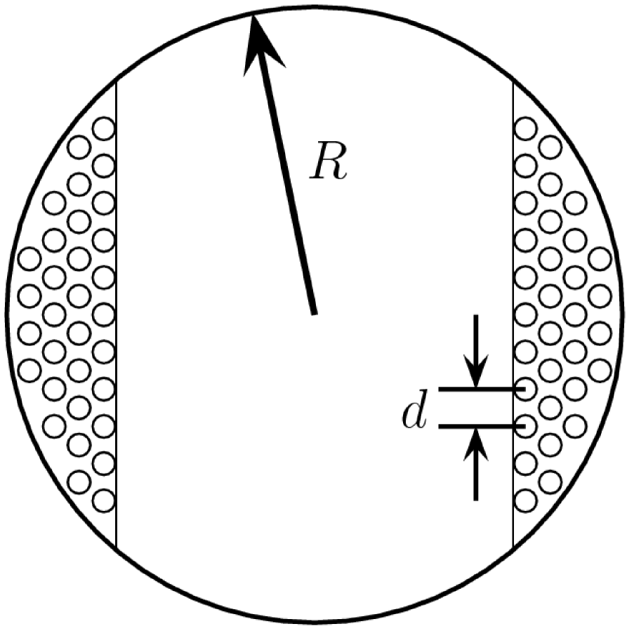
There exists different models to calculate the bending energy of a packaged DNA molecules in literature [Riemer and Bloomfield, 1978; Kindt et al., 2001; Purohit et al., 2005; Petrov, Lim-Hing, and Harvey, 2007; Petrov and Harvey, 2008]. In this paper, for simplicity, we employ the viral DNA packaging model used previously in Ref. Purohit et al., 2005, Riemer and Bloomfield, 1978, Kindt et al., 2001. In this model, the DNA viral genome are assumed to simply coil co-axially inward with the neighboring DNA helices forming a hexagonal lattice with lattice constant (Fig. 4). For a spherical capsid, this model gives:
| (15) |
where is the radius of the inner surface of the viral capsid.
To calculate the interaction energy between neighboring DNA segments inside the capsid, , we assume that DNA molecules are almost neutralized by the counterions (the net charge, of the DNA segment inside the capsid is much smaller than that of the ejected segment because the latter has higher capacitance). In the previous section, we have shown that for almost neutral DNA, their interaction is dominated by short range attraction forces. Hence, one can approximate:
| (16) |
Here, is the equilibrium interaxial distance of DNA bundle condensed by multivalent counterions. Due to the strongly pressurized viral capsid, the actual interaxial distance, , between neighboring DNA double helices inside the capsid is smaller than the equilibrium distance, , inside the condensate. The experiments from Ref. Rau and Parsegian, 1992 provided an empirical formula that relates the restoring force to the difference . Integrating this restoring force with , one obtains an expression for the interaction energy between DNA helices for a given interaxial distance :
| (17) |
where the empirical values of the constants and are pN/nm2 and nm respectively.
As we shown in the previous section, like the parameter , accurate calculation of is also very sensitive to an accurate determination of the counterion correlation energy, . Adopting the same point of view, instead of using the analytical approximation Eq. (11), we treat and as additional fitting parameters. In total, our semiempirical theory has three fitting parameters (, , ). From experimental data, we have three fitting constrains (the two coordinates of the minimum and the curvature of the curve in Fig. 1). Thus the theory does not contain unnecessary degrees of freedom.
IV Fitting of experiment of DNA ejection from bacteriophages and discussion
Equation (12) together with equations (13), (14), (15) and (17) provide the complete expression for the total energy of the DNA genome of our semi-empirical theory. For a given external osmotic pressure, , and a given multivalent counterion concentration, , the equilibrium value for the ejected DNA genome length, , is the length that minimizes the total free energy of the system, where
| (18) |
Here, is the volume of ejected DNA segments in aqueous solution. The specific fitting procedure is following. The energy of the DNA segment inside the capsid is minimized with respect to to acquire the optimal DNA-DNA interaxial distance for a given DNA ejected length, . Then, we substitute into Eq. (18) and optimize with respect to to obtain the equilibrium ejected length . By fitting with experiment data we can obtain the values for the neutralizing counterion concentration, , the Mg+2 mediated DNA-DNA attraction, , and the equilibrium DNA-DNA distance . The result of fitting our theoretical ejected length to the experimental data of Ref. Evilevitch et al., 2008 is shown in Fig. 1. In the experiment, wild type bacteriophages was used, so nm and m [Baker, Olson, and Fuller, 1999]. is held fixed at 3.5 atm and the Mg+2 counterion concentration is varied from 10 mM to 200 mM. The fitted values are found to be mM, per nucleotide base, and nm.
The strong influence of multivalent counterions on the process of DNA ejection from bacteriophage appears in several aspects of our theory and is easily seen by setting , thus neglecting the weak dependence of on and using Eq. (16) for DNA-DNA interactions inside the capsid. Firstly, the attraction strength appears in the expression for the free energy, Eq. (18), with the same sign as (recall that ). In other words, the attraction between DNA strands inside capsid acts as an additional “effective” osmotic pressure preventing the ejection of DNA from bacteriophage. This switch from repulsive DNA-DNA interactions for monovalent counterion to attractive DNA-DNA interactions for Mg+2 leads to an experimentally observed decrease in the percentage of DNA ejected from 50% for monovalent counterions to 20% for Mg+2 counterions at optimal inhibition (). Secondly, the electrostatic energy of the ejected DNA segment given by Eq. (13) is logarithmically symmetrical around the neutralizing concentration . This is well demonstrated in Fig. 1 where the log-linear scale is used. This symmetry is also similar to the behavior of another system which exhibits charge inversion phenomenon, the non-monotonic swelling of macroion by multivalent counterions [Skinner and Shklovskii, 2009].
It is very instructive to compare our fitting values for and to those obtained for other multivalent counterions. Fitting done for the experiments of DNA condensation with Spm+4 and Spd+3 shows to be 0.07 and 0.02 /base respectively [Rau and Parsegian, 1992, Nguyen, Rouzina, and Shklovskii, 2000]. For our case of Mg+2, a divalent counterion, and bacteriophage experiment, is found to be /base. This is quite reasonable since Mg+2 is a much weaker counterion leading to much lower counterion correlation energy. Furthermore, was found to be 3.2 mM for the tetravalent counterion, 11 mM for the trivalent counterion. Our fit of 64 mM for divalent counterions again is in favorable agreement with these independent fits. Note that in the limit of high counterion valency (), Eq. (9) shows that varies exponentially with [Shklovskii, 1999; Grosberg, Nguyen, and Shklovskii, 2002; Moreira and Netz, 2002]. The large increase in from 3.2 mM for tetravalent counterions to 11 mM for trivalent counterions, and to 64mM for divalent counterions is not surprising.
It is quantitatively significant to point out that our fitted value per base explains why Mg+2 ions cannot condense DNA in free solution. This energy corresponds to an attraction of per persistence length. Since the thermal fluctuation energy of a polymer is about per persistence length, this attraction is too weak to overcome thermal fluctuations. It therefore can only partially condense free DNA in solution [Hud and Downing, 2001]. Only in the confinement of the viral capsid can this attraction effect appear in the ejection process. It should be mentioned that computer simulations of DNA condensation by idealized divalent counterions [Lee, Le, and Nguyen, 2010, Lyubartsev and Nordenskiöld, 1995] show a weak short-range attraction comparable to our . The correlation induced DNADNA interaction obtained in the simulation of Ref. Lee, Le, and Nguyen, 2010 matches well with our value of . This suggests that in the presence of divalent counterions, electrostatic interaction are an important (if not dominant) contribution to DNADNA short range interactions inside viral capsid.
The phenomenological constants and depend strongly on the strength of the correlations between multivalent counterions on the DNA surface. The stronger the correlations, the greater the DNADNA attraction energy and the smaller the concentration . In Ref. Evilevitch et al., 2008, MgSO4 salt induces a strong inhibition effect. Due to this, for MgSO4 falls within the experimental measured concentration range and we use these data to fit our theory. MgCl2 induces weaker inhibition, thus for MgCl2 is larger and apparently lies at higher value than the measured range. More data at higher MgCl2 concentrations is needed to obtain reliable fitting parameters for this case. In fact, the value mM obtained from the computer simulation of Ref. Lee, Le, and Nguyen, 2010 is nearly twice as large as our semiempirical results. This demonstrates again that this concentration is very sensitive to the exact calculation of the counterion correlation energy . The authors of Ref. Evilevitch et al., 2008 also used non-ideality and ion specificity as an explanation for these differences. From our point of view, they can lead to the difference in , hence in the value . In the future, we plan to complimentary our phenomenological theory with a first principle calculation to understand the “microscopic” quantitative differences between MgSO4 and MgCl2 salts.
V Conclusion
In conclusion,this paper has shown that divalent counterions such as Mg+2 have strong effects on DNA condensation in a confined environment (such as inside bacteriophages capsid) similar to those of counterions with higher valency. We propose that the non-monotonic dependence of the amount of DNA ejected from bacteriophages has the same physical origin as the reentrant condensation phenomenon of DNA molecules by multivalent counterions. Fitting our semi-empirical theory to available experimental data, we obtain the strength of DNADNA short range attraction mediated by divalent counterions. The fitted values agree quantitatively and qualitatively with experimental values from other DNA system and computer simulations. This shows that in the problem of viral DNA package where DNA lateral motion is restricted, divalent counterions can plays an important role similar to that of counterions with higher valency. This fact should to be incorporated in any electrostatic theories of bacteriophage packaging. The strength of short-range DNA-DNA attractions mediated by MgSO4 salt is first obtained by the authors. It provides a good starting point for future works with DNA-DNA condensation in the presence of divalent counterions.
Acknowledgements.
We would like to thank Doctors Shklovskii, Evilevitch, Fang, Gelbart, Podgornik, Naji, Phillips, Rau, and Parsegian for valuable discussions. TTN acknowledges the hospitality of the Fine Theoretical Physics Institute and the Aspen Physics Center where part of this work was done. TTN acknowledges the support of junior faculty from the Georgia Institute of Technology. SL acknowledges financial support from Korean-American Scientists and Engineers Association (Georgia chapter).References
- Smith et al. (2001) D. E. Smith, S. J. Trans, S. B. Smith, S. Grimes, D. L. Anderson, and C. Bustamante, Nature 413, 748 (2001).
- Evilevitch et al. (2003) A. Evilevitch, L. Lavelle, C. M. Knobler, E. Raspaud, and W. M. Gelbart, Proc. Nat. Acad. Sci. USA 100, 9292 (2003).
- Castelnovo et al. (2003) M. Castelnovo, R. K. Bowles, H. Reiss, and W. M. Gelbart, Eur. Phys. J. E 10, 191 (2003).
- Petrov, Lim-Hing, and Harvey (2007) A. S. Petrov, K. Lim-Hing, and S. C. Harvey, Structure 15, 807 (2007).
- Letellier et al. (2004) L. Letellier, P. Boulanger, L. Plancon, P. Jacquot, and M. Santamaria, Front. Biosci. 9, 1228 (2004).
- Black (1989) L. W. Black, Annu. Rev. Microbiol. 43, 267 (1989).
- Murialdo (1991) H. Murialdo, Annu. Rev. Biochem. 60, 125 (1991).
- Purohit et al. (2005) P. K. Purohit, M. M. Inamdar, P. D. Grayson, T. M. Squires, J. Kondev, and R. Phillips, Biophys. J. 88, 851 (2005).
- Evilevitch et al. (2004) A. Evilevitch, M. Castelnovo, C. M. Knobler, and W. M. Gelbart, J. Phys. Chem. B 108, 6838 (2004).
- Evilevitch et al. (2008) A. Evilevitch, L. T. Fang, A. M. Yoffe, M. Castelnovo, D. C. Rau, V. A. Parsegian, W. M. Gelbart, and C. M. Knobler, Biophys. J. 94, 1110 (2008).
- Fang (2009) L. T. Fang, private communications (2009).
- Shklovskii (1999) B. I. Shklovskii, Phys. Rev. E 60, 5802 (1999).
- Grosberg, Nguyen, and Shklovskii (2002) A. Y. Grosberg, T. T. Nguyen, and B. Shklovskii, Rev. Mod. Phys. 74, 329 (2002).
- Moreira and Netz (2002) A. G. Moreira and R. R. Netz, Eur. Phys. J. E 8, 33 (2002).
- Besteman, Eijk, and Lemay (2007) K. Besteman, K. V. Eijk, and S. G. Lemay, Nature Physics 3, 641 (2007).
- Kanduč, Naji, and Podgornik (2010) M. Kanduč, A. Naji, and R. Podgornik, J. Chem. Phys. 132, 224703 (2010).
- Rau and Parsegian (1992) D. C. Rau and V. A. Parsegian, Biophys. J. 61, 246 (1992).
- Hud and Downing (2001) N. V. Hud and K. H. Downing, Proc. Nat. Acad. Sci. USA 98, 14925 (2001).
- Koltover, Wagner, and Safinya (2000) I. Koltover, K. Wagner, and C. R. Safinya, Proc. Nat. Acad. Sci. USA 97, 14046 (2000).
- Lee, Le, and Nguyen (2010) S. Lee, T. T. Le, and T. T. Nguyen, Phys. Rev. Lett. (Accepted for publication) (2010), arXiv:cond-mat/0912.3595 .
- Lyubartsev and Nordenskiöld (1995) A. P. Lyubartsev and L. Nordenskiöld, J. Phys. Chem. 99, 10373 (1995).
- Winterhalter and Helfrich (1988) M. Winterhalter and W. Helfrich, J. Phys. Chem. 92, 6865 (1988).
- Perel and Shklovskii (1999) V. I. Perel and B. I. Shklovskii, Physica A 274, 446 (1999).
- Bruinsma (1998) R. Bruinsma, Eur. Phys. J. B 4, 75 (1998).
- Gelbart et al. (2000) W. M. Gelbart, R. F. Bruinsma, P. A. Pincus, and A. V. Parsegian, Phys. Today 53, 38 (2000).
- Riemer and Bloomfield (1978) S. C. Riemer and V. A. Bloomfield, Biopolymers 17, 785 (1978).
- Kindt et al. (2001) J. Kindt, S. Tzlil, A. Ben-Shaul, and W. M. Gelbart, Proc. Nat. Acad. Sci. USA 98, 13671 (2001).
- Petrov and Harvey (2008) A. S. Petrov and S. C. Harvey, Biophys. J. 95, 497 (2008).
- Baker, Olson, and Fuller (1999) T. S. Baker, N. H. Olson, and S. D. Fuller, Microbiol. Mol. Biol. Rev. 63, 862 (1999).
- Skinner and Shklovskii (2009) B. Skinner and B. I. Shklovskii, Physica A 388, 1 (2009).
- Nguyen, Rouzina, and Shklovskii (2000) T. T. Nguyen, I. Rouzina, and B. I. Shklovskii, J. Chem. Phys. 112, 2562 (2000).