Effect of Bending Anisotropy on the 3D Conformation of Short DNA Loops
Abstract
The equilibrium three dimensional shape of relatively short loops of DNA is studied using an elastic model that takes into account anisotropy in bending rigidities. Using a reasonable estimate for the anisotropy, it is found that cyclized DNA with lengths that are not integer multiples of the pitch take on nontrivial shapes that involve bending out of planes and formation of kinks. The effect of sequence inhomogeneity on the shape of DNA is addressed, and shown to enhance the geometrical features. These findings could shed some light on the role of DNA conformation in protein–DNA interactions.
pacs:
87.15.-v, 87.15.La, 87.14.-g, 82.39.PjInteractions between DNA and proteins that cause deformations in the structure of DNA are essentially ubiquitous during many life processes inside cells Cell . For example, in eukaryotes the packing of DNA into nucleosome has been shown to lead to formation of sharp bends Luger97 . DNA packing in a viral capsid involves a high degree of confining and bending of viral genome inside a volume with dimensions that are comparable to the DNA persistence length Baker . DNA is also deformed by proteins during gene expression, when relatively short loops of DNA are formed Halford-andothers . It is known that the shape of DNA matters to its interaction with proteins such as RNA polymerase Hu-2006 , and that proteins locate their specific targets on DNA Cell . Therefore, it will be important to understand the role of mechanical effects such as tension, torsion, or bending and their couplings in determining the shape of DNA and their corresponding potential roles in positioning strategies Calladine . In addition, most cases of short genomes and plasmids have circular shapes in physiological conditions, which suggests that the exact shape of a circular short segment of DNA could be of significant biological implications Cell .
Conformational properties of relatively short DNA segments have been the subject of recent studies. These include experiments on loop formation and measurement of the persistence length in different scales Shore ; Cloutier04 ; Vologodskii05 ; Yuan08 , theoretical works on the probability of loop formation and efforts to interpret the findings of the experiments Shimada ; ZC-j ; Marko04 ; Ranjith05 ; Nelson05 ; Cocco05 ; Wiggins06 ; Spakowitz06 ; Popov07 ; Frey2007 , and molecular dynamics simulations lankas06 ; sarah . It is generally agreed that while modeling DNA as an isotropic elastic rod works perfectly for length scales larger the DNA persistence length ( nm), more elaborate models are needed to explain the conformational properties of shorter segments of DNA. These models should take account of various nonlinearities and structural properties of DNA elasticity that appear when shorter segments are subject to extreme constraints on bending and twisting Calladine ; sarah2 .
One of the nonlinear features that could affect the elasticity of DNA is the anisotropy in the effective bending rigidities corresponding to bending into the major and minor grooves. Such anisotropic bending elasticity models have been considered in studies of DNA segments of about 10 base pairs (bp) maha , and shown to predict formation of kinks and modulations in the curvature in 2D Farshid-03-05 . The anisotropy has also been shown to be responsible for some of the geometrical features observed in nucleosomal DNA Farshid-PRL05 . However, it is generally presumed to be unimportant when the DNA segment is long enough to have a few full helical turns, and in particular for segments of about 100 bp that have been the subject of recent controversy ZC-j ; Vologodskii05 .
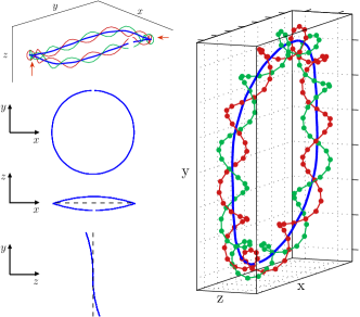
Here we study the three dimensional shape of short fragments of looped DNA using an elastic model that includes anisotropic bending rigidities. We show that unlike the commonly accepted picture, the anisotropy has a significant effect on the shape of DNA segments of about 100 bp. We find that the equilibrium shape of DNA loops could involve large changes in the local writhe and/or in the local curvature, which we generally call kinks. Figure 1 shows an example of a conformation with kinks (in the writhe). We also examine the effect of sequence dependence on the actual shape by incorporating varying bending rigidities within the region of permissible values found in recent molecular dynamics simulations. The sequence causes the DNA to soften/harden at some places, which offers the structure the possibility of lowering the overall energy by undergoing nontrivial deformations. The effect of the sequence-dependent conformations are probed using the distributions of local curvature, twist, and writhe, in the equilibrium shape.
To study the structure of looped DNA, we consider a simple model in which the molecule is represented as an anisotropic elastic rod. The rod is parametrized by the arclength , and at each point an orthonormal basis is defined with the unit vectors , , and , where corresponds to the direction from the minor groove to the major groove. The deformation of the double helix is characterized by the angular strains corresponding to bending in the plain perpendicular to and corresponding to twist. The elastic energy for the deformation of DNA in units of thermal energy () is written as Marko94 where and are the bending rigidities for the “hard” and “easy” axes of DNA cross section, is the twist rigidity, and is the intrinsic twist of B-DNA. In terms of Euler angles, we have , , and , where the dot denotes differentiation with respect to . In order to find the ground state of the system we should minimize this functional subject to the constraints in the system. For closed loops one of the constraints is to conserve the linking number, which can be expressed in terms of the Euler angles as Rudnick99 . Another constraint in the problem is a global vector constraint that guarantees a closed loop, and is written as . We also note that our Lagrangian is invariant under a translation by one half of the length of the loop. Therefore, we only focus on half of the loop, and demand that the tangent vectors at the two ends of the two halves are opposite to each other to ensure continuity. There is another important issue with regards to the linking number. In order to have a closed loop the two end base pairs should meet each other in phase, which means that the linking number for a closed loop is an integer. As a result, we might have to underwind or overwind the molecule to produce a loop. In the case of B-DNA, the pitch is nearly 10 bp long, and for example, a 94 bp fragment of DNA can form a loop if it is underwound by the spontaneous twist of 4 bp, or overwound by that of 6 bp, before the ends are joined up. It is not clear a priori which one is more favorable, and one should compare the energies of both solutions to decide that. Therefore we solve the Euler-Lagrange differential equations subject to the boundary conditions of , , , and , where is the linking number of the DNA or the number of DNA turns. Furthermore, we set just to fix the starting plane of the loop.
The equilibrium structure of the loop depends on the values of the elastic constants.While a direct experimental determination of the anisotropic bending rigidities is still lacking, a recent simulation suggests a range of values of nm and nm, depending on the sequence of the nucleotides lankas . In order to examine the effect of the anisotropy, here we consider a representative case of nm and nm, and compare it with the isotropic case with nm. Both of these choices will lead to a persistence length of nm, which can be calculated via LanLif . There are several suggested values for the DNA twist rigidity in the range of 50-110 nm Lankas2000 , and we consider a representative value of nm.
In order to decrease the elastic deformation energy, the bent anisotropic rod can explore non-planar configurations. To probe the 3D structure of the DNA, we calculate the values of the local curvature, defined as , the local writhe , and the local twist Rudnick99 . In Fig. 2, these geometric measures are plotted for half of the DNA loop for two lengths of 87 bp and 94 bp, for the anisotropic model. The curvature is normalized with the uniform curvature of an isotropic untwisted circled rod . Due to the anisotropy the local twist and the local curvature modulate around their mean values. Note that an increase in the curvature always coincides with a decrease in the twist, and vice versa. The local writhe achieves nonzero values, which indicates that the loop goes out of plane. The writhe could actually become very large at a singular point (owing to large values of ), which corresponds to the presence of a kink in the structure of the looped DNA, as shown in Fig. 1.
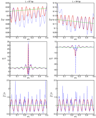
Sequence inhomogeneity of DNA causes variations in the local bending rigidities and along the DNA. The molecular dynamics simulation by Lankas et al. lankas suggests that sequence dependence can cause a change of 10-20 in the bending rigidities locally. To see the effect of sequence on the shape of short DNA loops, we consider a random sequence with a Gaussian distribution around the anisotropic bending rigidities used above with a width of 7 nm in the resulting persistence length around its mean value of 50 nm. In Fig. 2, the effect of the Gaussian random sequence on the local shape parameters has been shown. One can see that the curvature modulation in some places have been sharpened, presumably because there are softer places along the DNA available for accommodating the modulations that are needed for overall equilibrium. While some of the bends are sharp enough to be called kinks, the signature feature in the local writhe seems to be also present in a system with a random sequence. The bending kinks in the soft areas are accompanied by untwisting of the helix. This behavior can be important for some biological problems such as specific protein-DNA interactions, as it suggests that depending on the sequence, there might be a possibility for proteins to recognize the target location for an interaction. For example, the so-called AT-boxes are known to be relatively easier places for unwinding, and they are known to be the starting point for some protein functions. Our results suggest that the anisotropy can help the strands to be opened up more easily at softer sequences and can facilitate the searching process of proteins along the DNA.
| model | |||||||
|---|---|---|---|---|---|---|---|
| I | 87 | 8.91 | 0.09 | 8.90 | 1.01 | 1.03 | 36.6 |
| A | 87 | 8.90 | 0.10 | 8.88 | 1.04 | 1.36 | 36.4 |
| sd-A | 87 | 8.90 | 0.10 | 8.87 | 1.06 | 2.54 | 34.1 |
| I | 94 | 9.12 | -0.12 | 9.11 | 1.02 | 1.05 | 36.2 |
| A | 94 | 9.12 | -0.12 | 9.09 | 1.05 | 1.37 | 36.1 |
| sd-A | 94 | 9.12 | -0.12 | 9.08 | 1.07 | 2.17 | 34.6 |
It is also interesting to examine the effect of the anisotropy on the total writhe and the total twist of DNA. In our notation, they correspond to the average of their corresponding local values, namely, and , where the local quantities and are defined above. Table 1 shows the values for the total twist and writhe for two different DNA lengths of 87 and 94 bp. One can check that the sum of the writhe and the twist is constant for a given length, as required by White’s theorem White69 . Because in our examples the lengths of DNA are not integer multiples of the pitch, the molecule should be underwound or overwound to produce a loop. For the length of 94 bp, the energy of the underwound DNA is smaller than the overwound one. We note that the energy cost of changing the twist of the molecule is reduced by the formation of writhe, and that the absolute value of the total writhe becomes larger when the anisotropy increases. In other words, the system tries to minimize the energy by combining slight untwisting of the double strands with increased bending. Implementing this strategy locally causes harmonious modulations in the geometric measures of the DNA shape, as can be seen in Fig. 2. Table 1 also shows the extremal values of twist, and curvature for the homogeneous and random sequence cases, which shows that randomness can allow for significant enhancement in local geometrical features.
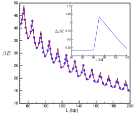
Figure 3 shows the total elastic energy of DNA loops of different lengths for the two choices of the bending elastic constants. The oscillations in the energy of the loop are because of the additional twist deformation that is needed to make a full loop (see above). We have calculated the energy for both over-twisted and under-twisted configurations in every case, and used the lower value in Fig. 3. For comparison, for the length of 94 bp the energies of over- and under-twisted states are found as and , respectively. One can see that the anisotropic system has a lower energy for all lengths, but the difference (as compared to the thermal energy ) is negligible. Introducing inhomogeneity in the bending rigidities (as in the example shown in Fig. 2) can lower the energy of the system significantly, as Table 1 shows. As the energy difference between the isotropic and anisotropic looped DNA is not large, we do not expect that anisotropy without sequence dependency can change the -factor significantly. In the example given in Fig. 2, sequence disorder can reduce the energy by , which could increase the -factor value by a factor of .
We find that both torsional and bending kinks will form in DNA loops to reduce the elastic energy cost by taking advantage of bending and torsion along the easy axis as much as possible. Atomistic modeling studies using MD simulations support the presence of kinks along highly deformed DNA lankas06 . The kink corresponds to a concentrated region of high curvature and/or torsion, and one wonders whether it might cause the hydrogen bonds to break in that area. We find that the maximum bending energy stored in a kink is per base pair for the sequence dependent anisotropic model, which is much smaller than the energy of the hydrogen bonds that is per base pair. Finally, we note that the elastic energy of the anisotropic model in the equilibrium conformation of the isotropic model is higher than its own ground state energy for the lengths used here. This means that the specific geometrical features of the anisotropic looped DNA are robust, and will not be blurred by thermal fluctuations.
In conclusion, we have studied the effect of bending anisotropy for bp segments of looped DNA, and found that while the anisotropy does not affect the overall elastic energy significantly, it can have drastic effects on the shape in the form of the emergence of kinks and unwindings. We also found a coupling between DNA sequence, conformation, and its energy, which could be important in the DNA cyclization probability and biological functions.
We are thankful to J. Gregory, M. Maleki, M. Kardar, G. I. Menon, and M. Rao for valuable discussions.
References
- (1) B. Alberts et al., Molecular Biology of the Cell, 4th ed. (Garland, New York, 2002).
- (2) K. Luger et al., Nature (London) 389, 251 (1997); T. J. Richmond and C. A. Davey, Nature (London) 423, 145 (2003).
- (3) T. S. Baker et al., Microbiol. Mol. Biol. Rev. 63, 862 (1999).
- (4) S. E. Halford et al., Annu. Rev. Biophys. Biomol. Struct. 33, 1 (2004); R. Schleif, Annu. Rev. Biochem. 61, 199 (1992); S. Adhya, Annu. Rev. Genet. 23, 227 (1989); H. G. Garcia et al., Biopolymers 85, 115 (2006).
- (5) T. Hu et al., Biophys. J. 90, 2731 (2006).
- (6) C. R. Calladine et al., Understanding DNA, the molecular & how it works (Elsevier Academic Press, 2004).
- (7) D. Shore et al., Proc. Natl. Acad. Sci. USA 78, 4833 (1981).
- (8) T. E. Cloutier and J. Widom, Molecular Cell 14, 355 (2004); Proc. Natl. Acad. Sci. USA 102, 3645 (2005).
- (9) Q. Du et al., Proc. Natl. Acad. Sci. USA 102, 5397 (2005).
- (10) C. Yuan et al., Phys. Rev. Lett. 100, 018102 (2008).
- (11) J. Shimada, H. Yamakawa, Macromolecules, 17, 689 (1984).
- (12) Y. Zhang and D. M. Crothers, Biophys. J. 84 136 (2003).
- (13) J. Yan and J. F. Marko, Phys. Rev. Lett. 93, 108108 (2004).
- (14) P. Ranjith et al., Phys. Rev. Lett. 94, 138102 (2005).
- (15) P. A. Wiggins et al., Phys. Rev. E 71, 021909 (2005).
- (16) N. Douarche and S. Cocco, Phys. Rev. E 72, 061902 (2005).
- (17) P. A. Wiggins and P. C. Nelson, Phys. Rev. E 73, 031906 (2006).
- (18) A. J. Spakowitz, Europhys. Lett. 73, 684 (2006).
- (19) Y. O. Popov and A. V. Tkachenko, Phys. Rev. E 76, 021901 (2007).
- (20) K. Alim and E. Frey, Phys. Rev. Lett. 99, 198102 (2007); Eur. Phys. J. E 24, 185 (2007).
- (21) F. Lankas et al., Structure 14, 1527 (2006).
- (22) S. A. Harris et al., Nucl. Acids Res. 36, 21 (2008).
- (23) T. B. Liverpool et al., Phys. Rev. Lett. 100, 238103 (2008).
- (24) A. Balaeff et al., Phys. Rev. Lett. 83, 4900 (1999).
- (25) F. Mohammad-Rafiee and R. Golestanian, Eur. Phys. J. E 12, 599 (2003); J. Phys. Condens. Matter 17, S1165 (2005).
- (26) F. Mohammad-Rafiee and R. Golestanian, Phys. Rev. Lett. 94, 238102 (2005).
- (27) J. F. Marko and E. D. Siggia, Macromolecules 27, 981 (1994); 29, 4820(E) (1996).
- (28) B. Fain, J. Rudnick, Phys. Rev. E 60, 7239 (1999); R. D. Kamien, Rev. Mod. Phys. 74, 953 (2002).
- (29) F. Lankas et al., Biophys. J. 85, 2872 (2003).
- (30) L. D. Landau and E. M. Lifshitz, Statistical Physics, Part 1 (Butterworth-Heinemann, Oxford, 1980).
- (31) F. Lankas et al., J. Mol. Biol. 299, 695 (2000); S. Neukirch, Phys. Rev. Lett. 93, 198107 (2004); V. Rossetto, Europhys. Lett. 69, 142 (2005).
- (32) J. H. White, Am. J. Math. 91, 693 (1969).