Rhythmogenic neuronal networks, pacemakers, and k-cores
Abstract
Neuronal networks are controlled by a combination of the dynamics of individual neurons and the connectivity of the network that links them together. We study a minimal model of the preBotzinger complex, a small neuronal network that controls the breathing rhythm of mammals through periodic firing bursts. We show that the properties of a such a randomly connected network of identical excitatory neurons are fundamentally different from those of uniformly connected neuronal networks as described by mean-field theory. We show that (i) the connectivity properties of the networks determines the location of emergent pacemakers that trigger the firing bursts and (ii) that the collective desensitization that terminates the firing bursts is determined again by the network connectivity, through k-core clusters of neurons.
pacs:
87.19.L-, 87.10.lj, 05.45.XtA neuronal network is a group of interconnected neurons functioning as a circuit. Each neuron receives electrical signals from a collection of tree-like dendrites, connected via synaptic junctions to the branched output terminals of other neurons. The neuron responds, based on some function of its input signals, by either doing nothing or by “firing,” i.e., by producing an action-potential output pulse that is received by other neurons Nelson:03 . In a network of excitatory “integrating” neurons, the electrical potential of the cell body of a neuron always increases by an amount when the cell receives a voltage input pulse. The potential of the cell effectively integrates the signals from other neuronal outputs. The firing probability of a neuron depends sensitively on the electrical potential of the cell, leading to threshold behavior in which the neuron can be considered to be either in a quiescent state characterized by sporadic firing if the cell potential is large and negative (“hyperpolarized”), or an activated state at higher potentials (“depolarized”), with more than an order of magnitude increase in firing rate over the quiescent state.
A classical example of an integrating neuronal network is the preBötzinger Complex (pBC) of about neurons located in the brain stem Feldman:90 ; Smith:91 . In this network, which collectively produces a rhythmic voltage signal that sets the timing of inspiration in mammals under resting conditions, each neuron is connected on average to one-sixth of the other neurons. The period of the current bursts is on the order of a second, which is about times longer than the time scale associated with repeated firing by activated individual neurons. The slow modulation is believed to be due to calcium-mediated “adaptation.” With each input pulse, the dentritic calcium concentration increases by an amount . The increase in calcium concentration leads to an increase in the leakage conductance between the dendrites and the surrounding medium, making the neuron’s somatic potential insensitive to incoming action potentials. When the somatic neuron potential drops below the threshold, it stops firing. After a recovery period during which the dendritic calcium relaxes back to its equilibrium value, the neuron once again begins integrating input signals.
Two different mechanisms have been proposed to explain the synchronization of the firing of the different neurons of the pBC. Neurons that in isolation can oscillate autonomously between cycles of firing and quiescence are known as pacemakers Butera:99 . In the individual pacemaker hypothesis, it is assumed that the pBC neurons are slaved to a small number of pacemakers believed to be present in the pBC. In the emergent pacemaker hypothesis (EPH), it is assumed that the oscillation is a collective property of a large group of neurons that would not oscillate in isolation DelNegro:08 . True pacemaker neurons, if present, only provide a back-up function and are not essential for the oscillation. The oscillations are in this case expected to disappear if the number of neurons drops below some threshold value. Indeed, when more than of these pBC neurons are destroyed in an in vitro experiment, the firing sequence changes from periodic oscillation to an increasingly complex pattern. This also happens when the excitability of the neurons is increased DelNegro:02 .
In this letter we show that the ability of a non-uniform neuronal network to collectively generate an oscillation, as assumed in the EPH, depends on general properties of the network connectivity, independent of the details of the model of neuron dynamics; moreover, these properties can be analyzed in terms of simple graph-theoretic methods Newman:03 ; Timme:06 . Specifically, we show that: (i) a network of identical but randomly connected neurons supports periodic synchronized bursts triggered by those neurons that are linked to a maximum number of other network neurons through a minimum number of links, and (ii) for highly excitable networks, the minimum number of neurons required for rhythmogenesis is determined a sequence of Magical Numbers, which are a function solely of the adjacency matrix.
We demonstrate these claims using a simple model for an excitatory neuronal network regulated by calcium-based adaptation. Each neuron is represented by two dynamical variables: the somatic potential and the calcium concentration of the neuron. The neurons fire according to the coupled non-linear rate equations:
| (1) | |||||
| (2) |
and are, respectively, the resting potential and the equilibrium calcium concentration of a neuron with (ms) and (ms) Koch:99 the respective equilibration times (the calcium concentration is thus the slow variable). Calcium-mediated adaptation is allowed for by assuming that drops rapidly when the calcium concentration exceeds a threshold . The time-sequence of firing events generated by a neuron is assumed to be a Poisson process with a voltage-dependent mean firing rate . For we will assume that if exceeds the threshold then increases from a basal rate of about five spikes per second to a high rate of about seventy-five spikes per second. Finally, the entries of the adjacency matrix are equal to one if the output of neuron is an input to neuron , and zero otherwise.
We start with the very simplest case of a homogeneous network where every neuron is linked to every other neuron in both directions: for all (known as a “clique”). If the initial potentials and calcium concentration also are the same for all neurons, then the rate equations reduce to a single pair that describes all neurons:
| (3) | |||||
| (4) |
which can be analyzed by the standard methods of dynamical systems Izhikevich:07 . The pair of equations can also be viewed as a “mean-field” approximation for more complex networks Bressloff:00 . The resulting dynamical phase diagram is shown in the upper panel of Fig. 1.
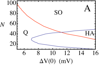
In the parameter regime marked SO (“stable oscillation”), the potential and calcium concentrations of the neurons undergo a stable limit-cycle oscillation. For lower values of the input voltage jump at zero calcium concentration , corresponding to weakly excitable neurons, the period of the oscillation increases and then diverges as the number N of neurons is reduced due to the appearance of an infinite-period saddle-node bifurcation Ermentrout:94 . A line of these bifurcations separates the SO phase from a quiescent phase, marked Q, where all neurons are permanently in a state of low activity. For higher values of , corresponding to highly excitable neurons, the unstable fixed point at the center of the limit cycle becomes stable as N is reduced. In the part of the phase diagram where this happens, marked HA, the neurons are permanently in a state of high activity. This mean-field HA phase does not show the complex firing pattern reported experimentally when the excitability was increased DelNegro:02 .
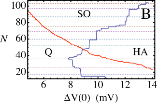
In actuality, each neuron of the pBC is believed to be linked to of the other neurons so the pBC network is not a clique. To describe this, we use an Erdős-Rényi random adjacency matrix Solomonoff:51 ; Erdos:59 , assigning zeros and ones as the entries of with probabilities and , respectively. Solution of the coupled rate equations on a single random graph produces the phase-diagram shown in the lower panel of Fig. 1. The heterogeneity of the network does not destroy its ability to produce robust, synchronized stable oscillations, though note that the SO section of the phase diagram has been reduced in area as compared to the mean-field case.
Unlike the mean-field case, the firing pattern now varies greatly from one neuron to the next. Superimposing the firing patterns of different neurons reveals an important feature (see Fig. 2A).
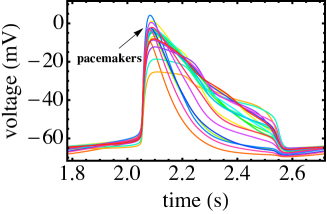
In the low-activity part of the cycle, the potentials of all neurons are below , but they rise more rapidly for a sub-population; these reach the firing threshold first. Their increased firing rate pushes sub-threshold neurons linked to them past the firing threshold as well. A chain reaction spreads over the network until all neurons are above threshold. Note that the least excitable neurons that crossed the firing threshold latest remain active over a longer period of time, producing a highly asymmetric pulse shape. Even though in our model all neurons are identical, and none can oscillate autonomously, a few neurons, selected through the network connectivity, are timing the oscillations. This subpopulation of spike leaders can be interpreted as the emergent pacemakers of the network. The other neurons effectively amplify their action.
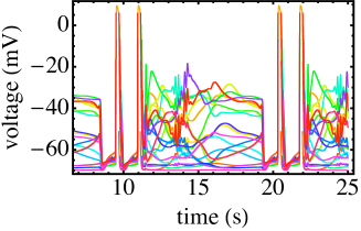
Network degradation, i.e., randomly knocking out neurons, leads to complex changes in the set of these emergent pacemaker neurons. It also takes longer for them to reach threshold as N is reduced so the oscillation period increases. For lower values of , i.e., weakly excitable networks, the period diverges along the phase-boundary between the SO and Q phases in Fig. 1B, which agrees with the predictions of mean-field theory. For higher values of the system enters the HA phase. Unlike in mean-field theory, the HA phase of the random network exhibits the complex dynamical behavior, with period doubling and deterministic chaos, that was reported experimentally DelNegro:02 . One example of this is shown in Fig. 2B where groups of high-activity bursts alternate with periods of deterministic chaos, forming a complex limit cycle.
The threshold curve has a surprising stair-case dependence on differing dramatically from the continuous curve predicted by the mean-field theory (see Fig. 1). These discontinuities define certain privileged numbers of neurons for which the network fails to support stable oscillations. The values of these ’s are independent of system parameters such as . The boundary of the SO regime makes discrete jumps between ’s as the parameters (, ) of the neuronal dynamical model are changed. In contrast, the phase boundary separating the SO and Q phases in Fig. 1B largely follows mean-field predictions.
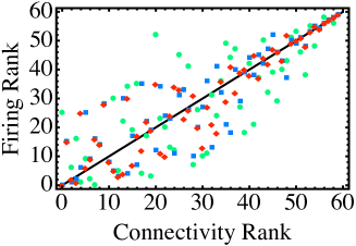
Both the selection of the pacemaker neurons and the values of the privileged number of neurons appear to be determined largely by the mathematical properties of the adjacency matrix independent of the details of our dynamical model. In Fig. 3 we show how one can identify the pacemaker neurons through their connectivity by ranking them according to the number of neurons connected to a given neuron by no more than three links. These pacemaker neurons do not, however, play a central role in the determination of the minimum number of network neurons able to support collective oscillations for highly excitable neuronal networks. While sufficiently active synaptic inputs to a pacemaker indeed will more quickly drive the dendritic calcium concentration past the threshold , so that it becomes desensitized and thus unable to spike, a small set of such desensitized and quiescent neurons will not drive an active network to collectively desensitize. Rather, one must have a system-spanning high-connectivity network capable of simultaneously desensitizing all of the neurons to quiet the inherently excitable system.
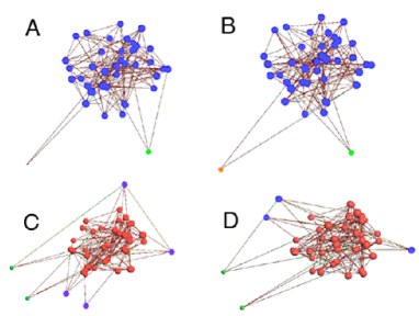
To quantify the size of a high-connectivity part of the network, it is useful to introduce the concept of a -core Dorogovtsev:06 . A -core of a graph (for integer ) is a subgraph in which all nodes (i.e. neurons) have at least inputs from other nodes in the subgraph. As the number of nodes increases in an Erdős-Rényi random network, -core clusters appear with larger values at sharply defined thresholds. As an example we show in Fig. 4A the -cores of a symmetric random adjacency matrix. Nearly all nodes form a single -core cluster. Adding one more node at random does not change this feature (Fig. 4B), but adding two nodes at random, so that produces a sharp transition in which the network is now dominated by a single, system-sized -core cluster (Fig. 4C). Adding an additional node to does not alter the dominance of the 5-core, as shown by Fig. 4D. For the random network used to generate Fig. 1B, these discontinuous transitions take place at , when a -core appears; at when a -core appears; at when a -core appears, and so on. The values of for this realization of the random network are represented by dotted lines in Fig. 1B. The locations of the discontinuities of the phase boundary as a function of agree well, though not perfectly, with the -core transition values . The discrepancies are presumably due to the fact that a member of a -core can have more than input links, including links from non -core neurons. We emphasize that the -core concept is inapplicable to the transition. Along the transition line, the few neurons with the highest connectivity are able to trigger an excitation wave that spreads through the whole system. These few emergent pacemakers are simply outliers having maximal connectivity and need not be part of a high -core.
In summary, we have presented a simple model for rhythmogenic neuronal networks, such as the pBC, using a combination of excitable integrate-and-fire neurons modified by a slower process of calcium-mediated desensitization. The most important conclusion of our work is that key features of the network dynamics - determination of the pacemaker neurons and determination of the minimal number of neurons that support stable oscillation - are determined (largely) by network connectivity. We also showed that in the phase diagram there is an asymmetry between the transition from the stable oscillation phase to the quiescent phase and the transition from the stable oscillation phase to the high activity phase. The first transition is well described by mean-field theory, while the staircase structure of the phase boundary of the second transition reflects the full nature of network connectivity. This asymmetry originates from the difference between the dynamics of a spreading wave of voltage-mediated excitation and collective, calcium-mediated desensitization. Tests of the model should be straightforward. The excitability of neurons can be increased in experiment, effectively controlling the size of the action potential in our model. Measuring the onset action potential for complex firing patterns as a function of the number of neurons should then directly reveal the predicted staircase structure of Fig. 1B.
We thank J. Feldman for enjoyable conservations and for sharing unpublished data on pBC dynamics.
References
- (1) P. Nelson, Biological Physics: Energy, Information, and Life, (W.H. Freeman & Co., New York, 2003).
- (2) J.L. Feldman, et al., Eur. J. Neurosci. 3, 171 (1990); J.L. Feldman and C.A. Del Negro, Nature Rev. Neurosci. 7, 232 (2006).
- (3) J.C. Smith, et al., Science 254, 726 (1991)
- (4) R.J. Butera Jr. et al., J. Neurophys. 82, 382 (1999).
- (5) C.A. Del Negro et al. in Integration in Respitory Control: From Genes to Systems eds. M.J. Poulin and R.J.A. Wilson (Springer, New York, 2008).
- (6) C.A. Del Negro, et al., Biophys. J. 82, 206 (2002).
- (7) M. Karoński, J. Graph Th., 6, 349 (1982); M.E.J. Newman, SIAM Rev. 45, 167 (2003).
- (8) M. Timme, Europhys. Lett. 76, 367 (2006).
- (9) C. Koch, Biophysics of Computation, (Oxford U. Press, New York, 1999); W.H. Nesse, et al., J. Comput. Neurosci. 25, 317 (2008).
- (10) E.M. Izhikevich, Dynamical Systems in Neuroscience: The Geometry of Excitability and Bursting (MIT Press, Boston, 2007).
- (11) P. Bressloff and S. Coombes, Neural Comp. 12, 91 (2000); G. Gigante, et al., Phys. Rev. Lett. 98, 148101 (2007).
- (12) G.B. Ermentrout and N. Kopell, SIAM J. Appl. Math. 54, 478 (1994).
- (13) R. Solomonoff and A. Rapoport, Bull. Math. Biol. 13, 1 (1951).
- (14) P. Erdős, A. Rényi, Pub. Math. 6, 290 (1959); P. Erdős, A. Rényi, Pub. Math. Inst. Hun. Acad. Sci. 5, 17 (1960); P. Erdős, A. Rényi, Acta Math. Si. Hun. 12, 261 (1961).
- (15) I. Alvarez-Hamelin, et al., Adv. Neural Inf. Process. Syst. 18, 41 (2006)
- (16) S.N. Dorogovtsev, et al., Phys. Rev. Lett. 96, 040601 (2006); S. N. Dorogovtsev et al., Rev Mod Phys. 80, 1276 (2008).