A model for the generation of localized transient elevations in vascular smooth muscle
Nicola Fameli,222Corresponding author. Address:
2176, Health Sciences Mall, Vancouver, B. C., Canada V6T 1Z3
tel 1-604-8226198; fax 1-604-8226012.
email: fameli@interchange.ubc.caKuo-Hsing Kuo, Cornelis van Breemen
Department of Anesthesiology, Pharmacology and Therapeutics
The University of British Columbia
Abstract
We present a stochastic computational model to study the mechanism of signalling between a source and a target ionic transporter, both localized on the plasma membrane (PM) and in intracellular nanometre-scale subplasmalemmal signalling compartments comprising the PM, the sarcoplasmic reticulum (SR), and transporters, and the intervening cytosol. We refer to these compartments, sometimes called junctions, as cytoplasmic nanospaces or nanodomains. In the chain of events leading to influx for SR reloading during asynchronous waves in vascular smooth muscle (VSM), the physical and functional link between non-selective cation channels (NSCC) and / exchangers (NCX) needs to be elucidated in view of two recent findings: the identification of the transient receptor potential canonical channel 6 (TRPC6) as a crucial NSCC in VSM cells and the observation of localized cytosolic transients following purinergic stimulation of these cells. Having previously helped clarify the signalling step between NCX and SERCA behind SR refilling, this quantitative approach now allows us to model the upstream linkage of NSCC and NCX. We have implemented a random walk (RW) Monte Carlo (MC) model with simulations mimicking a diffusion process originating at the NSCC within PM-SR junctions. The model calculates the average in the nanospace and also produces profiles as a function of distance from the source. Our results highlight the necessity of a strategic juxtaposition of the relevant signalling channels as well as other physical structures within the nanospaces to permit adequate build-up to provoke NCX reversal and influx to refill the SR.
Keywords: calcium oscillations, calcium signaling, sodium transient, vascular smooth muscle, sarcoplasmic reticulum, stochastic model, monte carlo, random walk, computational model, TRPC6, / exchanger.
1 Introduction
We are not used to thinking of ionic sodium () as a signalling ion, despite its known relevance for vascular disease. On the other hand, the importance of the second messenger in signalling cell function is undisputed. There is however recent experimental evidence supporting the idea that this important species has, at least in vascular smooth muscle cells (VSMC), an almost equally important signalling partner in [1, 2], particularly in signalling events that are allowed by the juxtaposition of signalplexes and transporter carrying membranes at nanometric distances from one another. This observation, and others outlined below, as well as a wealth of accumulated knowledge on and transporters in VSMC has prompted us to take a more quantitative look at the question of -related signalling in PM-SR cytoplasmic nanospaces (also referred to as nanodomains or junctions): nanometre-scale signalling compartments comprising the PM, the sarcoplasmic reticulum (SR), , and other ionic transporters (channels, exchangers and pumps) and relevant signalplexes therein, and the intervening cytosol.
In this article we present a quantitative model aimed at elucidating the mechanism of selective (or site-specific) signalling between a source ionic transporter and a target ionic transporter, both of which are localized on the same membrane and are part of a nanodomain. We developed this model using a stochastic method based on the simulation of ionic diffusion by random walks (RW) within nanospaces modeled according to experimental observations. Here we concentrate on the specific example of a transporter, typically a NSCC and a NCX as its target. Generally, during these events entry via a NSCC generates a large gradient, which, in turn, enables reversal of a NCX (presumably in the vicinity of the NSCC) and consequent NCX-mediated entry into the subplasmalemmal nanodomain. A few examples highlighting the biological importance of this intra-membrane system and its link to pathogenic mechanisms are (table 1 summarizes these cases): 1) A critical subplasmalemmal step in the signalling cascade giving rise to VSM cell contraction following G-protein coupled receptor adrenergic stimulation[3]—where blocking NCX reversal causes attenuation and elimination of oscillations due to impairment of SR refilling; replacement of oscillations with a tonic signal causes a dramatic decrease in smooth muscle force development[4]; in pulmonary artery SM, upregulation of the NCX and entry via reverse NCX action is considered one of the mechanisms behind elevated in idiopathic pulmonary arterial hypertension patients[5]; 2) Receptor activated entry may also induce NCX reversal in endothelial cells (EC) and this, in turn, gives rise to selective -stimulated eNOS activity and NO production; eNOS derived NO is an important physiological vasodilator agent and is accepted as an independent marker of vascular health[6, 7]; in EC NCX operating in forward mode is also important in regulating both and ; this suggests that local , besides the cell membrane potential and the equilibrium potential of the NCX, have a role in the regulation of forward NCX too[8, 9]; 3) In nerve terminals, a transient increase linked to a tetanic pulse of the action potential can reverse the NCX to induce presynaptic potentiation; this observation implies a role for the NCX in synaptic facilitation and has consequences for short term memory from reverse NCX malfunction[10, 2]; 4) In skeletal muscle NCX plays an important role in homeostasis, operating in forward as well as reverse mode in that function; / exchange is involved in the control of muscle fatigue and there are reports supporting the notion that the beneficial role of external in protecting slow-contracting soleus muscle against high-frequency fatigue depends mostly on influx through reversal of the NCX[11]; 5) although the role of NCX in heart is still poorly understood, both its -efflux and influx modes are observed in cardiac myocytes and and the latter is likely a consequence of subplasmalemmal elevations[12].
All of the above emphasize that modulation of signaling via and / exchange is of great clinical relevance in areas such as hypertension, chronic heart failure and possibly cerebral and skeletal muscle malfunction.
This study sheds new light on a few new scientifically interesting key factors regarding the “workings” of intracellular signalling nanospaces. We investigate the role, and importance, of having a confining surface, namely another lipid membrane, facing the membrane where the source and target transporters belong. This emerges as an important feature of nanodomains, as previous observations had hinted[13, 14], but it would appear that it alone cannot be entirely responsible for the generation of a sufficiently large local transient elevations. We find that to understand how sufficient can be built up within the nanodomain to trigger NCX reversal and signalling downstream, it is important to account for other factors such as the role of physical obstructions to diffusion and the possible organization of these obstructions.
| System | NCX mode/function | Function | References |
|---|---|---|---|
| VSM | rev/SR refilling | blood flow | [3] |
| during ACaW | |||
| PASM | rev/NCX upregulation | blood flow | [5] |
| EC | rev and fwd/eNOS activity | NO production regulation | [6] |
| nerve | rev/tetanic pulse | short memory | [10, 2] |
| of action potential | |||
| skeletal muscle | rev/ influx | muscle fatigue | [11] |
2 Methods
2.1 Electron microscopy
Details of the electron microscopy have been described previously[15]. The primary fixative solution contained 1.5% glutaraldehyde, 1.5% paraformaldehyde and 1% tannic acid in 0.1 M sodium cacodylate buffer that was pre-warmed to the same temperature as the experimental buffer solution (37 ∘C). The rings of rabbit IVC were fixed at 37 ∘C for 10 minutes, then dissected into small blocks, approximately 1 mm0.5 mm0.2 mm in size and put in the same fixative for 2 h at 4 ∘C on a shaker. The blocks were then washed three times in 0.1 M sodium cacodylate (30 min). In the process of secondary fixation, the blocks were post-fixed with 1% OsO4 in 0.1 M sodium cacodylate buffer for 1 h followed by three washes with distilled water (30 min). The blocks were then further treated with 1% uranyl acetate for 1 h (en bloc staining) followed by three washes with distilled water. Increasing concentrations of ethanol (25, 50, 70, 80, 90 and 95%) were used (10 min each) in the process of gradient dehydration. 100% ethanol and propylene oxide were used (three 10 min washes each) for the final process of dehydration. The blocks were infiltrated in the resin (TAAB 812) and then embedded in molds and polymerized in an oven at 60 ∘C for 8 h. The embedded blocks were sectioned on a microtome using a diamond knife at the thickness of 80 nm. The sections were then stained with 1% uranyl acetate and Reynolds lead citrate for 4 and 3 min, respectively. Images were obtained with a Phillips 300 electron microscope at 80 kV.
2.2 Simulations
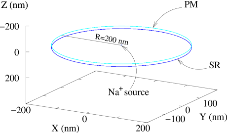
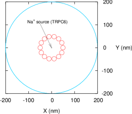
Simulations of diffusive motion from a source transporter are based on an implementation of a Monte Carlo random walk (RW), along the lines of methodology previously employed for transport simulations[14]. The model nanospace in which the simulations of diffusion take place arises from observed physical features and properties of the essential elements for nanodomain signalling. These are either obtained from our laboratory’s studies or from the available literature[16] and they are essentially the TRPC6 cytosolic ‘radius’ and [17], the typical dimensions of intracellular nanospace ultrastructure, estimates on the number of pillars, diffusivity in cytosol, expected necessary for NCX reversal (see section 3.1) and during localized transients (LNats) observed in [1]. Fig. 1 is a to-scale representation of the geometry of the model nanospace used in the simulations.
In the simulations, particles representing performs a RW on a cubic lattice with spacing nm; initially picked as an approximation to the expected mean free path in water (in turn, as an estimate to the mean free path in cytosol), we also carried out tests for effects of varying this parameter in a previous article and revealed no substantial influence on the results[14]. The RW time step was chosen by running several simulations letting particles cover a predetermined straight line distance , and recording the number of RW steps taken to cover . From diffusion theory, the total time taken by a random walker in three dimensions to cover the distance is , where is the measured diffusivity of in muscle cytosol[18]. The quantity was our choice for and its value is s.
The boundary conditions in our simulations are as follows. At the PM and SR membranes we implemented reflecting conditions: ions arriving at one of those surfaces during their RW are reflected back into the nanospace. At the edges of the model nanospace conditions are perfectly absorbing: ions reaching the lateral boundary of the junction are considered absorbed by the cytosol external to the nanospace, lost from the junctional population and no longer contributing to the PM-SR nanospace concentration, . As explained later, we also positioned a number of obstacles (we refer to them as pillars) to motion spanning the distance between the membrane in the nanospace. Pillars behave like elastic scatterers for .
Simulation code is written and tested in the C programming language on a computer running a Linux operating system. After testing and troubleshooting, programs are recompiled and run in one of the WestGrid computing nodes[19]. The pseudo-random number generator we used is the algorithm gsl_rng_m19937 of the GNU Scientific Library[20, 21], since it has sufficient randomness and quality requirements for our purposes. The plots with simulation results were produced using the “freely distributed plotting utility” gnuplot[22].
3 Results
3.1 Model foundation
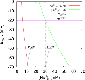
This laboratory has previously experimentally established that a transient from a NSCC enabled the generation within a PM-SR junctional nanospace of a sufficiently high gradient to cause NCX reversal[3]. A quantitative estimate of the level of such burst can be obtained by comparing the equilibrium potential of the NCX, , with the membrane potential, , using typical values for vascular smooth muscle cells. These quantities are linked by the equation (where ) between the electrochemical potential of (), of () and the membrane potential ()[9]. (In general, , where is the universal gas constant, the absolute temperature, is a -valent cation, and is Faraday’s constant.) NCX reversal occurs when . We can study this inequality for both resting and activated cell conditions by a plot like the one in Fig. 2. Using for an activated cell mV, mM, , mM, J/(mol K), K, and J/(V mol), we observe that, during activation, a transient of the order of 30 mM or greater is necessary to cause NCX reversal. In this exercise, we have used an estimated value for , as that is approximately the lowest value we expect from observations cited earlier[14].
Now, equipped with (a) the fundamental observation of localized elevation transients in full agreement with the values suggested by the study of Fig. 2[1], (b) better knowledge of the identity of NSCC as TRPC6[1, 23], (c) the basic idea that the presence of intracellular nanospaces is necessary for this signalplex to be complete, we propose a model to investigate the role of strategic placement of transporters with respect to each other, as well as of a confining membrane and other ionic diffusion limiting structures in the generation within these nanospaces of sufficiently high to permit NCX reversal.
The model nanospace used for the study is illustrated in Fig. 1. The dimensions expressed therein are based on high quality EM images showing that the PM-SR separation in these nanospaces is remarkably uniform and about 20 nm. Lateral extension of these closely apposed PM-SR regions is approximately 400 nm (see [14]).
3.2 Random walk simulations: bare PM-SR nanospace
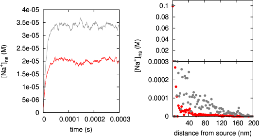
The simplest model nanodomain we studied consists of a shallow cylinder-shaped volume of height and radius , as in Fig. 1. entering the nanodomain via an NSCC are represented as particles doing a RW based on a given diffusivity , corresponding to that of in muscle cytoplasm as reported in [18]. For simplicity, there is only one source positioned at the centre of the PM side of the nanospace (in section 4 we will elaborate further on the issue of the number of sources). The simulation programs output the computed rise above resting level as a function of time, thereby giving an average increase in the nanospace. (From now on denotes the increase in cytoplasmic concentration in the nanodomain between PM and SR.) Results from this set of simulations are shown in Fig. 3, left panel.
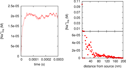
This plot helps establish the time scale after which we can consider that the has reached a steady-state level. We can observe from the graph that after approximately 100 the concentration has reached a plateau after having increased from zero during an initial transient, as we have explained in the previous section. Having established a time scale for the formation of the approximately maximum level of , we compute and study the concentration profile inside the junction, by plotting as a function of the distance from the source. The graph in the right panel of Fig. 3 illustrates a representative result.
3.3 Random walk simulations: randomly distributed obstacles in the nanospace
Evidently, this simplest incarnation of the model is inadequate to describe the generation of transients of the observed size[1], since at steady state elevation of hovers around M or about three orders of magnitude less than the observed values of 15–20 mM. We need to consider other nanospace features emerging from our ultrastructural images that may be responsible for a larger increase in the . Barring artificially changing the value of diffusivity, can be “forced” to increase if the ions were able to dwell longer in the nanospace than they are in the simple version of the system analyzed so far.
There is convincing evidence suggesting the existence of structures spanning the width of the nanospace and which could constitute an impediment to the free diffusion of [2, 24]. Our own observations confirm the existence of electron opaque “pillars” in transmission electron microscopy images like the one in Fig. 4. The size and abundance of these electron opaque structures compares well with the electron dense “bridges” observed by Devine and collaborators in the early ‘70s[24].

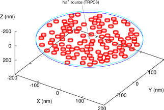
Keeping the overall geometry of the model nanospace the same (Fig. 1), we have therefore implemented a number of junction spanning structures in the form of cylindrical pillars, having estimated their size from several images like the one in Fig. 4. From those same images it is possible to approximate the percentage junctional volume occupied by those structures and, in turn, an approximate number of them expected per nanospace. In a series of simulations, we have represented up to 200 pillars randomly distributed within the nanospace (Fig. 5), and then simulated the diffusion by a random walk within the pillar-populated nanospace.
In this case too, we let the simulations run for a time sufficiently long to ensure that a steady-state level for the was established. Results are reported in Fig. 6. In all simulations involving random positioning of pillars, to minimize bias from the particular random pillar distribution, we have take average values of the computed over 10 different random pillar distributions. These are the values plotted in the graphs we present in this article.
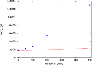
The results of this series of simulations indicate that having some form of impediment to ionic motion in the junction does produce the effect of increasing the and of changing its profile to one that decays more slowly with distance from the source. We ensured that this effect was not simply a consequence of the decreased nanospace volume due to the presence of the pillars by plotting the steady-state computed with different numbers of pillars in the junction (blue dots in Fig. 7) and comparing it with an increase in merely due to reducing the nanospace volume by the volume of the pillars (red line in Fig. 7). The plot in Fig. 7 demonstrates that ion collisions with pillars do indeed have a role in forcing to dwell longer in the nanospace.
3.4 Random walk simulations: non-randomly distributed obstacles
Clearly, the presence of obstacles to diffusion has an effect of increasing , however the values we obtain this way are not yet comparable with those measured during the local elevation transients. Other junctional features need to be accounted for in order to understand the mechanism giving rise to such high transients necessary to reverse the NCX and observed by Poburko and coworkers[1]. We considered the hypothesis that nature might place these obstacles “strategically” rather than randomly, in a neighbourhood of a source, so as to favour the generation of the gradients needed to drive the signalling chain. The rationale is that while a random set of pillars does show an ability to retain ions in the nanospace longer and therefore allow higher concentration build up, it does not do it efficiently enough to quantitatively account for the observed transients. We then ran some simulations in which pillars are placed in a circle around a source in such a way that we can control the porosity of this pillar fence to the passage of random walking ions. (Imagining to stand where the source is, the circle of pillars would appear as a 20-nm-high set of slabs surrounding the source itself, with thin rectangular gaps between the slabs; with this in mind, porosity is defined as the ratio of the entire surface area of the gaps to that of the slabs plus gaps.) The configuration of our model nanospace in this case is shown in Fig. 8, as a two dimensional ,-projection.
Representative results from these simulations are reported in Fig. 9.
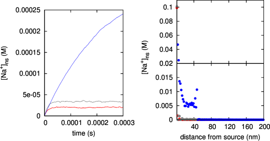
The dramatic increase in the in the vicinity of the source caused by this type of obstacle configuration is immediately evident. is in this case of the same order of magnitude as the observed localized transient elevation phenomenon[1]. Note furthermore that in this configuration the model suggests that the time for the to reach steady-state is much longer than in the case of random distribution of pillars. The more scattered data in the plot of the right panel in Fig. 9 are due to the fact that the system in this case has yet to reach equilibrium.
4 Discussion
The stochastic computational model presented herein attempts to give a quantitative description of the mechanism behind the observed localized transients observed in VSMC[1]. Based as much as feasible on experimental observations of the physical and physiological features of the intracellular nanospaces in which these transients are hypothesized to occur, the model results lead us to conclude that can build to sufficiently high values in PM-SR junctions to give rise to the observed transients.
Three main steps lead to the fundamental hypothesis behind this work. The first stems from earlier work by this laboratory elucidating the sequence leading to asynchronous oscillations in VSMC[1, 14]. Succinctly, a large external influx causes the reversal of the NCX and consequent entry to refill the SR after SR- release via IP3R channels upon cell stimulation provokes contraction. The second is the observation of two main features of the localized elevation transients[1]: they appear as a punctate pattern on the periphery of VSMC (with puncta having a given time course), and their peak values are comparable to the values necessary to cause NCX reversal (see Fig. 2 and relevant text). Thirdly, our previously published model supports the idea that NCX reversal-mediated entry in PM-SR nanospaces introduces sufficient to refill the SR during asynchronous waves[14]. Based on these three points, the hypothesis we set out to study with our model is that the observed localized transients occur in PM-SR nanodomains (or nanospaces or junctions), in other words, that in those junctions, due to influx via a TRPC6 channel, can attain levels, that cause entry via NCX reversal.
Simulation results from our simplest version of model (Fig. 3), namely, a PM-SR nanospace filled with cytosol (represented in our simulations by the diffusivity of ) with only one TRPC6 channel as source suggest that this simple view is not adequate to describe the formation of transients. This is mainly due to the large value of the diffusion coefficient of , which is about three times that of free in cytosol, and the fact that there is no observed buffering effect of in cytosol[18]. Recent measurements in isolated rabbit ventricular myocytes by Despa and Bers ([25, 26]) also suggest that the effective diffusivity of may be much slower than the one found by Kushmerick and Podolsky[18], although no explanation as to the mechanism behind it was suggested. In [25], among other things, Despa and Bers measured endogenous buffering in cytosol and found it negligible when compared to that of other important species like , for example. It is well known that buffering lowers its diffusivity dramatically[27, 28], thus possibly contributing to easier generation of gradients in confined spaces like the PM-SR nanospaces. Presuming the slight buffering effect observed in cardiac cell cytosol is mirrored in smooth muscle cells, it clearly could not provide sufficient slowing down of diffusivity to aid local transient creation. It seems instead ever more plausible that it must be some sort of physical obstruction to ionic motion that gives rise to a slower effective diffusivity for . We then considered that physical obstructions to ionic motion can have the overall effect of increasing the time spent by each in the nanospace thereby increasing the value of . Ever since first reported measurement of diffusivity in muscle cells[18], it was hinted that it is physical rather than chemical interactions that cause a retardation of the ionic (and non-ionic for that matter—sorbitol and sucrose specifically) intracellular diffusion. Our ultrastructural observations of nanospace spanning electron opaque structures support this idea (Fig. 4) and agree fully with earlier reports of similar structures in SMC[24]. The introduction of such physical obstruction in the simulations seems then more than warranted and very plausible. Moreover, if the “tortuosity factor”, introduced by Kushmerick, arising from physical interaction is indeed important in the bulk cytosol, then it would be even more so in the restricted PM-SR nanospaces. While the introduction of physical obstacles to ionic motion in our simulations does produce higher gains (Fig. 6 and 7), to obtain a quantitative agreement with the observed level of during the transients we need to hypothesize further that these junction spanning structures are not distributed at random, but rather form an organized barrier around a given source within the nanospace. Our simulation results in this case (Fig. 9) agree quantitatively with the observed transients, giving a of the same order of magnitude as the experimental measurements. The left panel plot in Fig. 9 also shows that the with this configuration of pillars will reach steady state with a time constant that is at least ten times longer than in the case of randomly distributed pillars. This is another feature supporting our hypothesis that these transients are formed in the PM-SR nanospaces with the aid of a relatively tight fence of pillars around the source. In fact, the measured transients are reported to be very long lived with a ramp up time to steady state of about 30 s, a time scale that the simpler versions of the model could not reproduce at all.
It therefore appears that two features are essential if these transients must occur within PM-SR nanospaces: there must be physical obstructions to motion, forming an organized barrier around each source (TRPC6 channels) and NCX must be localized near a TRPC6 within such barrier to be able to sense the high , reverse and allow entry. The second of these two features is suggested by a number of observations. There are studies supporting the idea that TRPC channels and exchangers are functionally and physically linked and form an important signalplex in several different systems[29]. Still other studies indicate that at least certain kinds of TRP channels are found in close proximity of caveolin[30, 31]. Combined with our earlier observations that NCX tend to crowd near the necks of caveolae[14], this makes a strong case for a physical association between TRP channels and NCX. The first of the above mentioned desirable features—patterns of physical obstructions within the nanodomains—is a harder matter to study experimentally and one of the current/future directions our laboratory is following.
To further study the issue of the localized transient duration requires more computationally demanding simulations, which we are currently tackling. We can however make a qualitative argument to suggest how the time scale of the observed transients can also be supported by our model. We have so far only implemented one source, or one TRPC6 channel. However, we have observed earlier that during activation of rabbit inferior vena cava SMC there could be about 16 NCX in the junctions[14]. If we conjecture that TRPC6 and NCX must be in physical proximity, as we speculated above, it is consistent to assume that there may be more than one TRPC6 in each junction functioning as a entry gate, and perhaps even as many as there are NCX.
Other transporters such as NCX and /K+ ATPases (NKA) certainly also play a role in shaping the PM-SR nanodomain profiles that drive NCX-mediated entry during VSMC activation. In the simple model we presented in this article, we focussed on the role of TRPC6 since it is by far the larger capacity transporter of the TRPC6, NCX, NKA trio (millions of ions per second, hundreds per second and tens per second, respectively). This first approximations quantitative model supports the idea that localized elevation transients take place at PM-SR nanodomains and strongly suggests that physical impediment to ionic mobility of is also a necessary factor for their generation.
Acknowledgments
The research was supported by grants from the Canadian Institute of Health Research and the Heart and Stroke Foundation of British Columbia and Yukon. This project has been enabled by the use of WestGrid computing resources, which are funded in part by the Canada Foundation for Innovation, Alberta Innovation and Science, BC Advanced Education, and the participating research institutions. WestGrid equipment is provided by IBM, Hewlett Packard and SGI.
References
- [1] Damon Poburko, Chiu-Hsiang Liao, Virginia S Lemos, Eric Lin, Yoshiaki Maruyama, William C Cole, and Cornelis van Breemen. Transient receptor potential channel 6-mediated, localized cytosolic [na+] transients drive na+/ca2+ exchanger-mediated ca2+ entry in purinergically stimulated aorta smooth muscle cells. Circ Res, 101(10):1030–1038, Nov 2007.
- [2] Damon Poburko, Nicola Fameli, Kuo-Hsing Kuo, and Cornelis van Breemen. Ca2+ signaling in smooth muscle: Trpc6, ncx and lnats in nanodomains. Channels (Austin), 2(1):10–12, 2008.
- [3] C. H. Lee, D. Poburko, P. Sahota, J. Sandhu, D. O. Ruehlmann, and C. van Breemen. The mechanism of phenylephrine-mediated [ca(2+)](i) oscillations underlying tonic contraction in the rabbit inferior vena cava. J Physiol, 534(Pt 3):641–650, Aug 2001.
- [4] H. T. Syyong, H. H C Yang, G. Trinh, C. Cheung, K. H. Kuo, and C. van Breemen. Mechanism of asynchronous ca(2+) waves underlying agonist-induced contraction in the rat basilar artery. Br J Pharmacol, 156(4):587–600, Feb 2009.
- [5] Shen Zhang, Hui Dong, Lewis J Rubin, and Jason X-J Yuan. Upregulation of na+/ca2+ exchanger contributes to the enhanced ca2+ entry in pulmonary artery smooth muscle cells from patients with idiopathic pulmonary arterial hypertension. Am J Physiol Cell Physiol, 292(6):C2297–C2305, Jun 2007.
- [6] M. Teubl, K. Groschner, S. D. Kohlwein, B. Mayer, and K. Schmidt. Na(+)/ca(2+) exchange facilitates ca(2+)-dependent activation of endothelial nitric-oxide synthase. J Biol Chem, 274(41):29529–29535, Oct 1999.
- [7] Magdalena M Szewczyk, Kim A Davis, Sue E Samson, Fiona Simpson, Patangi K Rangachari, and Ashok K Grover. Ca2+-pumps and na2+-ca2+-exchangers in coronary artery endothelium versus smooth muscle. J Cell Mol Med, 11(1):129–138, 2007.
- [8] M. A. Nazer and C. Van Breemen. A role for the sarcoplasmic reticulum in ca2+ extrusion from rabbit inferior vena cava smooth muscle. Am J Physiol, 274(1 Pt 2):H123–H131, Jan 1998.
- [9] M. P. Blaustein and W. J. Lederer. Sodium/calcium exchange: its physiological implications. Physiol Rev, 79(3):763–854, Jul 1999.
- [10] N. Zhong, V. Beaumont, and R. S. Zucker. Roles for mitochondrial and reverse mode na+/ca2+ exchange and the plasmalemma ca2+ atpase in post-tetanic potentiation at crayfish neuromuscular junctions. J Neurosci, 21(24):9598–9607, Dec 2001.
- [11] Elena Germinario, Alessandra Esposito, Menotti Midrio, Samantha Peron, Philip T Palade, Romeo Betto, and Daniela Danieli-Betto. High-frequency fatigue of skeletal muscle: role of extracellular ca(2+). Eur J Appl Physiol, 104(3):445–453, Oct 2008.
- [12] P. C. Levesque, N. Leblanc, and J. R. Hume. Role of reverse-mode na(+)-ca2+ exchange in excitation-contraction coupling in the heart. Ann N Y Acad Sci, 639:386–397, 1991.
- [13] Cheng-Han Lee, Kuo-Hsing Kuo, Jiazhen Dai, Joyce M Leo, Chun Yong Seow, and Cornelis van Breemen. Calyculin-a disrupts subplasmalemmal junction and recurring ca2+ waves in vascular smooth muscle. Cell Calcium, 37(1):9–16, Jan 2005.
- [14] Nicola Fameli, Cornelis van Breemen, and Kuo-Hsing Kuo. A quantitative model for linking na+/ca2+ exchanger to serca during refilling of the sarcoplasmic reticulum to sustain [ca2+] oscillations in vascular smooth muscle. Cell Calcium, 42(6):565–575, Dec 2007.
- [15] K. H. Kuo, L. Wang, P. D. Paré, L. E. Ford, and C. Y. Seow. Myosin thick filament lability induced by mechanical strain in airway smooth muscle. J Appl Physiol, 90(5):1811–1816, May 2001.
- [16] Cheng-Han Lee, Damon Poburko, Kuo-Hsing Kuo, Chun Yong Seow, and Cornelis van Breemen. Ca(2+) oscillations, gradients, and homeostasis in vascular smooth muscle. Am J Physiol Heart Circ Physiol, 282(5):H1571–H1583, May 2002.
- [17] A. Dietrich and T. Gudermann. Trpc6. Handb Exp Pharmacol, (179):125–141, 2007.
- [18] M. J. Kushmerick and R. J. Podolsky. Ionic mobility in muscle cells. Science, 166(910):1297–1298, Dec 1969.
- [19] http://westgrid.ca.
- [20] M. Galassi J. Davies J. Theiler B. Gough G. Jungman P. Alken M. Booth F. Rossi. GNU Scientific Libraries Reference Manual (2nd Ed.). 2006.
- [21] http://www.gnu.org/software/gsl/.
- [22] http://www.gnuplot.info.
- [23] Virginia S Lemos, Damon Poburko, Chiu-Hsiang Liao, William C Cole, and Cornelis van Breemen. Na+ entry via trpc6 causes ca2+ entry via ncx reversal in atp stimulated smooth muscle cells. Biochem Biophys Res Commun, 352(1):130–134, Jan 2007.
- [24] C. E. Devine, A. V. Somlyo, and A. P. Somlyo. Sarcoplasmic reticulum and excitation-contraction coupling in mammalian smooth muscles. J Cell Biol, 52(3):690–718, Mar 1972.
- [25] Sanda Despa and Donald M Bers. Na/k pump current and [na](i) in rabbit ventricular myocytes: local [na](i) depletion and na buffering. Biophys J, 84(6):4157–4166, Jun 2003.
- [26] Sanda Despa, Jens Kockskämper, Lothar A Blatter, and Donald M Bers. Na/k pump-induced [na](i) gradients in rat ventricular myocytes measured with two-photon microscopy. Biophys J, 87(2):1360–1368, Aug 2004.
- [27] N. L. Allbritton, T. Meyer, and L. Stryer. Range of messenger action of calcium ion and inositol 1,4,5-trisphosphate. Science, 258(5089):1812–1815, Dec 1992.
- [28] J. Wagner and J. Keizer. Effects of rapid buffers on ca2+ diffusion and ca2+ oscillations. Biophys J, 67(1):447–456, Jul 1994.
- [29] Petra Eder, Michael Poteser, Christoph Romanin, and Klaus Groschner. Na(+) entry and modulation of na(+)/ca(2+) exchange as a key mechanism of trpc signaling. Pflugers Arch, 451(1):99–104, Oct 2005.
- [30] T. Lockwich, B. B. Singh, X. Liu, and I. S. Ambudkar. Stabilization of cortical actin induces internalization of transient receptor potential 3 (trp3)-associated caveolar ca2+ signaling complex and loss of ca2+ influx without disruption of trp3-inositol trisphosphate receptor association. J Biol Chem, 276(45):42401–42408, Nov 2001.
- [31] So-Ching W Brazer, Brij B Singh, Xibao Liu, William Swaim, and Indu S Ambudkar. Caveolin-1 contributes to assembly of store-operated ca2+ influx channels by regulating plasma membrane localization of trpc1. J Biol Chem, 278(29):27208–27215, Jul 2003.