Half-life of the electron-capture decay of 97Ru: Precision measurement shows no temperature dependence
Abstract
We have measured the half-life of the electron-capture (ec) decay of 97Ru in a metallic environment, both at low temperature (19K), and also at room temperature. We find the half-lives at both temperatures to be the same within 0.1%. This demonstrates that a recent claim that the ec decay half-life for 7Be changes by under similar circumstances certainly cannot be generalized to other ec decays. Our results for the half-life of 97Ru, 2.8370(14) d at room temperature and 2.8382(14) d at 19K, are consistent with, but much more precise than, previous room-temperature measurements. In addition, we have also measured the half-lives of the -emitters 103Ru and 105Rh at both temperatures, and found them also to be unchanged.
I INTRODUCTION
Since the early days of nuclear science, nearly a century ago, it has been widely accepted that the decay constants of radioactive isotopes decaying by , or emission are independent of all physical or chemical conditions such as pressure, temperature and material surroundings. This belief was based on numerous measurements in the early 1900 s, some of which claimed remarkable precision (see Em72 for an interesting review): for example, Curie and Kamerlingh Onnes Cu13 in 1913 determined that the decay constant of a radium preparation did not change by more than 0.1% when cooled to 20K. In contrast, decays proceeding by internal conversion or electron capture (ec), to which atomic electrons contribute directly, were placed in a different category, being potentially susceptible to their chemical — though not physical — condition. There is a long history of 7Be decay measurements that demonstrate small but detectable effects on that isotope’s decay constant caused by its chemical environment.
Quite recently, however, measurements have been reported claiming relatively large changes in half lives for , , and ec decays depending on whether the radioactive parent was placed in an insulating or conducting host material, and whether the latter was at room temperature or cooled to 12K. Specifically, 210Po, an emitter, when implanted in copper was reported to exhibit a half life shorter by 6.3(14)% at 12K than at room temperature Ra07 ; the emitter 198Au in a gold host reportedly had a half-life longer by 3.6(10)% at 12K Sp07 ; 22Na, which decays predominantly (90%) by emission, was measured as having a 1.2(2)% shorter half life at 12K Li06 ; and 7Be, which decays by pure electron capture, apparently had a half-life longer by 0.9(2)% at 12K in palladium and by 0.7(2)% in indium Wa06 . The authors of these reports also proposed a theoretical explanation of their observations based on quasi-free electrons — a “Debye plasma” — causing an enhanced screening effect in metallic hosts. This would lead to host-dependent half-lives and a smooth dependence of half-life on temperature in a metal.
Needless to say, these claims led to considerable popular interest, not least because they could potentially have contributed to the improved disposal of radioactive waste Mu06 . Not remarked on at the time, though, was the impact that such a result would also have on all half-lives that have ever been quoted with sub-percent precision. Of greatest concern to us were the half-lives of superallowed transitions, essential to fundamental tests of the Standard Model Ha09 . Their precision has typically been quoted to less than 0.05%, well below the temperature and host-material dependence claimed by the new measurements Ra07 ; Sp07 ; Li06 ; Wa06 .
Based on this concern, we first repeated the measurement on the decay of 198Au ( = 2.7 d) in gold Go07 . While the original measurement by Spillane et al. Sp07 followed the decay for only a little over one half-life, we recorded the decay with much better statistics for over 10 half-lives at both room temperature and at 19K. Our results showed the half-lives at the two temperatures to be the same within 0.04%, a limit two orders of magnitude less than the difference claimed by Spillane et al. This null result was subsequently confirmed by two other measurements of 198Au, which set limits of 0.13% in a Al-Au alloy host Ku08 and 0.03% in gold Ru08 . The latter reference also reported a new 22Na decay measurement, which set an upper limit on the temperature dependence of that decay at 0.04%, again nearly two orders of magnitude below the earlier claim, in this case by Limata et al. Li06 . For decay, the 210Po measurement has not yet been repeated but low-temperature measurements on a variety of other emitters St07 ; Se07 have set upper limits of 1% on any possible temperature dependence in those cases. Though significantly lower than the temperature dependence claimed to have been observed in reference Ra07 , this 1% limit is considerably less stringent than the limits obtained for and decays.
The status of electron-capture decay is also less definitive. One new measurement of 7Be decay in copper Ku08 found no temperature dependence greater than 0.3% but another Ni07 actually found a small change in half-life — 0.22(8)% — depending on whether the host material was a conductor (Cu or Al) or an insulator (Al2O3 or PVC), both at room temperature. In neither case is the result as precise as has been achieved for and decays. Furthermore, since 7Be is known to show effects from its chemical environment, it is difficult to be certain about the cause of any observed effect and even more difficult to generalize its behavior to the electron-capture decay of other nuclei for which the -shell electrons are much better shielded from the external environment.
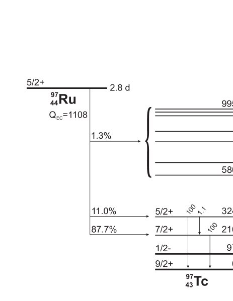
We thus set out to determine the temperature dependence for the ec-decay half-life of a nucleus with a that is considerably larger than that of 7Be. Our goal was to achieve a precision comparable to that obtained for and decays, i.e. 0.1%. For our measurement we sought a nucleus that decays entirely by electron capture with a few-day half-life and a delayed ray that can be cleanly detected. It also had to be producible by thermal-neutron activation so that we could obtain statistically useful quantities without serious contaminants. Although there are not a lot of candidates to choose among, we found 97Ru satisfied all our conditions. Its decay scheme appears in Fig. 1. We report here measurements of the half-life of 97Ru at room temperature and at 19K as measured via its 216-keV -delayed ray. We have found no temperature dependence in the results. Our upper limit is 0.1%, an order of magnitude below the effect claimed for 7Be Wa06 .
II Apparatus and set-up
We used the same set-up for both the cold and room-temperature measurements. As we did previously for our 198Au half-life measurement Go07 , we placed the ruthenium sample between two copper washers and fastened the assembly directly onto the cold head of a CryoTorr7 cryopump with four symmetrically placed screws. A 70% HPGe detector was placed facing the sample on the cryopump axis, just outside the pump’s coverplate, into which a cavity had been bored so that only 3.5 mm of stainless steel stood between the detector face and the sample. The total distance between the detector face and the ruthenium sample was 49 mm and remained unchanged throughout the experiment. We monitored the temperature of the sample with a temperature-calibrated silicon diode (Lakeshore Cryogenics DT-670) LS fastened in the same way as the ruthenium sample and placed right next to it on the head itself. The diode was connected to a Lakeshore Model 211 temperature monitor.
For the low-temperature measurement, we first used a roughing pump to bring the pressure down to about 9 mtorr, and then switched on the cryopump. Although the cold head, where the sample was located, is nominally expected to reach 12K, we measured its temperature to be between 18.2 and 20.8K, with an average value of 19K. The arrangement for the room temperature measurement was identical except that these pumping and cooling steps were omitted. Note that we did not alternate temperatures for a single source but rather made a complete decay measurement at one temperature with one source at a fixed geometry; then, with a fresh source, we made a similar dedicated measurement at the other temperature. Thus our results are entirely independent of any geometrical or source differences that might have occurred between the two measurements.
Our sample was a single crystal in the form of a circular disc, 8 mm in diameter and 1 mm thick, obtained from Goodfellow Corp. According to the supplier, the chemical purity of the material was 99.999%, with no identifiable impurities. For each measurement, the metal crystal was initially activated for 10 seconds in a flux of neutrons/cm2 s, at the Texas A&M Triga reactor. This activated crystal was then fastened directly to the cold head of the cryopump, ensuring a good thermal contact over the whole crystal area.
For the measurement itself, sequential -ray spectra were recorded from the HPGe detector. The detector signals were amplified and sent to an analog-to-digital converter, which was an Ortec TRUMPTM-8k/2k card OR controlled by Maestro software, which was installed on a PC operating under Windows-XP. During the entire period of the measurements, our computer clock was synchronized daily against the signal broadcast by WWVB, the radio station operated by the U.S. National Institute of Standards and Technology. For both the room- and low-temperature measurements, six-hour spectra were acquired sequentially for approximately one month. In each case, more than 110 -ray spectra were recorded.
The TRUMPTM card uses the Gedcke-Hale method Je81 to correct for dead-time losses. By keeping our system dead time below about 4% and recording all our spectra for an identical pre-set live time, we ensured that our results were essentially independent of dead-time losses. However, at a precision level of 0.1% or better, pile-up can also become an issue, so we carefully tested our system for residual rate-dependent effects, as reported in our previous article on 198Au Go07 . We first measured the 662-keV -ray peak from a 137Cs source alone, and then remeasured that source a number of times in the presence of a 133Ba source, which was moved closer and closer to the detector in order to increase the dead time and the number of chance coincidences. Each measurement was made for the same pre-set live time. We then obtained from each measurement the number of counts in the 662-keV peak and, from the decrease in that number as a function of increasing dead time, we determined that the fractional residual loss amounted to per 1% increase in dead time. At the count rates experienced during our 97Ru measurements, the required correction was never greater than 0.2% but it was nevertheless applied to all spectra.
III Results and Analysis
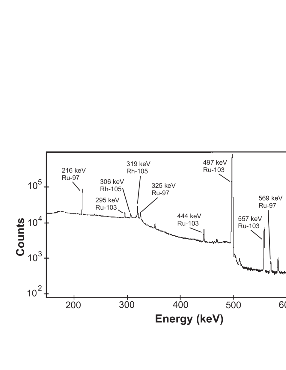
A typical -ray spectrum, one of the more than 220 obtained, is shown in Fig. 2. Apart from the weak peaks due to room background, the only observed rays are from the decays of 97Ru ( = 2.8 d), 103Ru (39 d) and 105Rh (35 h); the latter is the daughter of 105Ru (4.4 h), which had already decayed away by the time this spectrum was recorded. The appearance of these three ruthenium isotopes is consistent with their being produced by neutron activation of naturally occurring ruthenium. The 216-keV -ray peak from 97Ru is seen to be clear of any other peaks and to lie on a smooth, though rather high, background.
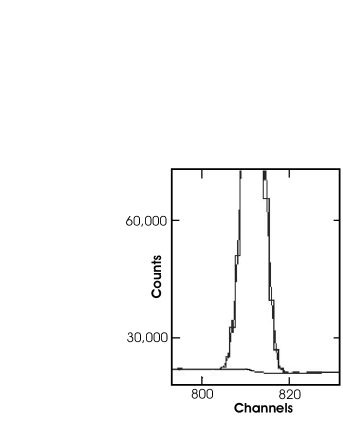
The 216-keV -ray peak in each recorded spectrum was analyzed with GF3, a least-square peak-fitting program in the RADware series Rapc . This program allowed us to be very specific in determining the correct background for a peak, and the 216-keV peak in each spectrum was visually inspected to this end. So far as possible, the same criteria were applied to each spectrum. Fig. 3 shows a sample peak and the fitted background, from which its area was determined.
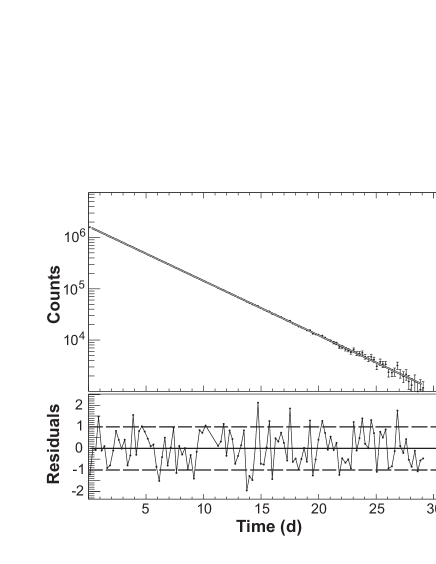
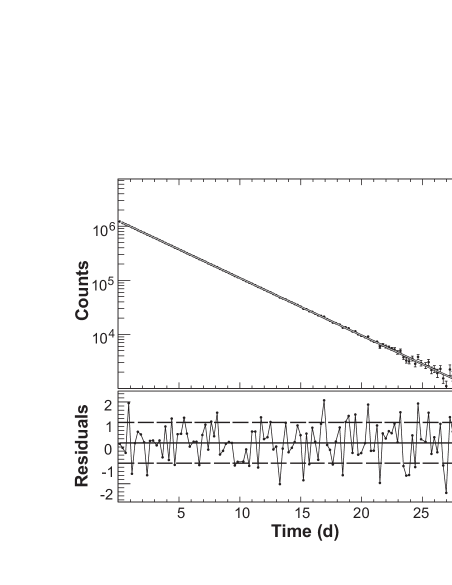
In total, 229 spectra were subjected to this careful analysis, and the counts recorded in the 216-keV peak for each were corrected for residual losses (see Sect. II). The results for the room temperature and 19K measurements are plotted as a function of time in Figs. 4 and 5. The decay curves were then analyzed by a maximum-likelihood fit to a single exponential. The code we used, which is based on ROOT Br97 , has previously been tested by us to 0.01% precision with Monte Carlo generated data. The data in Figs. 4 and 5 yield 97Ru half-lives (with statistical uncertainties only) of 2.8370(13) d for the room temperature measurement, and 2.8382(13) d for the one at 19K. The difference between these two results is 0.0012(18) d, which gives an upper limit of 0.0030 d, or 0.1%, on any temperature-dependent difference at the 68% confidence level.
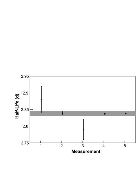
The half-life values taken from the computer fits incorporate the correction for residual losses described in Sect. II, but they do not yet include the uncertainty in that correction, since it is correlated for the two measurements and does not contribute to the difference between them. However, for our measurements to be compared with previous measurements of the 97Ru half-life, this systematic uncertainty is now incorporated, and yields the results 2.8370(14) d and 2.8382(14) d for the room temperature and 19K measurements, respectively. These values are compared with previous measurements of the 97Ru half-life in Table 1 and Fig. 6, where it can be seen that our results at both temperatures are much more precise than, but are entirely consistent with, the previous ones, all of which were presumably made at room temperature.
As a byproduct of our primary measurement on 97Ru we have also extracted from the same spectra half-lives at both temperatures for the nuclides 103Ru and 105Rh, both emitters. For 103Ru, we monitored the 497-keV peak in all 237 spectra, while for the shorter lived 105Rh there were only sufficient statistics for us to use 100 spectra to follow the 319-keV peak (see Fig. 2). These peaks were subjected to the same meticulous examination, fitting and analysis as just described for the 216-keV peak of Ru. Incorporating only statistical uncertainties, we obtained half-life values for 103Ru, of 39.210(16) d at room temperature, and 39.219(25) d at 19K, which are statistically the same within 0.1%. For 105Rh, our half-life values with statistical uncertainties only are 35.357(36) h at room temperature and 35.319(23) h at 19K, again the same, but in this case within 0.2%.
| Half-life (d) | Reference | Year |
|---|---|---|
| 2.8(3) | Sullivan et al. Su46 | 1946 |
| 2.8(1) | Mock et al. Mo48 | 1948 |
| 2.88(4) | Katcoff et al. Ka58 | 1958 |
| 2.9(1) | Cretzu et al. Cr66 | 1966 |
| 2.839(6) | Silvester et al. Si79 | 1979 |
| 2.79(3) | Kobayashi et al. Ko98 | 1998 |
| 2.838(6) | Weighted average | |
| This measurement: | ||
| 2.8370(14) | Room temperature | 2009 |
| 2.8382(14) | 19K | 2009 |
| 0.0012(18) | Difference | |
As we did when making the temperature comparison with 97Ru, we have so far quoted half-life values for 103Ru and 105Rh that do not yet include the (correlated) uncertainty attributable to residual losses (see Sect. II). We include that now in order to compare our results with previous half-life measurements. Our final half-life results for 103Ru then become 39.210(38) d at room temperature, and 39.219(35) d at 19K; and for 105Rh our results are 35.357(37) h at room temperature and 35.319(24) h at 19K. Note that the effect of residual losses on the uncertainty of the 103Ru half-life is much greater than it is for the 105Rh half-life. Since 103Ru is much longer lived, our data only encompass a little more than one half-life, during which time the overall count-rate in our detector has decreased significantly.
Unlike the situation for the other two radionuclides studied in this work, the half-life of 103Ru has been measured rather precisely in the past, with four of the previous results being of comparable precision to our current ones. Unfortunately, though, the earlier results are not particularly consistent with one another, as can be seen in Table 2. The normalized for the average of all previous measurements is 3.0, which results in our scaling up the uncertainty assigned to that average by a factor of 1.7. In comparison with this average value, our results are slightly low, though the discrepancy is not statistically very significant. Note also that our results are completely consistent with the 1981 value obtained by Miyahara et al. Mi81 .
| Half-life (d) | Reference | Year |
|---|---|---|
| 39.5(3) | Flynn et al. Fl65 | 1965 |
| 39.35(5) | Debertin De71 | 1971 |
| 39.254(8) | Houtermanns et al. Ho80 | 1980 |
| 39.214(13) | Miyahara et al. Mi81 | 1981 |
| 39.260(20) | Vaninbroukx et al. Va81 | 1981 |
| 39.272(16) | Walz et al. Wa83 | 1983 |
| 39.250(10) | Weighted average (scale factor, 1.7) | |
| This measurement: | ||
| 39.210(38) | Room temperature | 2009 |
| 39.219(35) | 19K | 2009 |
| 0.009(30) | Difference | |
There are only three previous measurements of the 105Rh half-life, none more recent than 1967; they are listed in Table 3. Strikingly, the earliest measurement Br62 has the tightest, 0.06%, uncertainty and a half-life value that disagrees completely with the two later measurements. The weighted average of all three measurements yields a normalized of 22 and, as shown in Table 3, its uncertainty consequently requires scaling by a factor of 4.7. Under the circumstances, it seems more reasonable not to use this average value, but simply to disregard the offending measurement and average the two remaining, mutually consistent, results Pi65 ; Ko67 . When compared with this new average, our results are a factor of two more precise and lie slightly lower. Considering that even the two previous measurements that have been retained are more than 40 years old and that the difference between their average and our recent results is less than two standard deviations, there seems little reason for concern.
| Half-life (h) | Reference | Year |
|---|---|---|
| 35.88(2) | Brandhorst and Cobble Br62 | 1962 |
| 35.4(1) | Pierson Pi65 | 1965 |
| 35.47(8) | Kobayashi Ko67 | 1967 |
| 35.84(9) | Weighted average (scale factor, 4.7) | |
| 35.44(6) | Weighted average of Pi65 and Ko67 | |
| This measurement: | ||
| 35.357(37) | Room temperature | 2009 |
| 35.319(24) | 19K | 2009 |
| 0.038(43) | Difference | |
IV Conclusions
We have measured the half-life of 97Ru in ruthenium metal at room temperature and at 19K, and have found the results to be the same within 0.1%. Since the maximum decay energy for any allowed transition from 97Ru is 892 keV, the nucleus must decay by pure electron capture. Three years ago, Wang et al. Wa06 reported half-life measurements of another pure electron-capture emitter, 7Be, situated in both palladium and indium metals, in which they observed differences of 0.9(2)% and 0.7(2)%, respectively, between room temperature and 12K. The same group also reported cases of temperature dependence for , and decay modes Ra07 ; Sp07 ; Li06 and interpreted them all as the result of a “Debye plasma,” which purportedly acts in any metal host and leads to a smooth dependence of half-lives on temperature. In that context, their result for 7Be decay was understood to be the indication of a generic property of all ec-decays rather than a unique property of 7Be.
Obviously we cannot comment on the validity of the 7Be measurement itself, but we can certainly refute any suggestion that the half-lives of ec-decays in general exhibit significant temperature dependence when the source is placed in a metal host. Wang et al. Wa06 used their model to calculate that the half-life of 7Be in a metal should change by 1.1% between T = 293 and 12K, a result that agrees reasonably well with their measured values. Using the same model, we calculate that the half-life change for the 97Ru decay should be 11.2% between T = 293 and 12K and 8.4% between T = 293 and 19K, the temperature we obtained. Our measured upper limit on any half-life change over this temperature range is nearly two orders of magnitude less than this model prediction. We have previously demonstrated that the Debye model has no validity for decay Go07 ; we can now state with equal confidence that it also does not apply to ec decay.
As a byproduct of this primary measurement, we also obtained half-life data for two emitters, 103Ru and 105Rh, at room temperature and 19K. These results, though slightly less precise than our measurements on the decay of 198Au Go07 , nevertheless confirm our previous conclusion for that decay mode. With any temperature dependence for decay also now ruled out at the 0.04% level Ru08 , it has become clear that there is no reason to doubt the accuracy of nuclear weak-decay half-lives that have been quoted over the past decades with sub-percent precision and without accounting for the host material or temperature. As has always been believed, those parameters indeed do not affect the result, at least not above the 0.1% level.
In all three cases, 97Ru, 103Ru and 105Rh, our measured half-lives are consistent with, and in two cases are substantially more precise than, previous measurements.
Acknowledgements.
We thank Prof. R. Watson for the generous loan of a cryopump. This work was supported by the U.S. Department of Energy under Grant No. DE-FG03-93ER40773 and by the Robert A. Welch Foundation under Grant No. A-1397.References
- (1) G.T. Emery, Ann. Rev. Nucl. Sci. 22, 165 (1972).
- (2) M. Curie and M. Kamerlingh Onnes, Le Radium 10, 181 (1913).
- (3) F. Raiola et al., Eur. Phys. J. A 32, 51 (2007).
- (4) T. Spillane et al., Eur. Phys. J. A 31, 203 (2007).
- (5) B. Limata et al., Eur. Phys. J. A 28, 251 (2006).
- (6) B. Wang et al., Eur. Phys. J. A 28, 375 (2006).
- (7) H. Muir, New Scientist vol. 192, issue 2574, p. 36 (2006).
- (8) J.C. Hardy and I.S. Towner, Phys. Rev. C 79, 055502 (2009).
- (9) J. R. Goodwin et al., Eur. Phys. J. A. 34, 271 (2007).
- (10) V. Kumar et al., Phys. Rev. C 77, 051304(R) (2008).
- (11) G. Ruprecht et al., Phys. Rev. C 77, 065502 (2008) and C 78, 039901(E) (2008).
- (12) N.J. Stone et al., Nucl. Phys. A 793, 1 (2007).
- (13) N. Severijns et al., Phys. Rev. C 76, 024304 (2007).
- (14) Y. Nir-El et al., Phys. Rev. C 75, 012801(R) (2007).
- (15) Evaluated Nuclear Structure Data File maintained by the National Nuclear Data Center, Brookhaven National Laboratory: http://www.nndc.bnl.gov; and G. Audi, A.H. Wapstra and C. Thibault, Nucl. Phys A 728, 337 (2003).
- (16) http://www.lakeshore.com/temp/sen/sd670_po.html.
- (17) http://www.ortec-online.com/trump.htm.
- (18) R. Jenkins, R.W. Gould and D. Gedcke, Quantitative X-ray Spectrometry (Marcel Dekker, New York, 1981) p. 266.
- (19) D. Radford, http://radware.phy.ornl.gov/main.html and private communication.
- (20) R. Brun and F. Rademakers, Nucl. Instrum. Methods A 389, 81(1997).
- (21) W.H. Sullivan et al., Phys. Rev. 70, 778 (1946).
- (22) D.L. Mock et al., Phys. Rev. 74, 1536 (1948).
- (23) S. Katcoff and D.C. Williams, J. Inorg. Nucl. Chem., 7, 189 (1958).
- (24) V.T. Cretzu et al., Annalen der Physik 472, 1 (1966).
- (25) D.J. Silvester et al., J. Labeled Compd. Radiopharm. 16, 226 (1979).
- (26) T. Kobayashi et al., Nucl. Phys. A 636, 367 (1998).
- (27) K.F. Flynn, L.E. Glendenin and E.P. Steinberg, Nucl. Sci. Eng. 22, 416 (1965).
- (28) K. Debertin, Z Naturforsch. 26a, 596 (1971).
- (29) H. Houtermans, O. Milosevic and F. Reichel, Int. J. Appl. Radiat. Isot. 31, 153 (1980).
- (30) H. Miyahara, T. Gotoh and T. Watanabe, Int. J. Appl. Radiat. Isot. 32, 573 (1981).
- (31) R. Vaninbroukx, G. Grosse and W. Zehner, Int. J. Appl. Radiat. Isot. 32, 589 (1981).
- (32) K.F. Walz, K. Debertin and H. Schrader, Int. J. Appl. Radiat. Isot. 34, 1191 (1983).
- (33) H.W. Brandhorst Jr. and J.W. Cobble, Phys. Rev. 125, 1323 (1962).
- (34) W.R. Pierson, Phys. Rev. 140, B1516 (1965).
- (35) Y. Kobayashi, J. Inorg. Nucl. Chem. 29, 1374 (1967).