Elucidating the bimodal acid-base behavior of the water-silica interface from first principles
Abstract
Understanding the acid-base behavior of silica surfaces is critical for many nanoscience and bio-nano interface applications. Silanol groups (SiOH) on silica surfaces exhibit two acidity constants—one as acidic as vinegar—but their structural basis remains controversial. The atomic details of the more acidic silanol site govern not just the overall surface charge density at near neutral solution pH, but also how ions and bio-molecules interacts with and bind to silica immersed in water. Using ab initio molecular dynamics simulations and multiple representative crystalline silica surfaces, we determine the deprotonation free energies of silanol groups with different structural motifs. We show that previously proposed motifs related to chemical connectivity or inter-silanol hydrogen bonds do not yield high acidity. Instead, a plausible candiate for pKa=4.5 silanol groups may be found in locally strained or defected regions with sparse silanol coverage. In the process, irreversible ring-opening reactions of strained silica trimer rings in contact with liquid water are observed.
I Introduction
Deprotonation of silanol (SiOH) groupsbook ; iler at water-silica interfaces is one of the most common and important, yet intriguing, interfacial chemical reactions. Silica (SiO2) is a major component of rocks and lines the channels of many nanofluidic devices.dekker1 ; baca ; yang Deprotonation governs dissolution rates,book affects lipid binding to silica nanostructures,baca creates negative surface charges that can be tuned with moderate changes in solution pH to perform desalination and ion gating,yang and may even hinder extraction of positively charged crude oil componentsoil from underground deposits. In particular, the atomic level structural details of deprotonated SiOH groups govern both the overall surface charge density and the binding of ions and molecules to immersed silica surfaces. In this work, we apply ab initio molecular dynamicscpmd (AIMD), which takes into account proton dynamics and hydrogen bond network fluctuations in liquid water essential to acid-base reactions in small moleculessprik ; klein ; parrin1 ; chandler as well as cooperative hydroxyl hydrogen bonding behavior specific to oxide surfaces,hass ; pore to investigate the enigmatic SiOH deprotonation equilibrium constant as a function of structural motifs.
Measurements of interfacial pKa (defined as Ka, where Ka is the acid dissociation constant) have been revolutionized by surface-sensitive second harmonic generation (SHG) and sum frequency vibrational spectroscopy (SFVS) techniques.eisenthal1 ; shen1 In 1992, Ong et al.ong demonstrated that 19% of silanol groups on fused silica surfaces exhibit a pKa of 4.5, about the same as vinegar (acetic acid), while 81% exhibit pKa=8.5. SFVS experiments on -quartz reached similar conclusions and further suggested that the low-acidity silanol groups reside in regions with strong water-water hydrogen bonds.shen05 A titration study on silica gel (amorphous silica)allen and X-ray photoelectron spectroscopy measurements on quartzduval also independently demonstrated the existence of SiOH groups with pKa between 4 and 5.5. Such qualitative agreement on different forms of silica is expected because liquid water is known to react with even crystalline silica to form an amorphous layer.iler ; shultz These measurements suggest that the earlier, single pK6.8 reported in amorphous silica titration experimentsschindler may reflect a composite of two types of SiOH.
(a)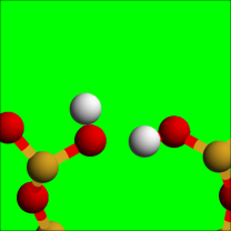
 (b)
(b)
(c)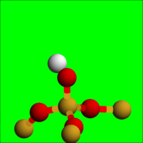
 (d)
(d)
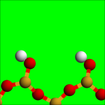
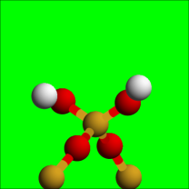
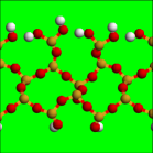
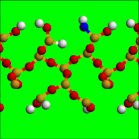
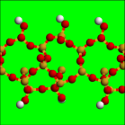
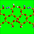
The acidities of surface silanol groups have been assigned to different chemical connectivities or inter-silanol hydrogen bonding. In accordance with the literature, we differentiate SiOH groups according to whether they are directly hydrogen bond to other SiOH (“H-bonded,” Fig. 1a), or are not so hydrogen-bonded (“isolated,” Fig. 1b); whether the 4-coordinated Si atom of the SiOH is part of 3 covalent Si-O-Si- linkages (“Q3,” Fig. 1c), or only part of 2 Si-O-Si (“Q2,” Fig. 1d). It has been suggested that the ratio of H-bonded to isolated SiOH is about 1 to 4, similar to the relative occurrence of pKa=4.5 and 8.5;ong thus pKa=4.5 has been ascribed to isolated silanol groups.ong ; dong ; lorenz ; fan1 On the other hand, the Q2:Q3 ratio has also been described as either approximately 1:4 or 4:1, which has prompted assignment of the pKa=4.5 SiOH group to either Q2 (Refs. mori1, ; nanosilica, ) or Q3 (Refs. others, ; shaw2, ; rosenholm, ). The conflicting estimates of the ratios of silanol groups with different structural motifsmori1 ; nanosilica ; others ; shaw2 ; rosenholm likely reflect the difficulty brought about by the tendency of liquid water to react with crystalline silica.shultz One indisputable experimental finding is that the silanol surface density is 4.6 nm-2 on well-soaked amorphous samples.iler ; zhuravlev On the theoretical side, static geochemical models,hiemstra ; bickmore which do not account for aqueous phase hydrogen bonding and dynamical proton motion, have also been applied, but they have not yet explained the two observed acidity constants,ong ; shen05 ; allen while quantum chemistry or DFT methods with a dielectric continuum treatment of the bulk water environment have been limited to calculating the pKa of small silica fragments.sahai2 ; rustad1 ; sefcik DFT modeling of amorphous silica slabs have also been considered,mauri but the pKa estimates therein often do not treat water explicitly or dynamically. (See Supporting Information (SI) for discussions of the significance of hydrogen-bond network fluctuations and excess proton hopping.)
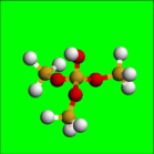
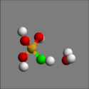
AIMD simulations have successfully reproduced the pKa of molecules in aqueous solutionsprik ; klein ; parrin1 and should be particularly well-suited for distinguishing relative SiOH pKa that are 4 pH units apart in different environments, provided we can demonstrate that reproducible pKa for chemically equivalent SiOH’s can be predicted. Coupled with static high-level quantum chemistry corrections, they provide the most rigorous predictions for liquid state reactions. As computing power has increased, AIMD modeling of liquid water-material interfaces has become viable,hass ; pore ; marx ; galli2 ; mundy ; car ; angelo although it remains costly because water dynamics is slower at interfaces.funel ; rossky Furthermore, investigation of multiple reaction sites and/or crystalline facets is often necessary when dealing with material surfaces. In this work, we study the bimodal acid-base behavior of silanol groupsong ; shen05 ; allen by performing AIMD simulations to directly calculate the pKa value. Given the absence of well-defined water-crystalline silica interfaces,shultz and the fact that the precise atomic structure of amorphous silica surfaces is unknown, we examine six distinct, representative silanol environments. These include hydroxylated -crystobalite (100) (Fig. 2a), hydroxylated -cristobalite (100) with one SiOH removed (Fig. 2b), reconstructed -cristobalite (100) (Fig. 2c), a molecular system (Fig. 2e), and two distinct SiOH on reconstructed quartz (0001) (Fig. 2f). They represent the SiOH motifs proposed to be responsible for pKa=4.5 or 8.5 in the literature (Figs. 1a-d).
(a) (b) (c)
(d) (e) (f)
II Computational Methods
AIMD simulations apply the Perdew-Burke-Ernezhof (PBE) functional,pbe the Vienna Atomic Simulation Package (VASP),vasp ; vasp1 a 400 eV energy cutoff, -point sampling of the Brillouin zone, deuterium mass for all protons, and a 0.375 fs time step at each Born-Oppenheimer dynamics time step. The trajectories are thermostat at T=425 K; elevated temperature is needed to represent liquid water properties when the PBE functional is applied and quantum nuclear effects are neglected, which is the case herein (SI, Sec. S1). Four to six umbrella sampling windows of 20 ps production trajectory length each are used per pKa calculation on hydroxylated -cristobalite (100) (Figs. 2a,b) and reconstructed -cristobalite (Fig. 2c) surfaces. These periodically replicated simulation cells measure 10.1710.1726 Å3. Their lateral dimensions are commensurate with simulation cells often used for AIMD studies of pure water structure or ion hydration. This is one of the reasons we have focused on the (100) rather than the (111) surface of -cristobalite, which actually has a similar to that of amorphous silica.iler The more computationally costly reconstructed quartz (0001) system has a cell size of 10.017.3224 Å3, and 12 to 18 ps trajectories are used per window. For each crystalline silica simulation cell, we always start from crystal slab structures optimized at zero temperature using Density Functional Theory (DFT) and the PBE functional.pbe Next we switch to the CHARMM SiO2 force fieldcharmm and the SPC/E model for water.spce The number of water molecules occupying the simulation cell is determined using these force fields, the Grand Canonical Monte Carlo (GCMC) technique, and the Towhee code.gcmc With this approach, the spaces between hydroxylated and reconstructed -cristobalite surfaces are filled with 58 and 63 water molecules, respectively, which amount to roughly six layers of water, while the reconstructed quartz simulation cell contains about 63 (about 4 layers of) water molecules. The (H3SiO)3SiOH pKa calculation utilizes a ( Å)3 cell with 57 H2O and 20 ps sampling trajectories. Finally, to check system size dependences, umbrella sampling simulations for a smaller reconstructed -cristobalite (100) simulation cell, 20 Å in the -direction and containing 44 (4 layers of) water molecules, are also conducted for at least 10 ps per window.
pKa has been reported for molecules in liquid water using the AIMD technique.sprik ; klein ; parrin1 It is related to the standard state deprotonation free energy via , where is and
| (1) |
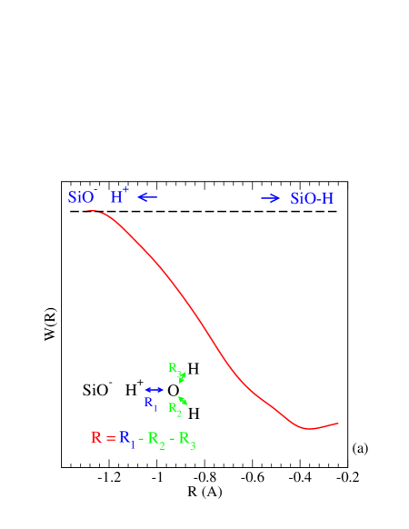
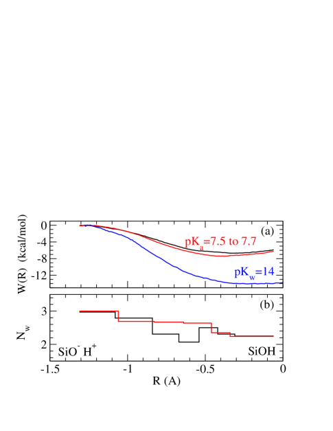
Here denotes 1.0 M concentration, is the reaction coordinate, is a phase space factor to be discussed below, is the cutoff distance delimiting the reaction and product valleys in the free energy landscape, and is the potential of mean force which provides the information needed to compute the free energy of deprotonation. Regardless of the reaction coordinate used,sprik ; klein generally do not exhibit turning points in the deprotonated region, and can be taken as the onset of the plateau where .
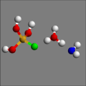
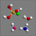
The umbrella sampling methodbook1 is used to compute the associated with SiOH deprotonation. A four-atom reaction coordinate (Fig. 3a) is found to work best under our simulation conditions. It controls what we call the “wandering proton” problem. We label the first, second and third neighbor H2O molecules of the SiO- oxygen (green sphere) shown in Figs. 3b-d “water 1” (O depicted red), “2” (blue), and “3” (pink). When Å (Fig. 3b), the SiOH bond is intact. As decreases to Å (Fig. 3c), the SiOH proton is transferred to a “water 1” which has been hydrogen-bonded to the SiOH group, yielding a SiO--H3O+ contact ion pair. As further decreases (Fig. 3d), a proton originally residing on “water 1” is now transferred to a second water molecule (“water 2”), creating a water-separated SiO-/H3O+ pair, at which point the deprotonation reaction is almost complete. This analysis appears consistent with insights from a transition path sampling AIMD simulation,chandler as follows. The Fig. 3d configuration, with the excess proton and the SiO- separated by two hydrogen bonds, form a possible free energy dividing surface between the intact and the deprotonated acid species, provided that the electric polarization between the two states, arising from the surrounding water molecules,chandler has sufficient time to equilibrate. Our simulation conditions allow such equilibration, and the excess proton is indeed observed to diffuse away if fluctuates to regions significantly more negative than Å. Umbrella potentials of the type are used to sample the reaction coordinate . Just as significantly, they ensure that Å and control the extent of proton transfer from “water 1” to all possible “water 2,” so that at most a water-separated ion-pair is obtained. Otherwise, if the excess proton is several hydrogen bonds removed from the SiO- (say if it spends a significant amount of time on “water 3” via proton transfer from “water 2”), it can start to diffuse (“wander”) through the simulation cell via the Grotthuss mechanism at O(1) ps time scale per proton transfer. With tens of H2O molecules in the simulation cell and 10-20 ps trajectories, once the excess H+ leaves the second hydration shell of the SiO- it does not return, and equilibrium sampling is not achieved. This “wandering” likely arises because the higher temperature and longer umbrella sampling trajectories than are generally used in the AIMD literature facilitate diffusion of the excess proton away from the SiO-. Other deprotonation reaction coordinates used in the literature are discussed in the SI (Sec. S2). They are found to give rise to wandering excess protons under our simulation conditions. As long as equilibrium sampling is achieved, the deprotonation free energy cost should not depend on the choice of coordinate.
To apply Eq. 1, we use a method similar to Ref. sprik, : finding the most probable optimal O-H+ hydrogen bond distance at each , thus locally converting to ; performing a spline fit to the resulting ; and integrating over with a volume element, which takes the place of the phase space factor in Eq. 1. Equation 1 assumes that the entropic factors such as rotations of the reactant and products about the O-H axis are adequately sampled in the AIMD trajectories; otherwise additional constraints and entropic factors are introduced.klein1 ; co2 SI Sec. S2 shows that such constraints do not affect water autoionization free energies, partly because the variation in needed to complete the deprotonation reaction is relatively small. Furthermore, we always reference predicted silanol pKa to that of water autoionizationklein computed using the same reaction coordinate and elevated temperature. This minimizes systematic error arising from the simulation protocol (SI Sec. S2), and phase space contributions approximately cancel out.
The metadynamics technique,meta1 ; meta2 ; meta3 a promising and powerful alternative to umbrella sampling, has been applied to calculate dissociation free energies on surfacesmarx and acid-base reactions of small molecules.parrin1 This method, not yet implemented in VASP, can potentially be used for efficient comparative study of pKa on other material surfaces after it has been adapted to deal with the wandering proton problem.
Gas-phase, high-level ab initio calculations are performed to check and correct chemical bonding energies predicted with the PBE functional used in the AIMD simulations. The Gaussian03 program suite is applied.g03 The pertinent sample chemical reaction is
| (2) |
Geometries are optimized, and harmonic vibrational frequencies computed, with density functional theory using the B3LYP methodlyp ; b3lyp and the 6-311++G() basis set. At the B3LYP geometries, energies are computed with the coupled-cluster singles and doubles method including a perturbative correction for triple substitutions, CCSD(T),CCSD(T) using the aug-cc-pVDZ basis set. Basis set incompleteness corrections are added to the CCSD(T) energies. Finally, zero-point vibrational energy corrections computed from the B3LYP/6-311++G() frequencies are added. Using this protocol, the high-level ab initio calculation yields a reaction energy of -30.51 for Eq. 2. Gas-phase VASP-based PBE calculations, conducted with an energy cutoff identical to that in AIMD simulations, predict -27.17 kcal/mol. These numbers do not include the 2.24 kcal/mol zero point energy (ZPE) corrections. Thus, the overall AIMD reaction energies should be corrected by a modest -1.10 kcal/mol. The basis set extrapolation procedure may have a systematic error larger than 1 pH unit (SI Sec. S3) but this does not affect the relative pKa of different SiOH groups.
(a) (b) (c)
See the SI for details about constraints introduced to prevent proton attacks on SiO-, the AIMD intialization protocol, further justifications for adopting the four-atom reaction coordinate, extrapolating quantum chemistry results to the infinite basis set limit.
III Results
Hydroxylated -cristobalite (100): chemically homogeneous SiOH. We first show that our simulation protocol predicts reproducible pKa for chemically equivalent SiOH groups on the hydroxylated -cristobalite (100) surface (Fig. 2a). This well-studied model crystalline surface exhibits =8 nm-2, larger than the experimental value of 4.6 nm-2 for amorphous silica.iler At zero-temperature, it features two types of Q2 silanol groups: alternating hydrogen bond donors and acceptors arranged in chainsmeng (Fig. 2a) not found in small molecules.sprik ; klein ; parrin1 This feature is dynamically preserved in our finite-temperature, aqueous-phase simulations (SI Sec. S4).
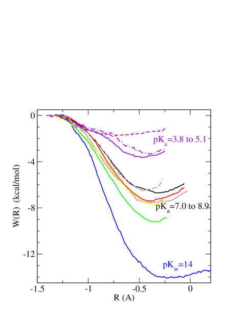
Figure 4a shows that two chemically equivalent, hydrogen bond-accepting SiOH groups on this surface are predicted to exhibit deprotonation within 0.5 kcal/mol of each other. At large negative values of the reaction coordinate , the deprotonated SiO- is stabilized with three hydrogen bonds (i.e., =3, Figs. 4b and 4c). At 0.8 Å, the SiO-H bond is only partially broken, 3, and the local is not sensitive to the slight difference in in the two simulations that arises from statistical noise. These observations appear consistent with a recent two-dimensional potential of mean force analysis of formic acid deprotonation.parrin1 Accounting for zero-point energy, correcting the AIMD functional with more accurate quantum chemistry methods, and referencing Eq. 1 to the water autodissociation constant pKw=14,klein we estimate pKa values of 7.5 and 7.7, close to the less acidic pKa value reported by Ong et al.ong The standard deviation is 0.3 pH unit. Multiple deprotonation on this surface is discussed in SI Sec. S5.
Heterogeneity: Isolated, H-bonded, Q2, and Q3 SiOH all exhibit pK 7. We next show that, contrary to previous hypotheses,ong ; lorenz ; fan1 ; mori1 ; nanosilica ; others ; shaw2 ; rosenholm isolated, H-bonded, Q2, and Q3 silanol groups all exhibit pK 7.0. We first create an isolated silanol group by replacing a hydrogen bond-donating SiOH group on the hydroxylated -cristobalite (100) surface with a SiH so that its neighboring SiOH group is no longer H-bonded (Fig. 2b). Figure 5 shows that this isolated SiOH exhibits pKa=8.9, and is less acidic by 1.2-1.4 pH unit than when the SiOH hydrogen donor is present (Fig. 2a). This is entropically reasonable because a hydrogen bond-donating SiOH partner stabilizes the neighboring SiO- alongside two water molecules, while three water molecules are required for an isolated SiO-. AIMD correctly accounts for this effect because it models H2O and SiOH on the same dynamical footing and because water-water and water-silanol hydrogen bond energies are similar.meng Hydroxyls on oxides with more ionic character than SiO2 form stronger hydrogen bonds, and indeed the relative abundance of inter-hydroxyl hydrogen bonding may partially be responsible for the crystal facet-dependent acidity of -Al2O3.fitts This will be the subject of future, comparative studies.
To compare Q2 and Q3 silanol groups, we reconstruct the hydroxylated -cristobalite (100) surface by condensing every other pair of H-bonded SiOH groups into a SiOH and a H2O molecule (Fig. 2c). This involves the elimination of a hydrogen-bond donating OH group plus the proton on its adjacent, hydrogen-bond accepting SiOH (Fig. 2d). The resulting undercoordinated Si and O atoms are joined together to form a covalent bond, in the process pulling apart the remaining H-bonded SiOH pairs so they are now isolated from one another. A similar structural motif has been considered in the literature.chuang This surface has =4 nm-2, with all SiOH groups being Q3, isolated, and residing on silica rings containing at least 5 Si atoms. Such rings should be unstrained, unlike 3-member (Si-O)3 rings discussed below. The pKa is found to be 8.1 (Fig. 5).
We also consider a (SiH3)3SiOH molecule featuring an isolated, Q3 SiOH group (Fig. 2e), which exhibits a comparable pKa=7.9. Thus, in general, Q3 and Q2 silanol groups do not exhibit pKa’s that differ by 4 pH units as previously proposed.lorenz ; fan1 ; mori1 ; nanosilica ; others ; shaw2 ; rosenholm Over the range 4 nm-2 8 nm-2, the precise value of has little effect on pKa.
(c) 
 (d)
(d)
To some extent, all our crystalline silica models are nano-slits with thin water slabs confined between the surfaces. As the water content decreases, the dielectric solvation of SiO- and H+ species should decrease, while intact SiOH groups should be weakly affected. Thus one expects a lower acidity and a higher pKa in strongly nano-confined aqueous media.yb To examine confinement effects, a pKa calculation is performed for a smaller reconstructed -cristobalite (100) simulation cell, 20 Å in the -direction, containing 4 layers of water. This system actually yields a lower pKa=7.00.4—but is lower only by 1.1 pH units (dashed brown line in Fig. 5). It is possible the unexpected pKa decrease between the 6- and 4-water layer models arises from anomalies in the hydrogen bonding network not apparent from visual inspection of water configurations. The water density is also affected by the confinement.garofalini2 In the 4-layer model, all water molecules are at most two layers away from the crystalline silica surfaces, and AIMD conducted with GCMC-predicted water content is found to yield a second layer H2O density that is 18% above 1.0 g/cc. However, this is unlikely to change the dielectric response sufficiently to lower the pKa by 1 unit. Assuming one can apply the Born hydration formula for excess proton hydration in this heterogeneous medium, where kcal/mol for the proton.tiss If the 6-layers of water have =80, the 4-layer system must exhibit =140 to make hydration more favorable by 1 pH unit when in fact confinement generally reduces the dielectric constant of water.pore In any case, the discrepancy in pKa is actually within two standard deviations and may simply arise from statistical uncertainties. This test suggests that confinement effects are not large for the slit pore geometrydekker1 down to about 1 nm slit widths. Therefore our reported pKa for 6-water layer systems should be good approximations of pristine crystalline silica surfaces in contact with bulk liquid water. We also stress that all slab geometries in Figs. 2a-c have been studied using 6-water layer simulation cells, and their relative pKa should be mostly free of system size effects. However, two-dimensional confinement in cylindrical amorphous silica nanoporespore ; lorenz ; schulten ; hartnig ; dipole may have a stronger impact on pKa.yb
High acidity and chemical reactions on strained reconstructed quartz surfaces. Finally, it seems imperative to demonstrate the possibility of an unusually acidic silanol group. The following “computational existence proof” is necessarily more speculative than the conclusions about Q2, Q3, isolated, and H-bonded SiOH pKa discussed above, but it emphasizes the likely role of defected regions when accounting for pKa=4.5.
Having considered =8 and 4 nm-2, we examine an even lower SiOH surface density. A recent experimental study has attached crystal violet dyes to deprotonated silanol groups. Based on the flat, 120 nm2 surface area of the dye molecule, it is proposed that strong acidity is correlated with local SiOH surface density 0.83 nm-2.dong In our DFT calculations, the optimal geometry of crystal violet when covalently bonded to (HO)3SiO- is not flat but is substantially distorted. However, this suggestion of low SiOH surface density being associated with the more acidic SiOH appears consistent with other experimental and theoretical observations discussed below.
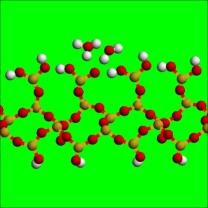
Since most cuts through crystalline forms of silica yield surfaces with substantial hydroxylation,nangia we investigate a reconstructed, completely dehydroxylated quartz (0001) model, featuring (Si-O)3 trimer rings, predicted to be metastable in vacuum.deleeuw1 ; deleeuw2 2.3 SiOH groups per square-nanometer are re-introduced by removing two surface Si atoms and performing further reconstruction and hydroxylation to keep all atoms fully coordinated. This yields two types of silanol groups which are hydrogen-bonded to each other; one member of the pair resides on a cyclic trimer while the other does not (Fig. 2f). Regions devoid of SiOH and dominated by siloxane (Si-O-Si) bridges are hydrophobic, and this model surface may therefore be consistent with hydroxyl groups in hydrophobic pores reported to be unusually acidic on other oxides.alumina2 We conduct one deprotonation umbrella sampling simulation of a silanol group residing on a cyclic trimer (Fig. 5, solid violet curve), and two simulations where the SiOH does not reside on a trimer (dashed and dot-dashed violet). Figure 5 indeed shows that these SiOH groups exhibit pKa , 4.8, and , respectively — close to the experimental value of 4.5. More significantly, their average pKa are separated from the median of all other SiOH groups previously examined in this work by 3.4 pH units.
Unlike cyclic silica tetramers or larger Si-O rings, cyclic silica trimers are strained.book ; strain ; wallace1 At zero temperature, in the absence of water, the Si atom of the SiOH group residing on a silica trimer ring exhibit Si-O-Si angles of 137.5o, 130.4o, and 132.0o. For the SiOH group not residing on a silica trimer, the angles are 154.9o, 131.8o, and 147.8o. A few of these angles deviate substantially from the ideal, unstrained Si-O-Si value of approximately 145o. This likely accounts for the low pKa computed for these SiOH groups. Other trimer rings not decorated with SiOH are also strained, and the slow spatial decay of their surface strain fieldsrickman may also contribute to the high acidity of SiOH group not residing on them.
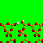
Within hours in moist air,book ; strain ; wallace1 cyclic trimer-containing surfaces are known to incorporate water and break open to reduce strain and increase the local . At the water-reconstructed quartz interface, during umbrella sampling deprotonation of the SiOH group residing on a 3-member ring (Fig. 2f), we indeed observe a water molecule forming a transient bond with another Si atom on a Si-O trimer 6 Å away from the tagged SiO- within picoseconds (Fig. 6b). The resulting 5-coordinated Si has been observed in simulationsdeleeuw2 ; garofalini1 ; garofalini and found to be the intermediate in the trimer ring-opening mechanism on wet silica amorphous surfaces in reactive force field and molecular orbital calculations.garofalini1 ; garofalini ; lasaga Our AIMD trajectories show that this mechanism remains operative at explicit liquid water-silica interfaces; proton hopping via the Grotthuss mechanism occurs readily, enabling the H2O adsorbed on the surface Si to lose a proton to bulk water, forming a new SiOH group within ps (Fig. 6c). Then, in 2 of the 4 sampling windows, a Si-O bond on the now 5-coordinated Si breaks to open the OH-incorporated (Si-O)3 ring and irreversibly introduce another new SiOH (Fig. 6d). Our trajectories thus differ from a recent molecular dynamics study of the liquid water-reconstructed quartz interface, where the adsorbed water molecule, not described by a reactive force field, ultimately desorbs from the 5-coordinated surface Si atom without inducing chemical reactions.deleeuw2
These irreversible side reactions prevent strict equilibrium sampling needed for calculations. Fortunately, for the SiOH residing on a cyclic trimer (Fig. 6), analysis of the pre- and post ring-breaking statistics reveals that the nearby chemical reaction has little effect on its pKa. We further analyze trimer ring-opening effects on the pKa of another SiOH group, this one not residing on a surface 3-member ring. A H2O incorporation reaction also occurs in the neighborhood of this tagged SiOH (Fig. 7). We split the sampling windows into two groups: (A) those without irreversible hydrolysis of a nearby silica ring (Fig. 7a); and (B) those with trimer ring breaking and formation of two new SiOH groups (Fig. 7b). Then two complete sets of sampling windows spanning the entire deprotonation pathway are spawned from these seed windows, yielding two pKa: case A, pKa=3.8 (Fig. 5, dashed violet curve); and case B, pKa=4.8 (Fig. 5, dot-dashed violet). The results show that H2O incorporation and a single ring-breaking event nearby does appear increase the pKa of the tagged SiOH not residing on a trimer ring. However, the increase is only 1.0 pH unit, almost within statistical uncertainties. In case (A), the three Si-O-Si angles on the silica trimer ring average to 121.1o, 134.0o, and 132.2o along the trajectory; the first refers to the angle where both Si are below the silica surface (“buried”). In case (B), this angle linking the buried Si atoms relaxes significantly to 140.5o. The second angle, which involves the surface Si and a buried Si, remains almost unchanged at 136.0o. (The third linkage is destroyed during ring opening.) Si atoms which no longer participate in strained Si-O-Si linkages should be more stable against H2O attack.
(a)  (b)
(b) 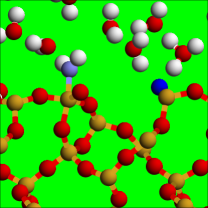
(c) 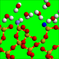 (d)
(d) 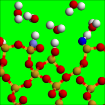
From these considerations, we conclude that, despite interference from H2O incorporation reactions, there remains a statistically significant difference in the pKa’s on this surface, and the pKa’s ranging from 7.0 to 8.9 for all other silanol groups investigated before (Fig. 5). This finding suggests that strain, low local silanol surface density, hydrophobicity, and low pKa are correlated on amorphous silica surfaces. Indeed, atomistic model surfaces with low local regions almost always exhibit 3-member rings.deleeuw1 ; garofalini1 ; singer These regions may be in dynamic equilibrium with solvated silica fragments in solution, constantly being dissolved/hydrolyzed and reconstituted when dissolved fragments re-nucleate on hydroxylated regions.deleeuw2 ; criscenti ; nangia1 ; meijer The dynamic equilibrium has recently been demonstrated in Monte Carlo simulationsnangia1 using a reactive silica force field.garofalini3
We have only observed one ring-opening reaction on each surface. The limited AIMD trajectory length does not conclusively allow us to predict how many trimer rings persist at the liquid water-amorphous silica interface as a function of time. Therefore we do not definitively assign this structure to the observed pKa=4.5 SiOH group, and instead pose it as a challenge to experimental work, including single molecule spectroscopy,ye to determine whether they are sufficiently abundant over time to account for the 19% of all silanol groups shown to exhibit high acidity.ong ; shen05 We also point out that, while cyclic silica trimers are well known to react with moist air, other popular crystalline silica model surfaces should also be hydrolytically unstable.shultz Thus, the hydroxylated -quartz (0001) and -cristobalite (100) surfaces, with 9 and 8 nm-2 respectively, are often used as models to study the interface between liquid water and generic silica solids in classical MD simulations. However, if these simulations permit chemical reactions over long enough times, we speculate that some of the SiOH groups on such surfaces may also react with watershultz ; nangia1 in a way to reduce the silanol surface density towards the amorphous silica =4.6 nm-2 observed in experiments.iler ; zhuravlev
(a) 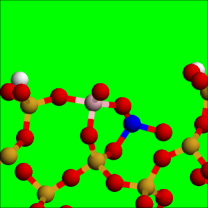 (b)
(b) 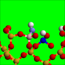
Discussions AIMD-based potential of mean force calculations simulations have been demonstrated to yield reproducible pKa for chemically equivalent silanol groups. The statistical uncertainties of our simulation protocol are estimated to be about 0.3-0.5 pH unit, consistent with explicit calculations on two chemically equivalent SiOH. Therefore these simulations should reliably distinguish relative pKa of heterogeneous SiOH groups 4 pH units apart. Resolving hydroxyl pKa on other surfaces may remain a challenge if the acidities are less widely separated.
Our pKa calculations suggest that comparative studies between SiO2 and other material surfaces will be extremely interesting. One intriguing candidate surface is a quartz surface densely functionalized with carboxylic acid groups. To our knowledge, this is the only other material surface which clearly exhibits bimodal pKa behavior.geiger1 Other candidates are the different facets of crystalline alumina, which may feature several pKa’s unresolved into distinct, measurable components.alumina2 ; alumina3 ; alumina4 Structural motifs such as chemical connectivity and inter-hydroxyl hydrogen bonding have also been invoked to explain the pKa in these systems; as mentioned in the text, inter-hydroxyl hydrogen bonding may affect the pKa of the more ionic Al2O3 surfaces more strongly than on any form of silica surfaces. See SI, Sec. S6, for more details on these material systems. Finally, SFVG spectrashen05 and time-dependent acid-base phenomena on quartzgeiger2 can also be investigated in the future.
IV Conclusions
In this paper, we have performed AIMD pKa calculations on five representative crystalline silica surfaces plus a molecular system exhibiting different silanol (SiOH) structural motifs. From the results, we have conclusively shown that the more acidic of the two pKa observed in experiments cannot, as previously proposed,dong ; lorenz ; fan1 ; mori1 ; nanosilica ; others ; shaw2 ; rosenholm be explained by the existence of silanol groups with certain chemical connectivities or inter-silanol hydrogen bonding. In fact, we find pK 4.5 silanol groups only on strained surfaces with sparse silanol coverage. While our demonstration of the existience of such low pKa SiOH groups is necessarily somewhat speculative, this study highlights the role of defected regions as the most promising candidate to explain the elusive bimodal acid-base behavior of silica surfaces.ong ; shen05 ; allen Assigning structural motifs to the more strongly acidic SiOH groups is particularly crucial in non-reactive force field-based modeling of silica nanofluidic channels, where the preferentially deprotonated SiOH sites at neutral pH have to be assigned in a static way.pore ; lorenz ; hartnig ; schulten ; schulten1 ; jctn In the process of studying the acid-base behavior, we also observe irreversible, water-assisted ring-opening reactions of strained silica trimer rings in contact with liquid water, The reaction was previously studied on wet silica surfaces;garofalini1 ; lasaga ; garofalini1 our AIMD simulations demonstrate that a similar mechanism is operative at liquid water-silica interfaces.
Acknowledgement
We thank Ron Shen, Steven Garofalini, Susan Rempe, Jeff Brinker, Dave Tallant, Ying-Bing Jiang, and Franz Geiger for discussions. This work was supported by the Department of Energy under Contract DE-AC04-94AL85000. Sandia is a multiprogram laboratory operated by Sandia Corporation, a Lockheed Martin Company, for the U.S. Deparment of Energy. LJC acknowledge support from the U.S. DOE Office of Basic Energy Sciences, Division of Chemical Sciences, Geosciences, and Biosciences.
Supporting Information Available
Further information are provided regarding details of the AIMD simulations, justification for the reaction coordinate used, quantum chemistry calculations (including all energies and optimized geometries), the dynamics of hydrogen bond fluctuations on silica surfaces, proton exchange due to multiple deprotonation, and a brief overview of acid-base behavior in alumina and carboxylate acid functionalized surfaces. This information is available free of charge via the Internet at http://pubs.acs.org/.
References
- (1) Brinker, C.J.; Scherer, G.W. Sol-Gel Science; Academic Press, London, 1990, Ch. 10.
- (2) Iler, R.K. The Chemistry of Silica: Solubility, Polymerization, Colloid and Surface Properties, and Biochemistry; Wiley, New york, 1979.
- (3) Stein, D.; Kruithof, M.; Dekker, C. Phys. Rev. Lett. 2004, 93, 035901.
- (4) Baca, H.K.; Ashley, C.; Carnes, E.; Lopez, D.; Flemming, J.; Dunphy, D.; Singh, S.; Chen, Z.; Liu, N.G.; Fan, H.Y.; Lopez, G.P.; Brozik, S.M.; Werner-Washburne, M.; Brinker, C.J. Science 2006, 313, 337.
- (5) Fan, R.; Huh, S.; Yan. R.; Arnold, J.; Yang, P.D. Nature Mater. 2008, 7, 303.
- (6) Kokal, S.; Tang, T.; Schramm, L.; Sayegh, S. Colloids and Surfaces A: Physicochemical and Engineering Aspects 1995, 94, 253.
- (7) Car, R.; Parrinello M. Phys. Rev. Lett. 1985, 55, 2471.
- (8) Sprik M. Chem. Phys. 2000, 258, 139.
- (9) Ivanov, I.; Chen, B.; Raugei, S.; Klein, M.L. J. Phys. Chem. B 2006, 110, 6365.
- (10) Park, J.M.; Laio A.; Iannuzzi, M.; Parrinello, M. J. Am. Chem. Soc. 2006, 128, 11318.
- (11) Geissler, P.L.; Dellago, C.; Chandler, D.; Hutter, J.; Parrinello, M. Science, 2001, 291, 2121.
- (12) Leung, K.; Rempe, S.B.; Lorenz, C.D. Phys. Rev. Lett. 2006, 96, 095504.
- (13) Hass, K.C.; Schneider, W.F.; Curioni, A.; Andreoni, W. Science 1998, 282, 265.
- (14) Eisenthal, K.B. Chem. Rev. 2006, 106, 1462.
- (15) Shen Y.R.; Ostroverkhov, V. Chem. Rev. 2006, 106, 1140.
- (16) Ong, S.W.; Zhao, X.L.; Eisenthal, K.B. Chem. Phys. Lett. 1992, 191, 327.
- (17) Ostroverkhov, V.; Waychunas, G.A.; Shen, Y.R. Phys. Rev. Lett. 2005, 94, 046102.
- (18) Allen, L.H.; Matijevic, E.; Meites, L. J. Inorg. Nucl. Chem. 1971, 33, 1293.
- (19) Duval, Y.; Mielczarski, J.A.; Pokrovsky, O.S.; Mielczarski, E.; Ehrhardt, J.J. J. Phys. Chem. B 2002, 106, 2937.
- (20) Li, I.; Bandara, J.; Shultz, M.J. Langmuir, 2004, 20, 10474.
- (21) von Schindler, P.; Kamber, H.R. Helv. Chim. Acta 1968, 51, 1781.
- (22) Dong, Y.; Pappu, S.V; Xu, Z. Anal. Chem. 1998, 70, 4730.
- (23) Lorenz, C.D.; Crozier, P.S.; Anderson, J.A.; Travesset, A. J. Phys. Chem. C 2008, 112, 10222.
- (24) Fan, H.-F.; Li, F.P.; Zare, R.N.; Lin, K.-C., Anal. Chem. 2007, 79, 3654.
- (25) Mori, T.; Kuroda, Y.; Yoshikawa, Y.; Nagao, M.; Kittaka, S. Langmuir 18, 1595 (2005).
- (26) Vance, F.W.; Lemon, B.I.; Ekhoff, J.A.; Hupp, J.T. J. Phys. Chem. B 1998, 102, 1845.
- (27) O’Reilly, J.P.; Butts, C.P.; I’Anson, I.A.; Shaw, A.M. J. Am. Chem. Soc. 2005, 127, 1632.
- (28) Fisk, J.D.; Batten, R.; Jones, G.; O’Reilly, J.P.; Shaw, A.M. J. Phys. Chem. B 2005, 109, 14475.
- (29) Rosenholm, J.M.; Czuryskiewicz, T.; Kleitz, F.; Rosenholm, J.B.; Linden, M. Langmuir 2007, 23, 4315.
- (30) Zhuravlev, L.T. Colloids Surf. A 2000, 173, 1.
- (31) See, e.g., Hiemstra, T.; De Wit, J.C.M.; Van Riemsdijk, W.H. J. Coll. Interface Sci. 1989, 133, 105, and references therein.
- (32) Bickmore, B.R.; Tadanier, C.J.; Rosso, K.M.; Monn, W.D.; Eggett, D.L. Geochim. Cosmochim. Acta 2004, 68, 2025.
- (33) Tossell, J.A.; Sahai, N. Geochim. Cosmochim. Acta 2000, 64, 4097.
- (34) Rustad, J.R.; Dixon, D.A.; Kubicki, J.D.; Felmy, A.R. J. Phys. Chem. A 2000, 104, 4051.
- (35) Sefcik, J.; Goddard, W.A. Geochmim. et Cosmochim. Acta 2001, 65, 4435.
- (36) See, e.g., Tielens, F.; Gervais, C.; Lambert, J.F.; Mauri, F.; Costa D. Chem. Mater. 2008, 20, 3336, and references therein.
- (37) Nair, N.N.; Schreiner, E.; Marx D. J. Am. Chem. Soc. 2008, 130, 14148.
- (38) Cicero, G.; Grossman, J.C.; Schwegler, E.; Gygi, F.; Galli, G. J. Am. Chem. Soc. 2008, 130, 1871.
- (39) Kuo, I.-F.W.; Mundy, C.J. Science 2004, 303, 658.
- (40) Kudin, K.N.; Car, R. J. Am. Chem. Soc. 2008, 130, 3915.
- (41) Liu, L.-M.; Krack, M.; Michaelides, A. J. Chem. Phys. 2009, 130, 234702.
- (42) Takahara, S.; Sumiyama, N.; Kittaka, S.; Yamaguchi, T.; Bellissent-Funel M.C. J. Phys. Chem. B 2005, 109, 11231.
- (43) Lee S.H.; Rossky, P.J. J. Chem. Phys. 1994, 100, 3334.
- (44) Perdew, J.P.; Burke, K.; Ernzerhof, K.M. Phys. Rev. Lett. 1996, 77, 3865.
- (45) Kresse G.; Joubert D. Phys. Rev. B 1999, 59, 1758.
- (46) Kresse G.; Furthmüller J. Phys. Rev. B 54, 11169-11186 (1996).
- (47) Lopes, P. E. M.; Murashov, V.; Tazi, M.; Demchuk, E.; MacKerell, A.D. J. Phys. Chem. B 2006, 110, 2782.
- (48) Berendsen, H.J.C.; Grigera, J.R.; Straatsma, T.P. J. Phys. Chem. 1987, 91, 6269.
- (49) Martin M.G.; Thompson, A.P. Fluid Phase Equil. 2004, 217, 105.
- (50) Chandler D. Introduction to Modern Statistical Mechanics; Oxford, New York, 1997, Ch. 6
- (51) Blumberger J.; Klein M. L. Chem. Phys. Lett. 2006, 422, 210.
- (52) Leung, K.; Nielsen, I.M.B.; Criscenti, L.J. J. Phys. Chem. B 2007, 111, 4453.
- (53) Laio, A.; Parrinello, M. Proc. Natl. Acad. Sci. USA 2002, 99, 12562.
- (54) Iannuzzi, M.; Laio, A.; Parrinello, M. Phys. Rev. Lett. 2003, 90, 238302.
- (55) Laio, A.; Gervasio, F.L. Rep. Prog. Phys. 2008, 71, 126601.
- (56) Frisch, M. J. et al., Gaussian 03 (Revision C.02), Gaussian Inc., Wallingford, CT, 2004.
- (57) Lee C.T.; Yang W.T.; Parr R.G. Phys. Rev. B 1988, 37, 785.
- (58) Becke A. D. J. Chem. Phys., 1993, 98, 5648.
- (59) Raghavachari, K.; Trucks, G.W.; Pople, J.A.; Head-Gordon, M. Chem. Phys. Lett. 1989, 157, 479.
- (60) Yang, J.; Meng, S.; Xu, L.F.; Wang, E.G. Phys. Rev. Lett. 2004, 92, 146102.
- (61) Fitts, J.P.; Shang, X.M.; Flynn, G.W.; Heinz, T.F.; Eisenthal, K.B. J. Phys. Chem. B 2005; 109, 7981.
- (62) Chuang, I.-S.; Maciel, G.E. J. Phys. Chem. B, 1997, 101, 3052.
- (63) Preliminary experimental measurements suggest that pH of zero charge increases inside narrow silica nanoporour membranes (Jiang, Y.-B., private communications).
- (64) Xu, S.; Scherer, G.W.; Mahadevan, T.S.; Garofalini, S.H. Langmuir 2009, 25, 5076, and references therein.
- (65) Tissandier, M.D.; Cowen, K.A.; Feng, W.Y.; Grunlach, E.; Cohen, M.H.; Earhart, A.D.; Coe, J.V.; Tuttle, T.R. J. Phys. Chem. A 1998, 102, 7787.
- (66) Hartnig, C.; Witschel, W.; Spohr, E.; Gallo, P.; Ricci, M.A.; Rovere, M. J. Mol. Liq. 2000, 85, 127.
- (67) Cruz-Chu, E.R.; Aksimentiev, A.; Schulten, K. J. Phys. Chem. B 2006, 110, 21497.
- (68) Leung, K. J. Am. Chem. Soc. 2008, 130, 1808.
- (69) Nangia, S.; Washton, M.M.; Mueller, K.T.; Kubicki, J.D.; Garrison, B.J. J. Phys. Chem. C 2007, 111, 5169.
- (70) Du, Z. M.; de Leeuw, N.H. Surf. Sci. 2004, 554, 193.
- (71) Du, Z. M.; de Leeuw, N.H. Dalton Trans. 2006, 22, 2623.
- (72) Braunschweig, B.; Eissner, S.; Daum, W. J. Phys. Chem. C 2008, 112, 1751.
- (73) Rickman, J.M.; Srolovitz, D.J. Surf. Sci. 1993, 284, 211.
- (74) Brinker, C.J.; Kirkpatrick, R.J.; Tallant, D.R.; Bunker, B.C.; Montez, B. J. Non-Cryst. Solids, 1988, 99, 418.
- (75) Wallace, S.; West, J.K.; Hench, L.L. J. Non-Cryst. Solids 152, 101 (1993).
- (76) Garofalini, S.H. J. Non-Cryst. Solids 1990, 120, 1.
- (77) Mahadevan T.S.; Garofalini, S.H. J. Phys. Chem. C 2008, 112, 1507.
- (78) Lasaga, A.C. Rev. Mineralogy 1990, 23, 17.
- (79) Hassanali A.A.; Singer, S.J. J. Phys. Chem. B 2007, 111, 11181; see Fig. 9.
- (80) The mechanism may be the reverse of the hydrolysis pathway examined by Criscenti, L.J.; Kubicki, J.D.; Brantley, S.L. J. Phys. Chem. A 2006, 110, 198.
- (81) Nangia, S.; Garrison, B.J. J. Am. Chem. Soc. 2009, 131, 9538.
- (82) Trinh, T.T.; Jansen, A.P.J.; van Santen, R.A.; Meijer, E.J. J. Phys. Chem. C 2009, 113, 2647.
- (83) Feuston, B.P.; Garofalini, S.H. J. Chem. Phys. 1988, 89, 5818.
- (84) Fu, Y.; Collinson, M.M.; Higgins, D.A. J. Am. Chem. Soc. 2004, 126, 13838.
- (85) Konek, C.T.; Musorrafiti, M.J.; Al-Abadleh, H.A.; Bertin, P.A.; Nguyen, S.T.; Geiger, F.M. J. Am. Chem. Soc. 2004, 126, 11754.
- (86) Zhang, L.; Tian, C.; Waychunas, G.A.; Shen, Y.R. J. Am. Chem. Soc. 2008, 130, 7686.
- (87) Stack, A.G.; Higgins, S.R.; Eggleston, C.M. Geochim. Cosmochim. Acta 2001, 65, 3055.
- (88) Gibbs-Davis, J.M.; Kruk, J.J.; Konek, C.T.; Scheidt, K.A.; Geiger, F.M. J. Am. Chem. Soc. 2008, 130, 15444.
- (89) Cruz-Chu, E.R.; Aksimentiev, A.; Schulten, K. J. Phys. Chem. C 2008, 113, 1850.
- (90) Leung, K.; Rempe, S.B. J. Theor. Comput. Nanoscience 2009, 6, 1948. 2008, 113, 1850.