Elastic energy of proteins and the stages of protein folding
Abstract
We propose a universal elastic energy for proteins, which depends only on the radius of gyration and the residue number . It is constructed using physical arguments based on the hydrophobic effect and hydrogen bonding. Adjustable parameters are fitted to data from the computer simulation of the folding of a set of proteins using the CSAW (conditioned self-avoiding walk) model. The elastic energy gives rise to scaling relations of the form in different regions. It shows three folding stages characterized by the progression with exponents , which we identify as the unfolded stage, pre-globule, and molten globule, respectively. The pre-globule goes over to the molten globule via a break in behavior akin to a first-order phase transition, which is initiated by a sudden acceleration of hydrogen bonding.
1 Introduction
The folding of a protein chain in water is driven mainly by the hydrophobic and hydrogen-bonding interactions [1]. The general shape of the fold, or tertiary structure, is designed to have hydrophobic side chains buried in the interior of the protein, in order to avoid direct contact with water. However, the side chains are attached to a backbone that welcomes exposure to water, because of their need for hydrogen bonding. These opposing tendencies reach mutual accommodation through the formation of secondary structures — alpha helices and beta sheets — which “use up” the hydrogen bonds on the backbone. This involves an intricate interplay between global geometry and local structure, and each protein seems to present special problems [2]. Proteins with different amino-acid sequences can invoke quite different folding mechanisms [3], while proteins with high sequence similarity can end up with very different folds [4, 5]. Nevertheless, universal aspects do emerge, if one overlooks details and concentrate only on a few general properties.
An overall characteristic of the tertiary structure is the radius of gyration , the root-mean-square separation between residues. It serves as a length scale, whose behavior can be studied experimentally [6, 7, 8]. Stages in the folding process are characterized by scaling relations of the form , where is the number of amino-acid residues. The “compactness index” can be measured through small-angle x-ray scattering [9]. It can be derived from physical models based on intuitive reasoning, as pioneered by Flory [10] and de Gennes [11] in the theory of homopolymers.
The unfolded protein chain, which is akin to a homopolymer, can be described by Flory’s model based on SAW (self-avoiding walk) [10]. One assumes a potential energy of the form where and are temperature-dependent coefficients, and is the spatial dimension. The first term is a stretching energy associated with random walk, for which scales like . The second term arises from the excluded volume effect, and is proportional to times the density. Minimizing the energy with respect to leads to , which gives for .
Hong and Lei [12] obtain for the native state by statistical analysis of data from PDB (the Protein Data Bank). They also derive it by generalizing Flory’s model. Through detailed arguments, they generalize Flory’s stretching energy to , where is the fractal dimension of the system. This leads to , which yields for Taking the fractal dimensions to be for polymer in good solvent, protein native state, polymer in poor solvent, respectively, one obtains for the respective indices. The first case reduces to Flory’s SAW model. For protein in the native state, the fractal dimension can be deduced from PDB, and the index implies Hooke’s law . We note that the native protein is less compact than the collapsed polymer, whose index is [11]. This is mainly because generally proteins are not fully hydrophobic. The proteins with hydrophobic residues do have indices close to [12]. Furthermore, native proteins are not well-packed because the secondary structures tend to create interior free volumes[13, 14].
Ptitsyn [15] has proposed a “molten globule” prior to the native state, in which the tertiary structure has taken shape, together with a large fraction of the final secondary structures. The main difference from the native state lies in orientations of side chains, which gradually lock into native contacts in a time scale of seconds. Thus the molten globule is almost as compact as the native state, and should have . This is supported by data from x-ray scattering [15]. In some proteins there is evidence for a pre-globule stage [16], which goes over to the molten globule in a sudden jump. Its observed index is [17].
In the present investigation, we try to verify these universal features, and understand them in a unified picture based on the twin actions of the hydrophobic force and hydrogen bonding. We do this by generalizing Flory’s potential energy to a universal elastic energy for proteins, via analysis and interpretation of data from computer simulations.
2 Protein folding in the CSAW model
We simulate the folding of several proteins using the CSAW (conditioned self-avoiding walk) model [18]. A conformation of the protein is specified by a set of torsion angles, and side chains are approximated by hard spheres. The unfolded chain is represented by SAW, and folding comes about through conditions that create a bias in SAW. The “self-avoidance” here means that all atoms on the backbone, as well as the side chains, are treated as hard spheres with appropriate diameters, and they are forbidden to overlap one another.
One begins with a SAW of steps. A trial update is generated by the pivot algorithm [19], and is accepted with a probability given by a Metropolis MC (Monte-Carlo) algorithm, based on the conformation energy
| (1) |
The two terms here correspond to the hydrophobic effect and hydrogen bonding, respectively. The quantity is a total hydrophobic shielding number, which measures how well hydrophobic side chains are shielded from water by neighboring protein atoms, and is defined as follows. The ’th residue has a hydrophobicity given by experiments [20], and a contact number , which is the number of nearest-neighbors of its side chain, in the existing conformation, and . Here, we adopt the HP model for the primary sequence, and assign for the hydrophobic residues (L, P, M, W, A, V, F, I), and for the others[21]. The quantity is the total number of internal hydrogen bonds, which are deemed to be in existence whenever two legitimate partners have relative positions that conform to the bond separation and angle. Other effects such as electrostatic and van der Waals interactions are ignored; but they can be easily included if desired. The MC updates eventually generate a sequence of conformations that form a canonical ensemble with respect to the conformation energy . The action of water is described entirely through the hydrophobic energy , and the random impacts implicit in random walk.
We assume that the parameters , take the same values for all proteins, which are determined in an earlier study of polyalanine [22]. The temperature is fixed, and implicit in the values of , , but not yet calibrated in the absolute scale.
| Protein name | ID | Structure | ||
| Polyalanine | ala20 | 1 alpha helix | ||
| Antimicrobial LCI | 2b9k | 1 beta sheet | ||
| Tedamistat | 3ait | 2 sheets | ||
| Myoglobin | 1mbs | 8 helices | ||
| Asparagine synthetase | 11as | 11 helices 8 sheets |
We simulate five proteins, as listed in Table 1. For each protein, folding starts from an unfolded state created by heating the native state to a high temperature, and then quenched to a fixed low temperature. The folding process runs for MC steps, with snapshots taken every 1000 steps. For each protein, the entire procedure is repeated 30 times to generate 30 folding trajectories, or an ensemble of conformations. In the case of myoglobin, folding is extended to a total of MC steps.
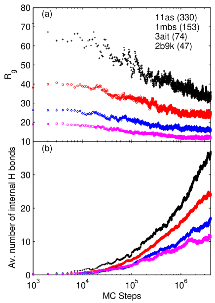
Fig. 1 shows the average radius of gyration and number of internal hydrogen bonds as a function of MC steps from the simulations of four globular proteins. From Fig. 1, we see that the radius of gyration decreases in the early stage, and form a plateau in a late stage (except the protein 11as whose simulation has not yet reached the plateau stage). This is consistent with experimental observations[6, 7, 8]. Existence of such stable collapsed states have been found in recent AFM experiments[23]. The number of internal hydrogen bonds shows a continuous increase during the whole process.
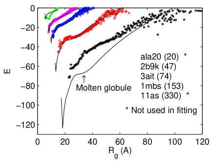
We compute an average potential energy , defined as follows. For each , we take the ensemble average of the model energy over all conformations along the folding trajectory that share the same value of . Thus, gives the pressure-force experienced by the protein as a function of radius, and can be observed in force spectroscopy experiments [24, 25]. In this sense we can call an “elastic energy”. The simulation results are shown in Fig.2.
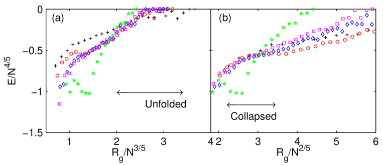
We can display universal features in the data by rescaling the variables (Fig. 3). The scales chosen are based on physical pictures, and their validity is to be judged by goodness of fit. First, we rescale the energy by a factor . This is chosen because in the native state the scaling laws and lead to Hooke’s law . On the horizontal axis of the plot, we rescale the radius by a factor to produce two separate plots, with to examine the unfolded region, and to examine the collapsed region. The rescaled graphs are shown in Fig.3(a) for the 3/5 scaling, and Fig.3(b) for the 2/5 scaling. As we can see, the data do exhibit universal behaviors in the respective regions, except for ala20, which fails to scale in the collapsed region. This exceptional protein has hydrophobic fraction , whereas the others have average . This exception, in fact, shows the relevance of hydrophobicity.
3 A universal elastic energy
We use physical arguments to suggest an analytical form of a universal elastic energy, and fit undetermined parameters to simulation data. Let us start from the unfolded chain, which is indicated in Fig.3(a). Assuming a power law , we have . In the hypothetical limit of a completely extended chain, we should have and . This determines and hence . This scaling law describes the hydrophobic energy, since hydrogen bonding is not significant in this stage. Now we fix the reference point of energy by taking for a completely extended chain, which is the convention used in CSAW simulations. Thus we arrive at the following potential energy in the unfolded region:
| (2) |
where and are parameters. As the chain folds, and decreases, the excluded volume effect becomes important. To take this into account, we add to a Flory term . Thus, we replace with
| (3) |
where , , are new parameters.
The energy has a local minimum corresponding to a metastable state, with scaling law , and . The index is consistent with the measured value for the natively unfolded proteins in pre-globule state [17]111Because of the hydrogen bonding attraction in the pre-globule, the experimental data shows an index closer to that for the molten globule (), rather than the ..
The universality in the collapsed region (Fig. 3(b)) suggests a power law , which implies . Fitting these forms to the pre-globule state, we find , and hence . This energy involves hydrogen bonding. We note that it has the same form as Flory’s excluded-volume energy, and thus is already taken into account in .
When a system of hard spheres on a chain collapses, overlaps can only be avoided through elaborate rearrangements, and the associated thermal energy depends on the intricate structure of the chain, and can only be treated phenomenologically. We do this by allowing the excluded volume to depend on and . Since the excluded volume effect should be universal for proteins at the molten globule stage, with scaling law , must depend on through the scaled radius . Thus we arrive at a universal elastic energy given by
| (4) |
Here, is a short-ranged potential, which we expand in an inverse power series of its argument:
| (5) |
In the power series (5), the terms with larger exponents describe higher-order short-range interactions, which depend on the detailed structure, and are chain specific. To obtain a universal elastic energy with the least number of parameters, we limit ourselves to odd powers . This gives rise 9 adjustable parameters, which are fitted to simulation data, excluding those from ala20, and 11as. The former is excluded because it is atypical, being 100% hydrophobic; the latter, because its simulation has not yet reached the pre-globule. With measured in angstroms, the parameters are as follows: , and for odd from to , {, , , , , , , }.
4 The stages of protein folding
Graphs of the elastic energy are shown in Fig.2. The fits are not ideal for ala20, presumably because the protein is all hydrophobic, nor for 11as, because the simulation in this case is far from complete.
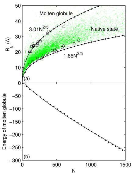
We refer to the curve of 11as for illustration. The region with large corresponds to the unfolded stage, which is under pressure to collapse, since . The only stable point on the curve is the lowest minimum, which corresponds to the most compact state. The states with smaller have rapidly increasing energy because of the excluded-volume effect. There is a flat shoulder corresponding to the molten globule, which can exist in neutral equilibrium. The radii of the molten globule and the most compact state are plotted in Fig.4(a), with for the most compact state, and for the molten globule. We can see from 4(a) that the above theoretical results agree well with the experimental data. The energy of the molten globule, shown in Fig.4(b), obeys , which implies . This agrees with the Hookes-law behavior in the collapsed state. In summary, the three folding stages — unfolded, pre-globule, molten globule — are characterized by the progression of the compactness index.
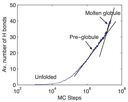
The rate of hydrogen bonding increases in the successive folding stages, as illustrated in Fig.5 from the simulation of myoglobin. The transition from the pre-globule to the molten globule is marked by a sudden acceleration of hydrogen bonding. This initiates the analog of a first-order phase transition. Like that in macroscopic matter, the volume is being compressed at constant temperature, and latent heat is released, since the transition connects two states of different energy. However, in a small system such as the protein, there is no clear separation of coexisting phases.
The subsequent evolution from the molten globule to the native state cannot be described in the present simulation, since it involves the locking of side chains, and we have approximate them with hard spheres. As we learn from experimental data, however, should remain unchanged.
5 Outlook
The elastic energy here is constructed at a fixed temperature. Work is in progress to extend it to a range of temperatures, with a view of obtaining a universal equation of state for proteins. Also under consideration is the use of the potential in a kinetic equation to compute the lifetimes of the various stages of protein folding. Although we concentrate on universal properties here, the CSAW model actually yields results for individual proteins, which could be analyzed for specificity. The CSAW model is highly flexible, and amenable to refinements, such as realistic simulation of side chains, and inclusion of other interactions not yet considered.
This work is supported in part by the National Natural Science Foundation of China (NSFC10601029).
References
- [1] Branden C. Tooze J. Introduction to Protein Structure, Garland Publishing, New York(1998).
- [2] Shakhnovich E. Chem. Rev. 69(2006):1559.
- [3] Daggett V. Fersht A. R. Trends Biochem Sci. 28(2003):18.
- [4] Alexander P.A., et al. Biochemistry 44(2005)(14045).
- [5] He Y., et al. Biochemistry 46(2005):14055.
- [6] Akiyama A., et al. Proc. Natl. Acad. Sci. USA 99(2002):1329.
- [7] Uzawa T., et al. Proc. Natl. Acad. Sci. USA 101(2004):1171.
- [8] Kimura T., et al. Proc. Natl. Acad. Sci. USA 102(2005):2748.
- [9] Huang K. Lectures on Statistical Physics and Protein Folding, World Scientific, Singapore (2005), Section8.4.
- [10] Flory P. Principles of Polymer Chemistry, Cornell University Press, London (1953).
- [11] de Gennes P. G. Scaling Concepts in Polymer Physics, Cornell University Press, Ithaca(1979).
- [12] Hong L. Lei J. J. Polymer Sci. B 47(2009):207.
- [13] Arteca, G. A. Phys. Rev. E. 51(1993):2600.
- [14] Liang, J. Dill, K. A. Biophys. J. 81(2001)751.
- [15] Ptitsyn O. B. Protein Folding T. E. Creighton T. E. Ed. W.H. Freeman & Co., New Yirk (1992).
- [16] Uversky V. N. Ptitsyn O. B. J. Mol. Biol. 255(1996):215.
- [17] Uversky V. N. Protein Sci. 11(2002):739.
- [18] Huang K. Biophys. Rev. Lett. 2(2007):139.
- [19] Li B., Madras N. Sokal A. D. J. Stat. Phys. 80(1995):661.
- [20] Kyte, J. Doolittle, R. A. J. Mol. Biol. 157(1982):105.
- [21] Garrett, R. H. Grisham, C. M. Biochemistry, Thomson(1999):84-85.
- [22] Lei J. Huang K. Eur. Phys. J. E 27(2008):197.
- [23] Garcia-Manyes, S., et al. Proc. Natl. Acad. Sci. USA 106(2009):10534.
- [24] Cecconi, C., et al. Science 309(2005):2057.
- [25] Fernandez, J. M. Li, H. Science 303(2004):1674.
- [26] Tcherkasskaya, O. Uversky, V. N. Proteins Struct. Funct. Genet. 44(2001):244.