Observation of atomic localization using
Electromagnetically Induced Transparency
Abstract
We present a proof-of-principle experiment in which the population of an atomic level is spatially localized using the technique of electromagnetically-induced transparency (EIT). The key idea is to utilize the sensitive dependence of the dark state of EIT on the intensity of the coupling laser beam. By using a sinusoidal intensity variation (standing-wave), we demonstrate that the population of a specific hyperfine level can be localized much tighter than the spatial period.
pacs:
32.80.Qk, 42.25.Kb, 42.50.GyIt is well-known that traditional optical techniques cannot resolve or write features smaller than half the wavelength of light. This barrier, known as the diffraction limit, has important implications for a variety of scientific research areas including biological microscopy and quantum computation. As an example, in a neutral-atom quantum computing architecture, the diffraction limit prohibits high-fidelity manipulation of individual atoms if they are separated by less than the wavelength of light. Recently, Agarwal and others Agarwal and Kapale (2006); Gorshkov et al. (2008); Yavuz and Proite (2007) have proposed to use the dark state of electromagnetically induced transparency (EIT) Harris (1997); Scully and Zubairy (1997) to address atoms at potentially nanometer spatial scales. This technique relies on the sensitive dependence of the dark state to the intensities of the driving probe and coupling laser beams. If a standing-wave coupling laser is used, the population of the excited Raman level can be very tightly localized near the intensity nodes, allowing for sub-wavelength control. In this letter, we present a proof-of-principle experiment that demonstrates the key ideas of this approach. By using ultracold Rubidium (Rb) atoms in a magneto-optical trap (MOT) and pulsed coherent transfer, we demonstrate atomic localization to spots much smaller than the spatial period of the coupling-laser intensity profile. Although due to imaging limitations we have used a large spatial period in this work (m), our results will likely scale to the sub-wavelength regime in the future.
Before proceeding, we cite important prior work leading up to this experiment. In their pioneering work, Thomas and colleagues have suggested and experimentally demonstrated sub-wavelength position localization of atoms using spatially varying energy shifts Thomas (1989); Stokes et al. (1991); Gardner et al. (1993). Zubairy and coworkers have discussed atom localization using resonance fluorescence and phase and amplitude control of the absorption spectrum Kiffner et al. (2008); Macovei et al. (2007); Kapale and Zubairy (2006). Knight and colleagues discussed localization via quantum interference at the probability amplitude of the excited electronic state Paspalakis and Knight (2001). Li et. al. have experimentally demonstrated probe narrowing beyond the diffraction limit using a spatially-varying coupling laser profile in a vapor cell Li et al. (2008). There has also been remarkable progress in utilizing the position dependent stimulated emission to achieve nanoscale resolution Hell (2007); Maurer et al. (2010). This last approach, also known as stimulated-emission depletion microscopy, is now a widely used technique in biological imaging. We note that our approach of using the dark state for atomic localization has the following key advantages: 1) For the ideal case of sufficiently slowly varying driving laser pulses, the dark-state technique has no population at the excited electronic state. As a result, the atomic localization can, in principle, be achieved without suffering from the detrimental effects of spontaneous emission. This is especially important for quantum computing applications Gorshkov et al. (2008) where coherent manipulation with little decoherence is essential. 2) The dark state can be prepared adiabatically by using a counter-intuitive pulse sequence. As a result, as discussed in detail in Ref. Yavuz and Proite (2007), the scheme is insensitive to many experimental fluctuations such as the intensity and the timing jitter of the driving laser pulses. 3) Since the scheme is coherent, localization can be achieved at faster time-scales at the expense of requiring more intense laser beams. Although in this work we use ns-long laser pulses, dark-state-based localization can easily be achieved at sub-ns time-scales by using more powerful laser beams.
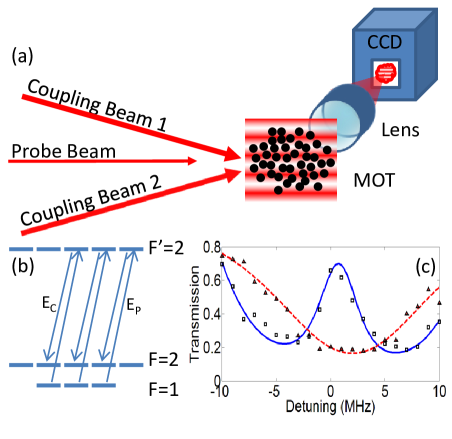
We next discuss the details of our experiment which can be viewed as a proof-of-principle demonstration of the suggestion by Lukin and colleagues Gorshkov et al. (2008). The experiment is performed inside a 14-port, stainless-steel ultrahigh vacuum chamber which is kept at a base pressure of about 10-9 torr. To construct the 87Rb MOT, we use three counter-propagating beam pairs that are locked to the cycling transition, each with a beam power of 100 mW and a beam size of 3 cm. The MOT lasers are obtained from an external cavity diode laser whose output is amplified with a tapered amplifier. We typically trap about 1 billion 87Rb atoms at a temperature of 150 K. The EIT beams are derived from a separate master diode laser which is saturated-absorption locked to the appropriate transition. The coupling laser beam is shifted by 6.8 GHz using high-frequency acousto-optic modulators and is amplified with a tapered amplifier Unks et al. (2007). As shown in Fig. 1, the probe and the coupling lasers are resonant with and transitions of the D2 line, near a wavelength of 780 nm. The beams have the same circular polarization and the experiment works in three parallel channels Braje et al. (2003). The coupling laser is split into two beams, which then reconverge at the MOT at an angle of 3 milliradians to form a vertical standing wave with a spatial period of m. We probe the localization by level-dependent fluorescence of the atomic cloud. The fluorescence signal is collected with a 2-inch achromatic-doublet outside of the vacuum chamber and is recorded with an electron-multiplying CCD camera.
Before proceeding further, we present a brief discussion of population localization using the dark state. Atoms distributed throughout the MOT will see different coupling laser intensities, based on where they are in the standing wave. Ignoring the complications due to parallel channels, the dark state of the atoms is given by Harris (1997); Gorshkov et al. (2008); Yavuz and Proite (2007):
| (1) |
where and are the Rabi frequencies of the respective beams. Here, for simplicity, we assume the probe beam to be uniform. The atoms can be prepared in the dark state of Eq. (1) by using the well-known counter-intuitive pulse sequence with coupling laser turning on before the probe laser beam. Once the laser beams are turned on, they can be turned-off simultaneously preserving the ratio of the Rabi frequencies Yavuz and Proite (2007). As a result, even after the laser pulses are turned-off, the atomic system is left in the state as determined by the probe and coupling laser Rabi frequencies at the temporal-peak of the pulses. Through this preparation, atoms will populate with a probability of . Due to the sensitive dependence to the coupling beam intensity, atoms located near a coupling field zero-crossing (intensity node) coherently transfer to with high probability. If we assume that is linear near a zero-crossing, then we expect the probability to be maximum at the coupling intensity node, and have an approximate spatial width of where is the peak coupling laser Rabi frequency Gorshkov et al. (2008); Yavuz and Proite (2007). As a result, with the probe laser intensity fixed, the population of level becomes more and more localized with increasing coupling beam power.
The experimental timing cycle is shown in Fig. 2. We begin the experiment by loading the MOT for one second and then turn off the MOT magnetic field gradient 50 ms prior to the EIT beams to reduce Zeeman splitting of the magnetic sublevels. All atoms are then initialized to by turning off the hyperfine repumping laser for the MOT. We drive the atoms to the dark state by using a 400 ns-long coupling laser and a 250 ns-long probe laser beam. After the EIT beams are turned-off, we probe the population of by fluorescing the atoms for 40 s via the cycling transition (). Due to sufficiently low atomic temperature, the motion of the atoms during fluorescence is negligible. In Fig. 2 we present two fluorescence images that show localization of the population as the coupling laser intensity is increased. Fig. 2(a) illustrates a case where we use a relatively weak coupling beam, where ( is the peak coupling intensity and is the probe intensity). The fringes align with the nodes of the coupling beam intensity and have wide profiles in the vertical dimension. In Fig. 2(b), we use a nearly 20 times more intense coupling laser beam such that . We observe the fringes to be vertically much more tightly confined to the coupling beam nodes. Both pictures use a probe intensity of 3.9 mW/cm2, and each picture is an average of 100 shots. Fig. 2(c) shows horizontally-averaged line profiles of each fluorescence image for more direct comparison.
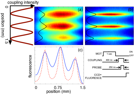
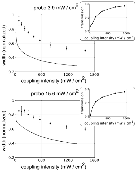
Figure 3 shows the normalized full-width-half-maximum (FWHM) of the fringes as the coupling beam intensity is varied for two values of probe laser intensity mW/cm2 and mW/cm2. Each data point is an average of 100 images and the error bars show the standard deviation of each set. For mW/cm2, we observe the population of level to localize by about a factor of two as the coupling beam intensity is increased. The solid lines in Fig. 3 are the results of numerical calculations without any adjustable parameters (i. e. each parameter that goes into the simulations are experimentally measured). Here, we include all relevant magnetic sub-levels and numerically solve the time-domain density matrix equations for the conditions of our experiment. We have experimentally measured the standing-wave interference of the coupling laser beam to be slightly imperfect with intensity contrast of 98%. This imperfection is included in our numerical calculations. The disagreement between theory and experiment is likely a result of 1) mechanical and interferometric fluctuations of the standing-wave intensity profile of the coupling laser beam, and 2) the Zeeman shift of the magnetic-sublevels due to a residual background magnetic field.
We next discuss the coherent nature of population localization. The insets in Fig. 3 show the integrated probe transmission through the atomic cloud as the coupling beam intensity is increased. We see better probe transmission with increased coupling beam intensity, demonstrating EIT for the exact conditions of each localization experiment. Furthermore, we have the ability to probe excited state fluorescence during the EIT process by collecting scattered photons for the duration of the coupling laser beam. We observe a reduction in the excited state fluorescence as the coupling laser intensity is increased, complementing the probe transmission data of the insets of Fig. 3. We also observe a strong increase in the excited state fluorescence when the coupling laser beam is turned-off (probe laser propagating alone through the cloud). This further confirms that the atoms are driven to a dark state with a small population at the excited electronic level.
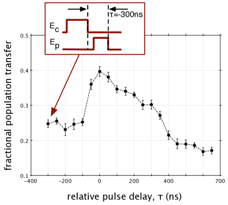
To further test the coherent nature of the population transfer, we have also performed a stimulated Raman adiabatic passage (STIRAP) experiment Bergmann et al. (1998). We measure the population transfer to at the intensity peaks of the coupling laser using a pulse sequence similar to above, but by changing the relative temporal overlap between the EIT beams. Noting Fig. 4, as expected, the maximum transfer to occurs when the probe and coupling pulses overlap, with coupling laser turning-on and turning-off before the probe laser beam. We observe a 20% increase in population transfer when the two pulses overlap, consistent within a factor of two of our density-matrix calculations. Near the intensity nodes of the coupling laser, we observe approximately 10% increase in population transfer when the pulses overlap (not shown in Fig. 4). As mentioned earlier, there is a coupling beam intensity offset of 2% of the peak at the nodes due to an imperfect interference profile. To increase contrast, the STIRAP experiments of Fig. 4 use beams that are 12 MHz detuned to the blue of the excited state. The intensities of the two beams are I = 130 mW/cm2.
To summarize, we have demonstrated localization of level population using EIT. As mentioned before, because our imaging system cannot resolve sub-wavelength spatial scales, we have performed this experiment with a small-angle between the two coupling-laser beams and therefore with a large spatial period of the standing-wave interference pattern. Future work will include extending this technique to the sub-wavelength regime and possibly demonstrate nanometer scale localization and addressing of neutral atoms. Furthermore, by using more powerful laser beams, we aim to explore atomic localization at much faster time-scales. If successful, the ability to address atoms at sub-ns time-scales with sub-wavelength resolution may provide a powerful tool for many challenging problems including initialization and addressability of a neutral-atom quantum register Urban et al. (2009); Ga tan et al. (2009).
We thank J. P. Sheehan for assistance with the experiment. This work was supported by the Air Force Office of Scientific Research (AFOSR).
References
- Agarwal and Kapale (2006) G. S. Agarwal and K. T. Kapale, J. Phys. B: At. Mol. Opt. Phys. 39, 3437 (2006).
- Gorshkov et al. (2008) A. V. Gorshkov, L. Jiang, M. Greiner, P. Zoller, and M. D. Lukin, Phys. Rev. Lett. 100, 093005 (2008).
- Yavuz and Proite (2007) D. D. Yavuz and N. A. Proite, Phys. Rev. A 76, 041802 (2007).
- Harris (1997) S. E. Harris, Phys. Today 50, 36 (1997).
- Scully and Zubairy (1997) M. O. Scully and M. S. Zubairy, Quantum Optics (Cambridge University Press, 1997).
- Thomas (1989) J. E. Thomas, Opt. Lett. 14, 1186 (1989).
- Stokes et al. (1991) K. D. Stokes, C. Schnurr, J. R. Gardner, M. Marable, G. R. Welch, and J. E. Thomas, Phys. Rev. Lett. 67, 1997 (1991).
- Gardner et al. (1993) J. R. Gardner, M. L. Marable, G. R. Welch, and J. E. Thomas, Phys. Rev. Lett. 70, 3404 (1993).
- Kiffner et al. (2008) M. Kiffner, J. Evers, and M. S. Zubairy, Phys. Rev. Lett. 100, 073602 (2008).
- Macovei et al. (2007) M. Macovei, J. Evers, C. H. Keitel, and M. S. Zubairy, Phys. Rev. A 75, 033801 (2007).
- Kapale and Zubairy (2006) K. T. Kapale and M. S. Zubairy, Phys. Rev. A 73, 023813 (2006).
- Paspalakis and Knight (2001) E. Paspalakis and P. L. Knight, Phys. Rev. A 63, 065802 (2001).
- Li et al. (2008) H. Li, V. A. Sautenkov, M. M. Kash, A. V. Sokolov, G. R. Welch, Y. V. Rostovtsev, M. S. Zubairy, and M. O. Scully, Phys. Rev. A 78, 013803 (2008).
- Hell (2007) S. W. Hell, Science 316, 1153 (2007).
- Maurer et al. (2010) P. C. Maurer, J. R. Maze, P. L. Stanwix, L. Jiang, A. V. Gorshkov, A. A. Zibrov, B. Harke, J. S. Hodges, A. S. Zibrov, A. Yacoby, D. Twitchen, S. W. Hell, R. L. Walsworth, and M. D. Lukin, Nat. Phys. advanced online publication (2010), 10.1038/nphys1774.
- Unks et al. (2007) B. E. Unks, N. A. Proite, and D. D. Yavuz, Rev. Sci. Instrum. 78, 083108 (2007).
- Braje et al. (2003) D. A. Braje, V. Balić, G. Y. Yin, and S. E. Harris, Phys. Rev. A 68, 041801 (2003).
- Bergmann et al. (1998) K. Bergmann, H. Theuer, and B. W. Shore, Rev. Mod. Phys. 70, 1003 (1998).
- Urban et al. (2009) E. Urban, T. A. Johnson, T. Henage, L. Isenhower, D. D. Yavuz, T. G. Walker, and M. Saffman, Nat. Phys. 5, 110 (2009).
- Ga tan et al. (2009) A. Gaetan, Y. Miroshnychenko, T. Wilk, A. Chotia, M. Viteau, D. Comparat, P. Pillet, A. Browaeys, and P. Grangier, Nat. Phys. 5, 115 (2009).