Paramecium swimming in capillary tube
Abstract
Swimming organisms in their natural habitat navigate through a wide array of geometries and chemical environments. Interaction with the boundaries is ubiquitous and can significantly modify the swimming characteristics of the organism as observed under ideal conditions. We study the dynamics of ciliary locomotion in Paramecium multimicronucleatum and observe the effect of the solid boundaries on the velocities in the near field of the organism. Experimental observations show that Paramecium executes helical trajectories that slowly transition to straight line motion as the diameter of the capillary tubes decrease. Theoretically this system is modeled as an undulating cylinder with pressure gradient and compared with experiments; showing that such considerations are necessary for modeling finite sized organisms in the restrictive geometries.
I INTRODUCTION
Microorganisms use a variety of propulsion mechanisms to swim around in their habitat for predator evasion Stocker1 or locomotion towards favorable gradients Berg1 . The locomotory behavior and performance is usually controlled by chemical or hydrodynamic cues arising due to interaction with the local environment. The behavior of organisms can be studied considering them as a single entity or in a group. Collective motion of organisms have revealed vivid characteristics; for example sperm swimming near surfaces execute circular trajectories and aggregation of organisms near the surfaces is an interesting phenomena Lauga1 . In addition, swimming velocity and direction, tumbling probability and turn angle in capillaries have been characterized Biondi1 ; Liu1 . Motility of cells and the morphological changes due to restrictive geometries are active area of interest Mannik1 ; Wang1 . In all of these systems organisms often interact with the nearby surfaces of organisms or the boundaries (geometrical constraints) imposed by the nature on the motion; which causes the organism to exhibit varied swimming characteristics as compared to its motion in ideal infinite fluid medium.
In many eukaryotic microorganisms coordinated motion of cilia helps in propagating metachronal waves; which propel the organismSleigh1 . Millions of ciliary hairs in mammals help in mucus transport and also function as sensory organelles that help in maintaining balanceSmith1 . The ciliary beat is an interesting physical phenomena and has been studied extensively from biological point of viewSleigh2 . For example, experiments have been conducted to gain insights into the electro-physiological changes in the ciliary beat Machemer3 and taxis of ciliates under various conditions have been categorized Dryl1 . The effect of high viscosities Machemer1 on the locomotory traits of Paramecium especially with regards to changes in wave velocities, amplitude of beat of the cilia, wavelength of the ciliary beat have been extensively documented. Experiments on swimming of ciliates in vertically aligned tapered glass tubes Winet1 have revealed interesting locomotory traits.
A 2D wavy sheet that can be used to model simplified swimming motion in many micro-organisms provided the theoretical framework for modeling microorganisms, by considering small amplitude expansions of the propagating waveTaylor1 . Further studies into the swimming of 2D sheet surrounded by the planar boundaries revealed propulsive advantages for specific beat patterns Katz1 . Phase locking in wavy sheets have been studied theoretically Lauga2 and flow patterns in the near-field of cilia have been investigated numerically Dauptain1 . Thousands of cilia in Paramecium beat just out of phase to propagate waves in fluid and hence can either be modeled as infinitely long cylinder Blake3 or sphere Blake4 with surface undulations or as a spheroid with slip velocity Keller1 . These models were also used to validate swimming patterns in variety of ciliates Brennen2 and to develop a boundary layer theory for predicting the near and far field velocities of ciliary micro-organisms in the unbounded fluid Brennen1 .
Previous experiments have involved measuring the average velocity; however various trajectories executed by Paramecium have not been considered. Theoretical studies involving swimming Paramecium have mostly focussed on infinite models without consideration of the boundary effects. We present a combined experimental and theoretical approach to reveal the locomotive patterns of Paramecium multimicronucleatum and rationalize the pressure gradient effects, on the swimming behavior in confined spaces. In section II, we explain the experimental methods of introducing the organisms in confined geometries like the capillary tubes and the techniques used to visualize their motion. A theoretical model with a pressure gradient is developed to understand the effect of boundaries in section III. In section IV, we compare the predicted swimming velocity with the experiments and discuss other important parameters that might affect locomotion of microorganisms close to the boundaries in section V.
II EXPERIMENTS
II.1 EXPERIMENTAL METHODS
Paramecium multimicronucleatum is a single-celled eukaryote commonly found in warmer regions of the freshwater ponds Sleigh1 . Cultures were reared in a double wheat medium and subcultures were placed every 11 days when they reach their peak population. Paramecium at the beginning of the their exponential growth curve were used for the experiments. The cultures were centrifuged and washed twice in a Buffer solution consisting 9 mM CaCl2, 3 mM KCl, 5 mM Tris-HCl (pH 7.2) to remove the debris and were allowed to equilibrate for 30 min. The equilibrated cells were then observed under the Leica DMI 3000 microscope at 5X, 20X and 40X with bright field or DIC optics and their motion was recorded using Redlake MotionXtra N3 camera.
The ciliary coordination in ciliates is often controlled by a complex collection of external cues that causes the organism to change the frequency or other parameters of wave propagation. However the difference in directions of the propagating metachronal wave and swimming direction causes the organism to move in helical path. Broadly the locomotory gait can be classified as forward(anterior) or backward(posterior) swimming; with the forward swimming exhibiting different helical modes. The anterior swimming direction and wave propagation direction are separated by 135∘ in right handed helix swimmers and by 225∘ in left handed helical swimmers Dryl1 . The ciliary reversal modes of locomotion are characterized by little or no helical motion and a coasting motion with lower swimming speed. The helical modes of swimming in contrast to the ciliary reversal modes allows us to better characterize the change in the locomotive pattern and hence will be used to study the effect of boundaries.
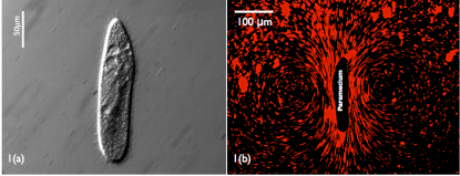
To investigate the flow-field around the organism suspensions of Polystyrene microspheres (5 m diameter, Thermo Scientific) prepared in EDC solution were introduced into the cultures. Small volume of the cultures 5 l were then placed on the glass slide which created a very thin film and allowed us to visualize the 2D flow field around the Paramecium. Figure 1(a) shows a Paramecium swimming in a thin film of liquid. We can see there are two strong vortices forming on the lateral sides of the organism; showing the strong tangential velocity of cilia on the far field of the organism.
The effect of confined geometries on the ciliary dynamics is examined by introducing the organisms in capillary tubes. Tubes required for this purpose are manufactured by attaching a dead weight to the end of the borosilicate glass pipettes and by heating their tips. By controlling the value of the dead weight and the intensity of the applied heat different diameters of capillary tubes ranging from 90250 m were manufactured. Some commercially available tubes with specific diameters(d=100,150, 200 m) made of borosilicate glass were ordered from Vitrotubes. The equilibrated cultures were then transferred to the extruded glass pipettes where they got pulled into the small constant cross section of the tube due to capillary forces.
II.2 EXPERIMENTAL OBSERVATIONS
Paramecium were found to have a long and slender structure with typical lengths around 21214 m and the width about 575 m (shown in Fig. 1(a)). The velocity of these micro-swimmers in unbounded fluid was found to be 106483 m/s. We then performed experiments with capillary tubes of different diameters. Paramecium swimming in buffer (isotonic solution) were put into the capillary tubes which caused them to be confined in small circular geometry. A generic code written in MATLAB was used to track the motion of these organisms and the velocities were computed.
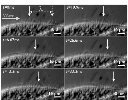
In order to measure the vital parameters for swimming; we captured the cilia motion with the high speed camera at 300 fps, which allowed us to visualize the metachronal wave propagation over the organism. Each cilium was found to be 1012 m in length and m in diameter and beats slightly out of phase compared to the nearby cilium, thereby causing a traveling wave to pass over the surface of the organism. The typical wavelengths of the metachronal waves measured from our experiments were 27 m, half peak to peak amplitude 4.2 m and the frequency of the beat being around 30 Hz Sleigh1 . We assume that the cilia are so closely packed that the fluid does not penetrate the material wave, thereby allowing no slip boundary condition to hold good.
Imaging of round capillaries under microscope caused optical distortions which leads to recording of altered amplitudes and velocities. We directly took images of the cross-section of various capillaries to get a relation between the true and the observed inner diameters of the tubes, which was further used to correct the observed amplitude and velocities of the organisms.
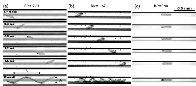
In tubes of extremely small diameter the swimming velocity of the organism was very low with almost a straight line motion with variable rotation rates. Whereas in tubes of larger diameter the organism was seen to move in a helical path instead of straight line motion. We applied a correction factor for the path of the swimming organisms, as the image analysis only revealed the 2D projection of the helix.
It is observed that the Paramecium swims slowly as the tube diameter is decreased. This can be attributed to the increased drag felt by the organism due to the proximity of the boundaries. For the capillary tubes whose diameter were very close to the diameter of the Paramecium, the swimming velocity was close to zero.
In tubes of smaller diameter () we observed that a backward (posterior) swimming Paramecium executed a helical swimming trajectory with small amplitude wavelengths. Such swimming gait have not been reported before Fukui1 ; Dryl1 . In this range of tube diameters and for the forward swimming Paramecium, little or no helical motion of the organism is observed.
III THEORETICAL MODEL
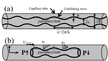
The governing equations for very low Reynolds number flows are the Stokes equations: . Since these organisms have a large length to diameter ratio; they can be effectively modeled as cylinders. As seen in the Figure 2 the cilia create synchronized motion to produce metachronal waves; to an observer this seems like a material wave propagating on the surface of the organism. Under the assumption that no fluid penetrates the wave of cilia tips since they are densely packed, the concept of envelop model can be applied for the physical system. The boundary in this case is the circular capillary tube; thus the problem reduces to modeling a cylinder with a wavy surface swimming inside a cylindrical geometry.
Figure 4 shows the schematic of the organism swimming the tube with a velocity . Paramecium has previously been modeled as an infinitely long cylinder so that there is no pressure gradient at the front and the back while swimming in the unbounded fluid. Similar infinite models can be developed for a Paramecium swimming inside a tube Blake3 . However, due to the presence of the confined spaces the finite length Paramecium experiences a pressure gradient at its ends that influences its locomotion within tubes. Considering the propagating wave to be of frequency ,wavelength , radial amplitude , transverse amplitude , and wave number ; any material point S on the undulating surface can be written as:
| (1) |
where is the phase difference.
We work in the frame in which the organism is swimming with a velocity . In the small amplitude limit, the boundary conditions of the surface of the organism at and on the solid walls at can be written as:
| (2) |
Thus the swimming problem can thus be envisioned as sum of pressure driven flow and shear flow in the narrow annulus that surrounds the Paramecium. We seek a solution in terms of the streamfunction such that where is the streamfunction corresponding to pressure driven flow and due to the the shear flow. Using cylindrical co-ordinates and axisymmetric potential theory we can write the velocity components to be ,. Solving for the pressure driven flow with stationary boundaries we can get the streamfunction to be:
| (3) |
For the shear flow part we can substitute the velocity components in the Stokes equation and by taking curl we end up with a equation of the form .
Rewriting coordinate systems as and and using separation of variables in and we can obtain a streamfunction solution of the form:
| (4) |
where,
and are the constants to be determined from the boundary conditions and and are modified Bessel functions of the first and second kind.
We seek perturbation expansions of the velocities and derived from the stream function to the and and compare the velocities with Equation 2. For calculations of the zeroth and first order the does not have any contribution and hence we are left to determine four unknowns for the problem.
Coupled with the pressure driven flow this gives us a set of five equations and six unknowns . Also from the flux condition of the swimming Paramecium we have . This gives us a condition between and . Thus we have a set of five equations and five unknowns which can be solved to find out the constants and the swimming velocity of the organism. The expression for swimming velocity of the Paramecium comes out to be:
| (5) |
The above expression shows that the swimming velocity is directly dependent on and also the pressure gradient terms. For the infinite boundaries case with no pressure gradient the above equation reduces to the following expression:
| (6) |
which is same as inBlake3 .
IV RESULTS
The motivation of the study was to rationalize the behavior of the organisms in close proximity to the boundaries. As the Paramecium swims inside the tube it traces out a helical path with the length of the body being aligned in the swimming direction. Figure 5 shows the variation of the amplitude of the organism as it swims in capillary tube of different diameters.
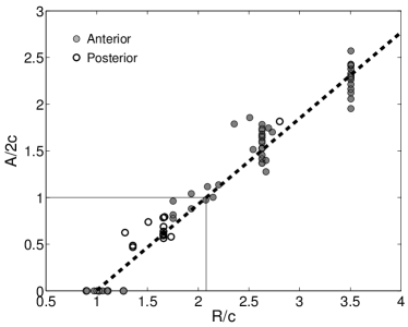
Theoretically we considered the small amplitude expansions for the waves and hence the expression derived for the swimming velocity would only be valid for smaller capillary tubes in which there is very less off axis movement. In larger tubes the organism does remain very close to the glass surfaces; but there is asymmetry in the boundary conditions that need to be applied. From the plot above and the constraints on the amplitude of swimming of Paramecium we can conclude that for our experimental and theoretical results would remain valid. This gives us corresponding value of the non-dimensional radius of the capillary tube .
Figure 6 shows the variation of velocity with the radius of the capillary tube. The dotted line shows the swimming velocity when no pressure gradient effects are considered and the solution converges very quickly to the case for which the boundaries are at infinity. In the experiments we observe a slowly increasing trend of velocity. For a finite size Paramecium swimming inside a restrictive geometry there is a finite pressure gradient at the front and the back. The plot of the swimming velocity considering finite pressure gradient is shown by the solid line. It shows a slowly increasing trend and finally converges to the infinite boundary and no pressure gradient case for very large values.
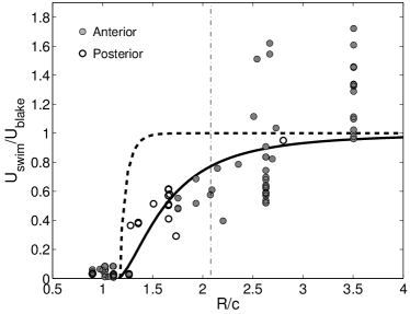
It can be seen that for both the experiments and theory show an increasing trend and predictions match quite well. For the larger we see that the velocities are much larger as opposed to that predicted value for the infinite boundary case. From the experiments we see that the organism follows the surface of the glass capillary as it swims in helical path in tubes of larger diameter. This situation can be thought of as Paramecium swimming close to a single solid wall and hence explains the observation of larger swimming velocities for tubes of larger diameter; where it constantly swims close to the wall. This reflects the large errors corresponding to the swimming case in larger tubes.
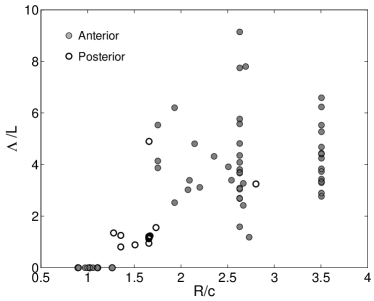
Figure 7 shows the variation of wavelengths of the helical path while they are swimming in the tube. The wavelengths also show a linearly increasing trend for the limit . In this limit the helices of the swimming are well defined and periodic wavelengths are observed. While swimming in tubes of larger diameter() it was observed many times that the organism did not execute a full helix.
V DISCUSSION
We investigated the locomotion of ciliary organisms in confined geometries. As the boundaries close on the organism, more viscous effects are felt by the cilia. The swimming velocity of Paramecium decreases due to the effect of close boundaries. Also in such confined spaces a finite sized organism feels a pressure gradient across the ends. This pressure gradient affects the swimming velocity and needs to be considered while modeling similar self propelling objects in restrictive geometries. Our experiments confirm this observation which show a slowly increasing trend of velocities.
Many interesting questions arise from this study especially about the amplitude of the waves propagated by the Paramecium. In a restrictive channel the beat of the waves is limited by the dimensions of channel and the size of organism. Due to the proximity, do the amplitudes of the beat change to provide a locomotory advantage?
The helical path traced out by the Paramecium with the anterior portion of the body aligned towards the local swimming direction is also an interesting locmotory trait. It was also observed that as the radii of the capillary tube increased the radius of the swimming helix also increased. It might be possible that being close to the curved surface provides a propulsive advantage. It was also found that anterior swimming Paramecium can execute well defined helical paths when put inside capillary tubes of certain diameter.
The study revealed the interesting locomotory traits in presence of solid wall. Future work would involve investigating the hydrodynamic effects of different textured boundaries on the swimming characteristics of the organism.
Acknowledgements.
The author Soong Ho Um was supported by NCRC program of the Korean Science and Engineering Foundation (Grant No. R15-2008-006-02002-0) and the National Research Foundation of Korea (NRF/MEST) (No. 20100007782; Mid-career Researcher Program).References
- (1) R. Stocker and W. M. Durham,“Tumbling for Stealth? ” Science 325 (5939), 400-402 (2009).
- (2) H. C. Berg and L. Turner, “Chemotaxis of bacteria in glass capillary arrays. Escherichia coli, motility, microchannel plate, and light scattering”, Biophysical Journal 58 (4), 919-930 (1990).
- (3) E. Lauga, W. R. DiLuzio, G. M. Whitesides and H. A. Stone,“Swimming in Circles: Motion of Bacteria near Solid Boundaries.” Biophysical Journal 90 (2), 400-412 (2006).
- (4) S. A. Biondi, J. A. Quinn and H. Goldfine, “Random motility of swimming bacteria in restricted geometries”. AIChE Journal 44 (8), 1923-1929 (1998).
- (5) K. P. Z Liu,“Unidirectional motility of Escherichia coli in restrictive capillaries.” Appl. Environ. Microbiol. 61, 3567-3572 (1995).
- (6) J. Mannik, R. Driessen, P. Galajda, J. E. Keymer and C. Dekker, “Bacterial growth and motility in sub-micron constrictions.” Proceedings of the National Academy of Sciences 106 (35), 14861-14866 (2009).
- (7) W. Wang, L. M. Shor, E. J. LeBoeuf, J. P. Wikswo and D. S. Kosson “Mobility of Protozoa through narrow channels” Appl. Environ. Microbiol. 71 (8), 4628-4637 (2005).
- (8) M. Sleigh, “Cilia and flagella.” (Academic Press, 1974).
- (9) D. J. Smith, E. A. Gaffney and J. R. Blake, “Mathematical modelling of cilia-driven transport of biological fluids.” Proc. R. Soc. Lond. A, 465 (2108), 2417-2439 (2009).
- (10) M.A. Sleigh,“Adaptations of ciliary systems for the propulsion of water and mucus”, Comparative Biochemistry and Physiology Part A: Physiology 94 (2), 359-364 (1989).
- (11) H. Machemer and K. Sugino, “Electrophysiological control of ciliary beating: A basis of motile behaviour in ciliated protozoa.” Comparative Biochemistry and Physiology Part A: Physiology 94 (2), 365-374 (1989).
- (12) S. Dryl and A. Grebecki, “Progress in the study of excitation and response in ciliates.” Protoplasma 62 (2), 255-284 (1966).
- (13) H. Machemer, “Ciliary Activity and the Origin of Metachrony in Paramecium: Effects of Increased Viscosity.” J Exp Biol 57 (1), 239-259 (1972).
- (14) H. Winet, “Wall Drag on Free-Moving Ciliated Micro-Organisms.” J Exp Biol 59 (3), 753-766 (1973).
- (15) G. Taylor, “Analysis of the swimming of microscopic organisms.” Proceedings of the Royal Society of London. Series A, Mathematical and Physical Sciences, 447-461 (1951).
- (16) D. F. Katz “On the propulsion of micro-organisms near solid boundaries.” Journal of Fluid Mechanics, 64 (01), 33-49 (1974).
- (17) E. Lauga and T. Powers, “The hydrodynamics of swimming microorganisms.” Reports on Progress in Physics 72, 096601 (2009).
- (18) A. Dauptain, J. Favier and A. Bottaro,“Hydrodynamics of ciliary propulsion.” Journal of Fluids and Structures 24 (8), 1156-1165 (2008).
- (19) J. Blake, “Infinite models for ciliary propulsion.”Journal of Fluid Mechanics 49 (02), 209-222 (2006).
- (20) J. R. Blake, “A spherical envelope approach to ciliary propulsion.” Journal of Fluid Mechanics 46 (01), 199-208 (1971).
- (21) S. R. Keller and T. Y. Wu “A porous prolate-spheroidal model for ciliated micro-organisms.” Journal of Fluid Mechanics, 80 (2), 259-278 (1977).
- (22) C. Brennen and H. Winet, “Fluid mechanics of propulsion by cilia and flagella.” Annual Review of Fluid Mechanics 9 (1), 339-398 (1977).
- (23) C. Brennen, “An oscillating-boundary-layer theory for ciliary propulsion.” Journal of Fluid Mechanics 65 (04), 799-824 (1974).
- (24) K. Fukui and H. Asai,“Spiral Motion of Paramecium caudatum in a Small Capillary Glass Tube.” Journal of Eukaryotic Microbiology 23 (4), 559-563 (1976).