A strategy on prion AGAAAAGA amyloid fibril molecular modeling
Jiapu Zhang
School of Sciences, Information Technology and Engineering, University of Ballarat,
Mount Helen, VIC 3350, Australia, j.zhang@ballarat.edu.au, Ph: (61)423487360
Abstract:
X-ray crystallography and nuclear magnetic resonance (NMR) spectroscopy are two powerful tools to determine the protein 3D structure. However, not all proteins can be successfully crystallized, particularly for membrane proteins. Although NMR spectroscopy is indeed very powerful in determining the 3D structures of membrane proteins, same as X-ray crystallography, it is still very time-consuming and expensive. Under many circumstances, due to the noncrystalline and insoluble nature of some proteins, X-ray and NMR cannot be used at all. Computational approaches, however, allow us to obtain a description of the protein 3D structure at a submicroscopic level.
To the best of the author’s knowledge, there is little structural data available to date on the AGAAAAGA palindrome in the hydrophobic region (113–120) of prion proteins, which falls just within the N-terminal unstructured region (1–123) of prion proteins. Many experimental studies have shown that the AGAAAAGA region has amyloid fibril forming properties and plays an important role in prion diseases. However, due to the noncrystalline and insoluble nature of the amyloid fibril, little structural data on the AGAAAAGA is available. This paper introduces a simple molecular modeling strategy to address the 3D atomic-resolution structure of prion AGAAAAGA amyloid fibrils. Atomic-resolution structures of prion AGAAAAGA amyloid fibrils got in this paper are useful for the drive to find treatments for prion diseases in the field of medicinal chemistry.
Keywords Prion AGAAAAGA palindrome, amyloid fibril, molecular modeling, prion dieseases.
1 Introduction
Prion diseases are invariably fatal and highly infectious neurodegenerative diseases affecting humans and animals. The neurodegenerative diseases such as Creutzfeldt-Jakob disease (CJD), variant Creutzfeldt-Jakob diseases (vCJD), Gerstmann-Straussler-Scheinker syndrome (GSS), Fatal Familial Insomnia (FFI), Kuru in humans, scrapie in sheep, bovine spongiform encephalopathy (BSE or mad-cow disease) and chronic wasting disease (CWD) in cattle belong to prion diseases. By now there have not been some effective therapeutic approaches or medications to treat all these prion diseases.
Prion diseases are amyloid fibril diseases. The normal cellular prion protein (PrPC) is rich in -helices but the infectious prions (PrPSc) are rich in -sheets amyloid fibrils. The conversion of PrPC to PrPSc is believed to involve a conformational change from a predominantly -helical protein (42% -helix, 3% -sheet) to a protein rich in -sheets (30% -helix, 43% -sheet) [9].
Many experimental studies such as [2, 3, 4, 10, 11, 12, 13, 14, 18] have shown two points: (1) the hydrophobic region (113-120) AGAAAAGA of prion proteins is critical in the conversion from a soluble PrPC into an insoluble PrPSc fibrillar form; and (2) normal AGAAAAGA is an inhibitor of prion diseases. Furthermore, we computationally clarified that prion AGAAAAGA segment indeed has an amyloid fibril forming property [20, 21, 22]. However, laboratory experiences have shown that using traditional experimental methods is very difficult to obtain atomic-resolution structures of AGAAAAGA due to the noncrystalline and insoluble nature of the amyloid fibril [16, 23]. By introducing novel mathematical canonical dual formulations and computational approaches, in this paper we may construct atomic-resolution molecular structures for prion (113 -120) AGAAAAGA amyloid fibrils.
Many studies have indicated that computational approaches or introducing novel mathematical formulations and physical concepts into molecular biology can significantly stimulate the development of biological and medical science. Various computer computational approaches were used to address the problems related to “amyloid fibril” [5, 6, 7, 8, 17, 19]. Here, we would like to use the simulated annealing evolutionary computations to build the optimal atomic-resolution amyloid fibril models in hopes to be used for controlling prion diseases.
The atomic structures of all amyloid fibrils revealed steric zippers, with strong van der Waals (vdw) interactions between -sheets and hydrogen bonds (HBs) to maintain the -strands [15]. The vdw contacts of atoms are described by the Lennard-Jones (LJ) potential energy:
| (1) |
where is the depth of the potential well and is the atom diameter; these parameters can be fitted to reproduce experimental data or deduced from results of accurate quantum chemistry calculations. The term describes repulsion and the term describes attraction. If we introduce the coordinates of the atoms whose number is denoted by and let be the reduced units, the form (1) becomes
| (2) |
where , is the coordinates of atom , . The minimization of LJ potential on (where ) is an optimization problem:
| (3) |
Similarly as (1), i.e. the potential energy for the vdw interactions between -sheets:
| (4) |
the potential energy for the HBs between the -strands has the formula
| (5) |
where are given constants. Thus, the amyloid fibril molecular modeling problem is deduced into well solve the optimization problem (3).
This paper is organized as follows. In Section 2, we first describe how to build the prion AGAAAAGA amyloid fibril molecular models, and then explain how the models with 6 variables only are built and can be solved by any optimization algorithm. At the end of Section 2 the models are done a little refinement by Amber 11 [1]. At last, we conclude that when using the time-consuming and costly X-ray crystallography or NMR spectroscopy we still cannot determine the protein 3D structure, we may introduce computational approaches or novel mathematical formulations and physical concepts into molecular biology to study molecular structures. This concluding remark will be made in the last section.
2 Prion AGAAAAGA amyloid fibril models’ Molecular Modeling and Optimizing
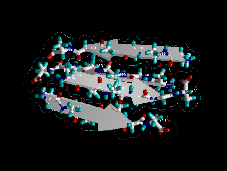
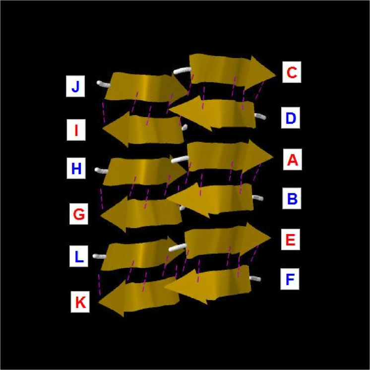
Constructions of the AGAAAAGA amyloid fibril molecular structures of prion 113–120 region are based on the most recently released experimental molecular structures of human M129 prion peptide 127–132 (PDB entry 3NHC released into Protein Data Bank (http://www.rcsb.org) on 04-AUG-2010). The atomic-resolution structure of this peptide is a steric zipper, with strong vdw interactions between -sheets and HBs to maintain the -strands (Figure 1).
In Figure 1 we see that G (H) chains (i.e. -sheet 2) of 3NHC.pdb can be obtained from A (B) chains (i.e. -sheet 1) by
| (6) |
and other chains can be got by
| (7) |
| (8) |
where is the 3-by-3 identity matrix. Basing on the template 3NHC.pdb from the Protein Data Bank, three prion AGAAAAGA palindrome amyloid fibril models - an AAAAGA model (Model 1), a GAAAAG model (Model 2), and an AAAAGA model (Model 3) - will be successfully constructed in this paper. AB chains of Models 1-3 were respectively got from AB chains of 3NHC.pdb using the mutate module of the free package Swiss-PdbViewer (SPDBV Version 4.01) (http://spdbv.vital-it.ch). It is pleasant to see that almost all the hydrogen bonds are still kept after the mutations; thus we just need to consider the vdw contacts only. Making mutations for GH chains of 3NHC.pdb, we can get the GH chains of Models 1-3. However, the vdw contacts between A chain and G chain, between B chain and H chain are too far at this moment (Figure 2).
Seeing Figure 2, we may know that for Models 1-3 at least two vdw interactions between A.ALA3.CB-G.ALA4.CB, B.ALA4.CB-H.ALA3.CB should be maintained. Fixing the coordinates of A.ALA3.CB and B.ALA4.CB, letting the coordinates of G.ALA4.CB and H.ALA3.CB be variables, we may get a simple LJ potential energy minimization problem (3) just with six variables. For solving this six variable optimization problem, any optimization computational algorithm can be used to solve this low-dimensional problem; for example, in this paper we may use the hybrid discrete gradient simulated annealing method [22]. Setting the coordinates of G.ALA4.CB and H.ALA3.CB as initial solutions, running the hybrid discrete gradient simulated annealing optimization algorithm, for Models 1-3 we get
| (9) |
By (9) we can get close vdw contacts between A chain and G chain, between B chain and H chain (Figure 3).
Furthermore, we may employ the Amber 11 package [1] to slightly optimize Models 1-3 and at last get Models 1-3 with stable total potential energies (Figure 4). The other CDIJ and EFKL chains can be got by parallelizing ABGH chains in the use of mathematical formulas (7)-(8).
3 Conclusion
X-ray crystallography is a powerful tool to determine the protein 3D structure. However, it is time-consuming and expensive, and not all proteins can be successfully crystallized, particularly for membrane proteins. Although NMR spectroscopy is indeed a very powerful tool in determining the 3D structures of membrane proteins, it is also time-consuming and costly. Due to the noncrystalline and insoluble nature of the amyloid fibril, little structural data on the prion AGAAAAGA segment is available. Under these circumstances, the novel simple strategy introduced in this paper can well do the molecular modeling of prion AGAAAAGA amyloid fibrils. This indicated that computational approaches or introducing novel mathematical formulations and physical concepts into molecular biology can significantly stimulate the development of biological and medical science. The optimal atomic-resolution structures of prion AGAAAAGA amyloid fibils presented in this paper are useful for the drive to find treatments for prion diseases in the field of medicinal chemistry.
Acknowledgments: This research was supported by a Victorian Life Sciences Computation Initiative (VLSCI) grant number VR0063 on its Peak Computing Facility at the University of Melbourne, an initiative of the Victorian Government.
References
- [1] Case, D.A., T.A. Darden, T.E. Cheatham, III, C.L. Simmerling, J. Wang, R.E. Duke, R. Luo, R.C. Walker, W. Zhang, K.M. Merz, B.P. Roberts, B. Wang, S. Hayik, A. Roitberg, G. Seabra, I. Kolossv ry, K.F. Wong, F. Paesani, J. Vanicek, J. Liu, X. Wu, S.R. Brozell, T. Steinbrecher, H. Gohlke, Q. Cai, X. Ye, J. Wang, M.-J. Hsieh, G. Cui, D.R. Roe, D.H. Mathews, M.G. Seetin, C. Sagui, V. Babin, T. Luchko, S. Gusarov, A. Kovalenko and P.A. Kollman. 2010. AMBER 11, University of California, San Francisco.
- [2] Brown D.R. (2000) Prion protein peptides: optimal toxicity and peptide blockade of toxicity. Mol. Cell. Neurosci. 15: 66 -78.
- [3] Brown D.R. (2001) Microglia and prion disease. Microsc. Res. Tech. 54: 71- 80.
- [4] Brown D.R., Herms J., Kretzschmar H.A. (1994) Mouse cortical cells lacking cellular PrP survive in culture with a neurotoxic PrP fragment. Neuroreport 5: 2057–2060.
- [5] Carter D.B., Chou K.C. (1998) A model for structure dependent binding of Congo Red to Alzeheimer beta-amyloid fibrils. Neurobiol. Aging 19: 37–40.
- [6] Chou K.C. (2004) Insights from modelling the tertiary structure of BACE2. J. Proteome Res. 3: 1069–72.
- [7] Chou K.C. (2004) Review: structural bioinformatics and its impact to biomedical science. Curr. Med. Chem. 11: 2105–34.
- [8] Chou K.C., Howe W.J. (2002) Prediction of the tertiary structure of the beta-secretase zymogen. Biochem. Biophys. Res. Commun. 292: 702–8.
- [9] Griffith J.S. (1967) Self-replication and scrapie. Nature 215: 1043 -4.
- [10] Holscher C., Delius H., Burkle A. (1998) Overexpression of nonconvertible PrPC delta114-121 in scrapie-infected mouse neuroblastoma cells leads to trans-dominant inhibition of wild-type PrPSc accumulation, J. Virol. 72: 1153–9.
- [11] Jobling M.F., Huang X., Stewart L.R., Barnham K.J., Curtain C., Volitakis I., Perugini M., White A.R., Cherny R.A., Masters C.L., Barrow C.J., Collins S.J., Bush A.I., Cappai R. (2001) Copper and zinc binding modulates the aggregation and neurotoxic properties of the prion peptide PrP 106–126, Biochem. 40: 8073–84.
- [12] Jobling M.F., Stewart L.R., White A.R., McLean C., Friedhuber A., Maher F., Beyreuther K., Masters C.L., Barrow C.J., Collins S.J., Cappai R. (1999) The hydrophobic core sequence modulates the neurotoxic and secondary structure properties of the prion peptide 106–126, J. Neurochem. 73: 1557–65.
- [13] Kuwata K., Matumoto T., Cheng H., Nagayama K., James T.L., Roder H. (2003) NMR-detected hydrogen exchange and molecular dynamics simulations provide structural insight into fibril formation of prion protein fragment 106–126, Proc. Natl. Acad. Sci. USA 100, 14790–5.
- [14] Norstrom E.M., Mastrianni J.A. (2005) The AGAAAAGA palindrome in PrP is required to generate a productive PrPSc–PrPC complex that leads to prion propagation, J. Biol. Chem. 280: 27236–43.
- [15] Sawaya M.R., Sambashivan S., Nelson R., Ivanova M.I., Sievers S.A., Apostol M.I., Thompson M.J., Balbirnie M., Wiltzius J.J., McFarlane H.T., Madsen A., Riekel C., Eisenberg D. (2007) Atomic structures of amyloid cross-beta spines reveal varied steric zippers, Nature 447: 453–7.
- [16] Tsai H.H.G. (2005) Understanding the biophysical mechanisms of protein folding, misfolding, and aggregation at molecular level (in Chinese), Chem. (The Chinese Chem. Soc. of Taipei) 63: 601- 12.
- [17] Wang J.F., Wei D.Q., Li L., Chou K.C. (2008) Review: Drug candidates from traditional Chinese medicines, Curr. Top. Med. Chem. 8: 1656–65.
- [18] Wegner C., Romer A., Schmalzbauer R., Lorenz H., Windl O., Kretzschmar H.A. (2002) Mutant prion protein acquires resistance to protease in mouse neuroblastoma cells, J. Gen. Virol. 83: 1237–45.
- [19] Wei D.Q., Sirois S., Du Q.S., Arias H.R., Chou K.C. (2005) Theoretical studies of Alzheimer’s disease drug candidate [(2,4-dimethoxy) benzylidene]-anabaseine dihydrochloride (GTS-21) and its derivatives, Biochem. Biophys. Res. Commun. 338: 1059–64.
- [20] Zhang J.P. (2009) Studies on the structural stability of rabbit prion probed by molecular dynamics simulations, J. Biomol. Struct. Dyn. 27: 159 -62.
-
[21]
Zhang J.P. (2011) Optimal molecular structures of prion AGAAAAGA amyloid fibrils formatted by simulated annealing, J. Mol. Model. 17: 173 -9. Reported by Crystallography Time, Volume 3, No. 1, January 2011:
http://www.rigakumsc.com/downloads/newsletter/LifeSciencesV03N01.html
and VerticalNews 2011 FEB 1:
http://technology.verticalnews.com/articles/4831360.html -
[22]
Zhang J.P., Sun J., Wu C.Z. (2011) Optimal atomic-resolution structures of prion AGAAAAGA amyloid fibrils, J. Theor. Biol. J. Theor. Biol. 279(1) (2011) 17- 28. PMID: 21420420. arXiv:1012.2504v6. Selected by Nuclear Energy Research Today Volume 7 Issue 4, April 2011:
http://nuclearenergy.researchtoday.net/archive/7/4/4311.htm - [23] Zheng J., Ma B.Y., Tsai C.J., Nussinov R. (2006) Structural stability and dynamics of an amyloid-forming peptide GNNQQNY from the yeast prion Sup-35, Biophy. J. 91: 824- 33.
![[Uncaptioned image]](/html/1012.5306/assets/x2.png)
![[Uncaptioned image]](/html/1012.5306/assets/x3.png)

![[Uncaptioned image]](/html/1012.5306/assets/x5.png)
![[Uncaptioned image]](/html/1012.5306/assets/x6.png)
![[Uncaptioned image]](/html/1012.5306/assets/x8.png)
![[Uncaptioned image]](/html/1012.5306/assets/x9.png)