The mechanics of a microscopic mixer: microtubules and cytoplasmic streaming in Drosophila oocytes
Abstract
Large scale motion of cytoplasm called cytoplasmic streaming occurs in some large eukaryotic cells to stir the cell’s constituents. In Drosophila oocytes, microtubules have been observed to undergo undulating motion, curving to form travelling waves during cytoplasmic streaming. Here we show that this wave-like motion can be understood physically as due to the hydrodynamic drag of streaming impellers attached to kinesin motors moving toward the plus-ends of microtubules whose minus ends are anchored to the cell cortex. The tangential forces applied to such microtubules by kinesin give rise to bending and leads to chiral symmetry breaking causing the microtubules to propagate long travelling waves. The waves are reminiscent of those seen in flagellar motion but of a much longer time scale and by a different physical mechanism. We show how kinesin movement can produce a bulk flow of cytoplasm surrounding a microtubule with the range of flow greatly enhanced by the effect of hydrodynamic coupling between impellers. That is, a relatively small number of motors can move a large amount of fluid. The chaotic nature of the fluid motion of cytoplasm caused by kinesin movement along constantly changing microtubule trajectories is important as it greatly enhances the efficiency of mixing. Existing data on in vitro microtubule gliding assays also show this chiral instability in two dimensions and an analysis of this gives quantitative estimates for the forces exerted by motors and the drag coefficient.
I Introduction
Microtubules are flexible hollow polymers of tubulin subunits that serve many critical functions in eukaryotic cells. They are utilized in structural contexts, because of their relatively stiff, yet flexible, mechanical properties. They also act as directional highways through the viscous cytoplasm. Molecular motor proteins carry cellular constituents along the microtubules with kinesin moving toward their fast growing “plus-ends” and dynein moving toward their slow growing “minus-ends”. Many studies have focused on motor driven transport processes that generate asymmetric distributions of specific cytoplasmic constituents; asymmetries that are essential for complex cellular functions. In this letter we will analyze a surprising role for microtubules and kinesin in a mass transport process called cytoplasmic streaming that has evolved to accomplish just the opposite; efficient homogeneous mixing of the contents of a cell SerbusSaxton and see there Supplemental Movie 13 Movie13 .
Vigorous streaming is initiated during the final stages of development of Drosophila oocytes to disperse asymmetrically distributed mRNA particles, protein complexes, and membranous organelles. The mixing process is important for subsequent embryonic development, and cannot be accomplished by diffusion alone. For example a yolk-filled vesicle in the cytoplasm, assuming a viscosity 8 times that of water LubyPhelps would take approximately a week to diffuse the length of an oocyte. This is far too long to satisfy the need for the mixing of yolk-filled oocyte cytoplasm with the mass of yolkless nurse cell cytoplasm that floods the anterior of the oocyte near the end of its development.
The problem of mixing in small systems, such as in microfluidics chambers, has been the subject of much investigation Squires . The way that a fluid at low Reynolds number is stirred has a profound effect on the efficiency of homogenization. For example, the steady state flow fields generated by a single stir bar inside a closed chamber are far less efficient than the more chaotic flows generated when several stir bars are used Aref ; Aref2000 . Rigorous analyses in two dimensions show that mixing is most efficient when topological chaos is created by three or more stirrers, as can be shown by application of the Thurston-Nielsen classification theorem Thurston ; Fathi ; Handel . With this in mind, it is interesting to note that during fast cytoplasmic streaming in Drosophila oocytes, microtubules appear to be locally aligned along dynamically changing curved paths that produce travelling waves SerbusSaxton . The streaming cytoplasmic fluid moves along those paths, in patterns reminiscent of flowing water and seaweed. Yolk particles in the cytoplasm have a speed of roughly . The particles within a region stream for the most part in one direction, but with a non-negligible deviation in that direction over time. The fluctuating directions, which parallel the curved paths of the microtubules in the same region, serve to stir cytoplasm in a chaotic manner that, as the preceding paragraph suggests, is important for efficient mixing.
At first sight, it might appear that the time-dependent wave-like motion is due to turbulence of the surrounding fluid. However at such minuscule Reynolds numbers, inertial effects are negligible and turbulence is impossible BergRandomWalksinBiology . Therefore one is left with a mystery of the relationship of fluid and filament and how such chaotic patterns could come about. The plus-end directed motor kinesin-1 plays a crucial role in this, as shown by genetic mutations in its force producing subunit (Khc) that prevent streaming and mixing SerbusSaxton . The speed of unloaded kinesin along the microtubule has been measured to be in the range SvobodaBlock ; MeyhoferHoward . This is higher than the fluid speeds measured during fast cytoplasmic streaming SerbusSaxton and is also consistent with the role of kinesin in powering the mixing. Inhibition of the opposing minus-end directed motor protein, dynein, has a complementary effect, actually stimulating fast cytoplasmic streaming SerbusSaxton . Microscopy studies suggest that microtubules participating in this motion have their minus ends attached to the cortex and their plus ends away from the cortex in the interior of the oocyte SerbusSaxton ; ChaSerbus .
The explanation that we analyze for the streaming phenomena is very simple: the mass motions of cytoplasm and the microtubule undulations are complementary physical consequences of kinesin moving cargo toward the plus ends of microtubules whose minus ends are in contact with the cortex. The cargoes serve as impellers that both drive the fluid motion away from minus-ends and generate tangential forces that move plus-ends toward minus-ends causing bends in the microtubules. This has been suggested previously to explain cytoplasmic streaming, but without a physical model of the mechanism SerbusSaxton . The analysis below shows that long range hydrodynamic forces couple individual impellers, resulting in an effective mechanism for drag-induced bulk movement of cytoplasm. We then show that an instability in the dynamics leads to chiral symmetry breaking giving rise to wave-like motion of microtubules, and show that the time and length scales predicted are in good agreement with the previous experimental results.

II Enhancement of Streaming Due to Hydrodynamics
Consider impellers to be objects each with maximum linear dimension , and with a mean spacing of arranged in a straight line as shown in Fig 1. These impellers are pictured as spheres, but hydrodynamics is not sensitive to the exact shape of an impeller, as it depends mainly on its maximum linear dimension BergRandomWalksinBiology . We will now analyze the amount of streaming due to motion of these impellers moving on a single microtubule. We initially consider the impellers to be much larger than the kinesin molecules so that they dominate the hydrodynamical response of the fluid.
If spherical impellers were close-packed along the microtubule, that is , then this problem would be equivalent to a single rod of length moving at constant velocity in the fluid in a direction parallel to its long axis. In that case, the velocity field for distances has only a weak logarithmic dependence of , meaning that up to a correction of order , the velocity field is only weakly dependent on distance and of order . (Here we take the velocity of the fluid to go to zero far from the rod.) This is related to the well known result that the drag on a rod of length is of the same order as that of a sphere of diameter despite the latter’s much greater volume BergRandomWalksinBiology . Such a system of densely packed impellers would be very efficient at driving fluid motion and only a low density of microtubules would be needed for cytoplasmic streaming.
Now consider the more realistic case in which the impeller size is less than the spacing between them, which is much less than the length of a microtubule, that is . At a distance , the velocity field will behave just as in the closed-packed case except appear to have a diminished impeller velocity. That is for , the magnitude of the velocity , where contains all the distance dependence of the velocity field, and is how the velocity scales with the ratio of . In the limit where is very small, the system becomes dilute and the velocity between impellers will decay to zero. We recover the motion of isolated impellers in this case, where it is well known (e.g. Stokes’ drag) that the velocity field is proportional to . Therefore for small argument , is linear (in other words, the fluid velocity is proportional to .)
Using this general argument we conclude that, independent of the exact shape of the impellers, the flow velocity is reduced from the closed-packed case by a factor . The effect of the impellers only starts decreasing substantially at a distance of order the length of the microtubule , below which it should only have a weak logarithmic dependence. In other words, for a spherical region just enveloping a microtubule, the fluid velocity is slowly varying and reduced from the kinesin motor velocity by a factor of order .
Of course there are many microtubules in these cells. To understand how this affects the above analysis, consider all space filled with an infinite forest of them all oriented in the same direction. First if we ignore the microtubules and just consider the spherical impellers, then if we move to a reference frame moving with the impeller velocity, the system is static and the velocity everywhere is zero. Therefore in the original reference frame, the fluid is also moving uniformly at the impeller velocity. This is not correct because we have ignored the hydrodynamic drag of the microtubules represented by the line going through the spheres in Fig. 1. To estimate their effect on the fluid velocity, denote the drag coefficient on an impeller by and that of a section of microtubule length by . Then we go to a reference frame velocity such that the total force acting on the combined system of impellers and microtubules is zero, so that , or . Because the net force acting on this system is zero in this frame, is the velocity of the fluid far from the microtubules. Because within logarithmic corrections, the drag coefficients are proportional to the maximal linear dimensions, then a conservative estimate of this speed is of order . The exact formula depends on the shape of the impellers. This argument assumes an infinite volume of microtubules but the corrections to this due to the finite nature of the system are not important for the estimates we are making.
The identities of impellers in this system are still unknown but there are many possible candidates. Anything with large linear dimensions in at least one direction will give a large hydrodynamic radius BergRandomWalksinBiology . The other requirement is that it can attach to kinesin. In fact it is possible that the impellers in this situation could themselves be microtubules that are not attached to the cortex WangRiechmann ; Seeger . Experimental estimates of the cytoplasmic streaming velocity are approximately , about to the typical velocity of a kinesin molecule. This suggests that is or greater. For example, if we take the maximum dimensions of an impeller to be , this predicts a spacing between impellers of or less.
Another important biological issue is the necessity to have some microtubules in direct physical contact with the cortex of the oocyte, for example by tethering or by frictional forces. A free floating microtubule with kinesin moving on it will apply zero net force to the fluid. This is a simple consequence of Newton’s third law, or equivalently, conservation of momentum. This does not contradict the fact that bacteria are able to swim: the force propelling the bacterium forward is countered by an equal and opposite force on the environment, leading to velocity fields that are dipolar at large distances. Unlike the case analyzed above, this will not lead to long range hydrodynamic motion of the fluid and will not lead to efficient cytoplasmic streaming by relatively few motor proteins. Contact with the cortex is crucial as it allows for transfer of force from outside of the oocyte to the cytoplasm enabling fast streaming of the bulk to be powered by a much smaller volume of kinesin driven impellers.
III Travelling Wave Instability of Microtubules
We now show how the kinesin generated tangential forces on microtubules give rise to travelling wave conformations and calculate their angular and spatial frequency. A microtubule has a configuration parameterized by an arclength and, at long enough length scales, can be modeled as being inextensible, that is with an elastic bending constant . The inextensibility is enforced by a position dependent tension . There is also a force acting on the microtubule as a result of kinesin walking along it. The magnitude of the force is proportional to the local speed and the size of the kinesin-driven impeller and the direction of the force is tangent to the microtubule, which, we will see, has the effect of making it buckle. We denote this with a force per unit length of . We also include a force due to the cytoplasm streaming at a velocity which we take to be in the direction away from the minus end and this force tends to straighten the microtubule. This leads to the equation
| (1) |
where is a hydrodynamic drag coefficient per unit length.
We first implemented this equation numerically for a range of parameters and enforced boundary conditions that tethered the minus end against the cortex, while the plus end was free. Starting from random initial conditions, the equation rapidly goes to a steady state that typically is described by a curve that asymptotically becomes helical for large and rotates uniformly at constant angular velocity. The results of a steady state configuration are shown in Fig. 2. The supplemental movies SupplMovies discussed below, show microtubule solutions for different parameters. Supplemental movie 1 shows the full time dependence SupplMovies . The chirality of the helix depends on its initial conditions. Therefore the direction of rotation is random but stable once steady state is reached.
As the microtubule is made longer, the angular velocity and radius of the helix go to a constant limit. However the tethered part is not helical but nevertheless rotates in synchrony with the rest of the microtubule. These dynamics we analyzed in detail (see the supplemental information) to find the form of the solution to this equation. In particular we show that this equation supports travelling waves and find the relationship between the angular velocity of rotation and the asymptotic radius of the helix, and the external velocity fields . In the case where , the relationship simplifies to
| (2) |
independent of chain length.
It is interesting to note that for travelling waves, solutions can be at any scale. There is a continuous family of solutions all with the same shape but different scale factors. In the case of a helix, many different radii are solutions to these equations.
What determines the value of that is selected? As with other problems in pattern formation such as the “geometric model” Kessler or the full dendrite problem Barbieri , it is the boundary conditions that are responsible for the unique value of that is selected. In this case, the microtubule minus end is tethered, but the plus end is free. The solution will only exist for discrete values of . Numerical analysis gives, . This implies that
| (3) |
This analysis was extended to non-zero and as shown in the supplemental materials SupplMat , does not appreciably change the estimate we will give below for microtubule wave parameters in fast streaming oocytes.
When the microtubule is tethered to an impenetrable surface (in this case, the oocyte cortex) and the external cytoplasmic streaming velocity is parallel to that surface, the form of the solution changes considerably. Numerical results show that the microtubule becomes completely two dimensional, lying close to the surface. For low enough , travelling wave solutions are close to prolate cycloids, meaning that the microtubule periodically loops back on itself (see supplemental movie 2 SupplMovies . This can be understood analytically. For sufficiently large it transitions to other states finally becoming two dimensional and looking close to a sinusoid (see supplemental movie 3 SupplMovies . This is analyzed in detail in the supplemental materials SupplMat .
We have not included hydrodynamic and steric interactions between different microtubules except in the approximate way of giving rise to a constant cytoplasmic streaming velocity. Given the microtubule density in the oocyte, we expect these interactions to be substantial. However the wave like motion that we find is quite robust. Attaching microtubules together or considering additional forces still leads to periodic or sometimes chaotic motion, still at the same characteristic time and spatial scales. Therefore despite the simplicity of the model, we expect that the basic length and time scales that we predict should be quite robust.
IV Comparison with Experiment and Discussion
We now check to see whether the above model is consistent with experimental results.
Estimates of the microtubule elastic constant vary considerably Felgner ; GittesRigidity but range mostly within to . We estimate the force due to the kinesin per unit length, to be its velocity times the cytoplasmic viscosity. We will assume as suggested by our above analysis, that is between and . We will take the kinesin velocity to be approximately SvobodaBlock ; MeyhoferHoward . The effective viscosity of the cytoplasm for small particles has been studied extensively and appears to vary depending on the type of cell on the length scale LubyPhelps . Particles of different sizes diffusing in the cytoplasm diffuse as if the medium had a different viscosity. Its viscoelastic properties will depend on many factors such as the state of gelation of actin filaments YinStossel . We will assume that during fast cytoplasmic streaming, the cytoplasmic actin is mainly in a ”sol” state allowing fast streaming to more readily take place. If this is not the case, the effective viscosity could be much higher LubyPhelps but the dissipation would increase proportionally requiring a higher energy input. For small particles of radius approximately , the effective viscosity as measured by diffusion is approximately 8 times that of water LubyPhelps and we will use this value for comparison with experiment. This gives in the range to . Plugging these numbers into Eq. 3 gives in the range to . We can estimate the angular velocity using Eq. 2 (which ignores cytoplasmic streaming). To get the largest range of times, we assume that the hydrodynamic drag coefficient is due solely to the impellers, so that . This is equivalent to or a period of which ranges from to .
Serbus and colleagues have observed microtubule behavior in living Drosophila oocytes using GFP-tubulin and confocal fluorescence microscopy SerbusSaxton . Time-lapse movies showed bright fibrous fluorescence, representing groups of microtubules, in a background of diffuse fluorescence from non-polymerized GFP-tubulin. The microtubule patterns changed over time, as did the patterns of motion of organelles that either excluded the GFP-tubulin or were themselves auto-fluorescent. Bends in the microtubules were at times visible in the optical plane and some remained visible within that plane for , allowing measurement of a radius of curvature that we expect to approximately correspond to the in the above analysis. was measured to be with measurements (standard error ), and the wave velocity was with measurements. Because these waves appear roughly sinusoidal, we can also estimate the period . Assuming a wavelength of then the measured velocity gives a period of with an even larger error bar considering the fact that this assumes a waveform that is probably not accurate. Nevertheless it suggests a characteristic time.
The motion studied here can be contrasted with ciliary motion Kennedy which has a period on the order of . Clearly the two mechanisms have fundamentally different explanations. The experimental time scale points to the mechanism described here rather than ciliary motion. The agreement found with experiment in our above analysis is to some extent fortuitous, given the large uncertainties in the experimental system. But nevertheless, it provides evidence that the simple mechanism proposed is the origin of the behavior seen in cytoplasmic streaming. The experimental evidence SerbusSaxton that dynein inhibits streaming and that minus ends of microtubules are in contact with the cortex ChaSerbus , both support this hypothesis as well.
More generally, the above analysis elucidates, at a qualitative level, the phenomena seen in experiment SerbusSaxton , of strong cytoplasmic streaming occurring concomitantly with wave-like motion of microtubules. The movement of a relatively few number of kinesin motors on a microtubule couples to the fluid causing a bulk hydrodynamic flow. The force a kinesin exerts on a microtubule sets the microtubule into motion causing it to execute wave-like motion. This in turn causes the flow lines around microtubules to be time dependent, making the microtubules act as stirrers for the surrounding fluid, which leads to chaotic flows and strong mixing Aref ; Aref2000 .
(a)  (b)
(b)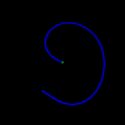
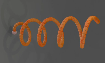
Can such motion be directly observed in an in vitro experiment? In fact it has been seen frequently as an unwanted artifact of sample preparation. Microtubule gliding assays were pioneered more than two decades ago ValeSchnappEtAl and have become a standard technique to better understand the motion of kinesin motors. When a solution microtubules and ATP is placed on a glass surface on which there is a high density of kinesin molecules, the kinesins propel the microtubules with their minus ends leading. Occasionally the minus end sticks, perhaps to damaged kinesins, and continued forces of the trailing portion of the microtubule cause flexing and curve generation.
Microtubule gliding assay videos of high quality MaloneyHerskowitzKoch provide an excellent source of data for investigating the two dimensional version of the problem studied here MaloneyKochVideo . The microtubule is being pushed by kinesin molecules that on average, apply a force tangent to the microtubule. When an end is tethered by sticking to a kinesin molecule, as noted above, this leads to a physical situation that is well described by Eq. 1. It should be emphasized that the values of the tangential force and the drag coefficient will be much larger than in the oocyte case. Consequently, sometimes microtubules flex,, forming a G-shaped curve that rotates around the anchored minus end of the microtubule. Fig. 3 shows a comparison of snapshots of the microtubule with that of a simulation of Eq. 1. The resemblance is quite striking and provides evidence that the theoretical modelling of this bending instability is valid. Note that the shape obtained is independent of the values of model parameters. From measuring the radius of the G-like configuration and its angular velocity of rotation, we can compute both and from Eqs. 3 and 2, giving and . Measurements give and the period . Therefore and , both estimates are accurate within a factor of 2 assuming that has been well determined. The force exerted by a kinesin motor in this situation is close to the experimentally determined stall value of approximately MeyhoferHoward . Assuming that each motor attached can exert this force, this gives an average distance between motors of . This assumes that the attachment of the motors is flexible enough to allow microtubule interactions capable of exerting this stall force. If this is not the case, we expect the kinesins to be spaced more closely but by no more than half this amount. This distance seems reasonable given the high density of kinesin molecules attached to the glass, and the size of the tubulin dimer which is approximately in length. Thus the above considerations can give a quantitative measure of the forces and drag in these gliding assays.
Nevertheless, it would also be of interest to verify the travelling wave solutions experimentally in three dimensions for isolated microtubules and kinesin, for example, by utilizing a single molecule optical trap, or by some other means, allowing for a more rigorous test of the predictions made here.
It is of interest to speculate on the reason for this microscopic mixing mechanism. If the microtubules were attached from the plus end, there would be no dynamical instability from kinesin generated forces but the cytoplasmic streaming would be more efficient, with greater transfer of force from outside the oocyte, because the tension at the tether point would increase linearly with the length of the microtubule, whereas the tension saturates to a constant value in the case considered above. However as discussed earlier, complex stirring patterns are expected to be far more efficacious in mixing than steady-state flows. This agitation could then serve an important biological function.
In summary, we have found a very simple set of conditions for generating efficient mixing in large eukaryotic cells. If microtubules are aligned so that their minus ends are in contact with the cell perimeter and dynein is suppressed, this will lead (a) to cytoplasmic streaming in the bulk of the cell and (b) wave-like motion of microtubules. The latter will promote efficient mixing of the cell’s contents by inducing chaotic flows. The efficiency of the cytoplasmic streaming is due to the long range nature of hydrodynamic coupling and the indirect linkage of microtubules to the oocyte surroundings via the cortex, leading to momentum transfer. The wave-like motion is due to an instability in the dynamics which breaks chiral symmetry and is a consequence of the tangential forces exerted by motor proteins on the elastic microtubules. Given the uncertainties in the experimental system and approximations made in the analysis, The wave-like motion seen in experiments agrees well with our theoretical predictions suggesting these effects represent a robust phenomenon. We are now engaged in experimentation to gather more accurate data on microtubule behavior in vivo and in vitro under various conditions in order to further understand the mechanisms for fast cytoplasmic streaming discussed here.
The authors would like to thank Ian Carbone, and Bill Sullivan for useful discussions. We also would like to gratefully acknowledge Andy Maloney and Steven J. Koch for their permission to use a snapshot of their video, Fig. 3(a) MaloneyKochVideo of their experimental results MaloneyHerskowitzKoch . This was generously distributed to us by them KochLab through the practice of Open Notebook Science OpenNotebookScience This material is based upon work supported by National Institutes of Health GM046295 (to W.M.S.), National Science Foundation CCLI Grant DUE-0942207 (to J.M.D.), the Defense Threat Reduction Agency basic research grant HDTRA-1-09-1-0018 (to Steven J. Koch), and the National Science Foundation IGERT Grant DGE-0549500 (to Steven J. Koch).
Note: After the completion of this work, we came across work of Bourdieu et al. Bourdieu95 which analyzed their experimental results on spirals in mysosin and kinesin gliding assays using the two dimensional version of Eq. 1, obtaining the identical scaling, and simulating it using a nearly identical approach. This provides further evidence that the mechanism we have proposed for cytoplasmic streaming is physically viable.
References
- (1) L.R. Serbus, B.J. Cha, W.E. Theurkauf, and W.M. Saxton, “Dynein and the actin cytoskeleton control kinesin-driven cytoplasmic streaming in Drosophila oocytes.” Development 132, 3743-3752 (2005).
- (2) See ref. SerbusSaxton Movie 13.
- (3) K. Luby-Phelps, “Cytoarchitecture and physical properties of cytoplasm: volume, viscosity, diffusion, intracellular surface area.” Int. Rev. Cytol. 192 189–221 (2000). fluorescence redistribution after photobleaching.” Journal of Cell Biology. 99 2165-2174 (1984).
- (4) T. M. Squires and S. R. Quake, “Microfluidics: Fluid physics at the nanoliter scale.” Rev. Mod. Phys., 77 977-1026 (2005)
- (5) H. Aref, “Stirring by chaotic advection.” J Fluid Mech. 143, 1-21 (1984).
- (6) P. L. Boyland, H. Aref and M. A. Stremler, “Topological fluid mechanics of stirring” J. Fluid Mech. 403 277-304 (2000),
- (7) W. Thurston, “On the geometry and dynamics of diœmorphisms of surfaces”, Bull. Am. Math. Soc. 19, 417-431 (1988).
- (8) A. Fathi, F. Laudenbach, and V Poenaru, “Travaux de Thurston sur les surfaces.” Asterique, 66 (Seminaire Orsay) (1979).
- (9) M. Handel, “Global shadowing of pseudo-Anosov homeomorphisms.” Ergod. Theor. Dyn. Syst. 5, 373-377 (1985).
- (10) Berg H. C. Random Walks in Biology (Princeton University Press, Princeton, NJ, 1983).
- (11) K. Svoboda and S. M. Block, “Force and velocity measured for single kinesin molecules.” Cell 77, 773-784 (1994).
- (12) E. Meyhöfer and J. Howard, “The force generated by a single kinesin molecule against an elastic load.” Proc. Natl. Acad Sci. USA. 92 574-578 (1995).
- (13) B J Cha, L R Serbus, B S Koppetsch, W E Theurkauf, “Kinesin I-dependent cortical exclusion restricts pole plasm to the oocyte posterior.” Nat Cell Biol. 4 592 (2002).
- (14) Y. Wang and V. Riechmann, “Microtubule anchoring by cortical actin bundles prevents streaming of the oocyte cytoplasm” 125 142-152 (2008).
- (15) M. A. Seeger and S. E. Rice, “Microtubule-associated Protein-like Binding of the Kinesin-1 Tail to Microtubules”, J. Biol. Cheme. 285 8155-8162 (2010).
- (16) The movies can be seen here.
- (17) D. A. Kessler, J. Koplik, H. Levine, “Geometrical models of interface evolution. III. Theory of dendritic growth” Phys. Rev. A 31 1712 (1985).
- (18) A. Barbieri, D. C. Hong, J. S. Langer, “Velocity selection in the symmetrical model of dendritic crystal growth.” Phys. Rev. A 35 1802 (1987).
- (19) J.M Deutsch, M. E. Brunner, and W.M. Saxton, “Analysis of microtubule motion due to drag from kinesin walkers” to be published.
- (20) H. Felgner, R. Frank and M. Schliwa, “Flexural rigidity of microtubules measured with the use of optical tweezers”, J. Cell Science 109, 509 (1996).
- (21) Gittes, F., B. Mickey, J. Nettleton, and J. Howard. 1993. “Flexural rigidity of microtubules and actin filaments measured from thermal fluctuations in shape.” J. Cell Biol. 120:923-934.
- (22) H. L. Yin and T. P. Stossel, “Control of cytoplasmic actin gel−sol transformation by gelsolin, a calcium-dependent regulatory protein” Nature 281, 583-586 (1979)
- (23) J. R. Kennedy and K. E. Duckett, “The study of ciliary frequencies with an optical spectrum analysis system”, Experimental Cell Research 135 147 (1981).
- (24) R.D. Vale, B.J. Schnapp, T.S. Reese, and M.P. Sheetz,“Identification of a novel force-generating protein, kinesin, involved in microtubule-based motility” Cell 40, 559-569 (1985).
- (25) A. Maloney, L. J. Herskowitz and S. J. Koch, “Effects of surface passivation on gliding motility assays”” Nature Precedings hdl:10101/npre.2010.5278.1 (2010).
-
(26)
Video is available at http://www.kochlab.org/ and
http://www.youtube.com/watch?v=y0QCkObJIto - (27) See the Koch Lab Web Page.
- (28) See the wikipedia page on Open Notebook Science.
- (29) L. Bourdieu, T. Duke, M.B. Elowitz, D.A. Winkelmann, S. Leibler, A. Libchaber, “Spiral Defects in Motility Assays: a Measure of Motor Protein Force” Phys. Rev. Lett 75 176-179 (1995).