Pseudogap behavior in Pr0.5Sr0.5MnO3: A photoemission study
Abstract
The valence band electronic structure of Pr0.5Sr0.5MnO3 has been investigated across its paramagnetic metallic (PMM) - ferromagnetic metallic (FMM) - antiferromagnetic insulator (AFMI) transition. Using surface sensitive high resolution photoemission we have conclusively demonstrated the presence of a pseudogap of magnitude meV in the near Fermi level electronic spectrum in the PMM and FMM phases and finite intensity at the Fermi level in the charge ordering (CO)-AFMI phase. The pseudogap behavior is explained in terms of the strong electron-phonon interaction and the formation of Jahn Teller (JT) polarons, indicating the charge localizations. The finite intensity at the Fermi level in the insulating phase showed a lack of charge ordering in the surface of the Pr0.5Sr0.5MnO3 samples.
pacs:
74.25.Jb, 75.47.GkI INTRODUCTION
The perovskite manganese oxides exhibit diversity in electronic, magnetic and structural transition. A notable feature of charge ordering (CO) resulting from the dominance of on-site coulomb interaction over the kinetic energy of the charge carriers shenoy1 ; shenoy2 has been observed for the half-doped manganites. It is quite fascinating due to the interesting basic physics underlying the phenomena. Although, in general, it is believed that the phenomena is based on strong electron - phonon coupling that results in polaron formations millis1 ; millis2 ; aliaga along with the double exchange (DE) interactions zener ; anderson , the detailed theoretical explanation is still under debate. In a microscopic scenario the polaron effect is considered to be intricately coupled to the CO phenomenon ahn . The prominent role of CO in manganites originates from the occurrence of a well pronounced metal-insulator transition associated with the magnetic phase transitions.
Among the half-doped manganites, Pr0.5Sr0.5MnO3 not only exhibits CO state across its ferromagnetic metallic (FMM) to antiferromagnetic insulating (AFMI) transition but also multiple magnetic phase transitions upon cooling resulting in alluring structural changes llobet . Moreover the magnetic phase transitions are expected to modify the near Fermi level (EF) electronic structure of these materials, especially when accompanied by a change in the lattice symmetry llobet . This motivates the study of electronic structure of the energy scales involved in the CO phenomena and the associated changes in the near EF electronic structure.
Photoemission spectroscopy is one of the most powerful experimental techniques to extract information in the low energy spectral weight changes near the Fermi level (EF). Pr0.5Sr0.5MnO3 has been studied earlier using photoemission spectroscopy kurmaev ; chainani ; pal1 . However the majority of these studies are emphasized on bulk sensitive angle integrated photoemission which just determine the bulk spectral density of states (DOS), but not the surface spectral DOS. By using high resolution surface sensitive ultraviolet photoemission spectroscopy (UV-PES) we have found the formation of a pseudogap of energy scale meV in the PMM and FMM phases and a finite intensity at the Fermi level in the CO-AFMI phase. The UV-PES (h eV) spectra with a short electron mean free path mainly reflect the surface electronic structures, which deviate from the bulk Mn states. In the surface, the CO state competes with the ferromagnetic fluctuations owing to which the photoemission results are quite different from those of bulk states at low temperatures. So far no measurements are available showing the formation of pseudogap in the PMM and FMM phase and a gapless excitations in the CO-AFMI phase using surface sensitive PES. Our studies, presented in this paper, show some new as well as improved results on the electron energy scales which will have substantial importance to the understanding of the lack of CO in the first few layers from the surface.
II EXPERIMENT
The single-crystal samples of Pr0.5Sr0.5MnO3 were prepared by the floating zone method in a mirror furnace. The compositional homogeneity of the crystal was confirmed using energy-dispersive spectroscopy analysis. Room temperature powder X-ray diffraction pattern confirmed the tetragonal structure (space group I4/mcm). Temperature dependence of magnetization and resistivity were measured to confirm the multiple transitions. From a paramagnetic metallic (PMM) state at room temperature, this composition turns to a FMM state below TC ( K) and finally to a CO - AFMI state at TN ( K) kawano . The PMM - FMM phase transition is isostructural (tetragonal with c/a ) while the FMM - AFMI transition leads to monoclinic change in lattice symmetry.
The high resolution photoemission (PES) measurements were performed on the Pr0.5Sr0.5MnO3 samples at I511 beamline, MaxLab, Lund, Sweden, using a Scienta analyzer. Photoelectrons were collected with an angle-integrated mode, with an acceptance angle of 19∘ around the normal to the sample surface. The photon energy eV was used for the PES measurements. The binding energy was calibrated by using the Fermi-edge of the gold reference sample and the energy resolution is set at meV for eV photons. The base pressure of the chamber was mbar. Temperature of the sample was measured using a calibrated silicon diode sensor and was controlled using a local heater. To check reproducibility of temperature dependent data, we scraped the sample at room temperature and then measured in a temperature cycle from to K and back to K. Scraping was repeated until negligible intensity was found for the bump around 9.5 eV, which is a signature of surface contamination or degradation sarma . The spectra were also obtained on several fractured samples cut from the same ingot. It is important to stress that no significant changes are observed in the valence band region cleaned by in-situ scraping or fracture. The spectra were collected within h of scraping or fracture and no changes in the spectra were observed due to surface degradation or contamination within this period.
III RESULTS AND DISCUSSION
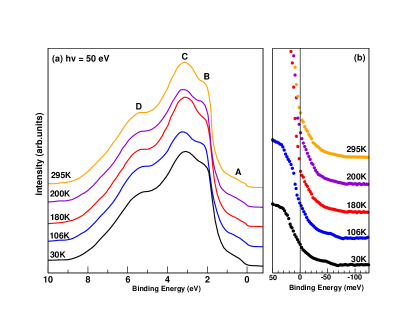
Fig. 1(a) shows the valence band photoemission spectra of the Pr0.5Sr0.5MnO3 sample obtained by using a photon energy of eV across its PMM-FMM-AFMI transitions. The spectra exhibit four distinct features marked A ( eV), B ( eV), C ( eV) and D ( eV). Earlier experiments pal1 ; dalai ; ebata ; pal2 and band structure calculations monodeep have shown that the feature A originates from the eg states and B and C are from the strongly mixed Mn 3d t2g, O 2p, and Pr 4f states. Feature D has a dominant O character with a small Mn contribution. The spectra from the PMM and FMM phases show a clear Fermi edge. It is also clear from the figure that the width of the feature A (which corresponds to the eg states) decrease as we go down in temperature across the FMM-AFMI transition. Further, the intensity of the near EF was found to decrease as the material goes through this transition [Fig.1(b)]. This could be due to the localization of the eg electrons in the CO-AFMI phase. Further, a small but finite intensity at EF was observed in the insulating state of the sample. The electron mean free path has a minimum at about eV electron kinetic energy. The spectra were also obtained using eV. We note that with eV (below the Mn - threshold) the intensity at Fermi level is more than that of the spectra measured at eV of photons. This confirm that the intensity at EF in the electronic structure increases in the surface and decreases systematically in the bulk. Considering the difference in the electron mean free paths between the bulk and surface sensitive photoemission spectra, our results indicate that the surface Mn states are somewhat different from that of the bulk Mn states in the Pr0.5Sr0.5MnO3. Bulk photoemission studies exhibit a CO gap at low temperature kurmaev ; chainani whereas surface sensitive photoemission measurements indicate a finite photoemission intensity at EF in the CO phase. It is to be noted that in-situ scraping or fracture will remove one out-of-plane oxygen (Mn-O-Mn bond) in the MnO6 octahedra. This structural change in the surface can lead to an increase in the other out-of-plane Mn -O hybridization strength due to one short bond and a consequent increase in the one electron band width (W). Thus it is justified to believe that a ferromagneic fluctuation of itinerant metallic nature of the Mn eg band in the surface competes with the local coulomb interaction. Consequently, the CO state in the surface is suppressed from that of the bulk and the coulomb interactions were found quite ineffective in the surface at low temperatures. This scenario is further supported by the observation of significant intensity at the Fermi level. The finite intensity at the Fermi level suggests the rotation and distortion of the MnO6 octahedra in the insulating phase giving rise to the changes in the crystal structure which is reflected strongly on its surface. We propose that the structure of first two layers from the surface may be different due to reduced atomic coordinations, which is responsible for the observed finite intensity at EF at the low temperature CO phase. In this regard it is important to mention that the earlier studies on related manganites using UV-PES also reported a finite intensity at EF in the insulating state wadati .
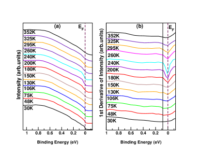
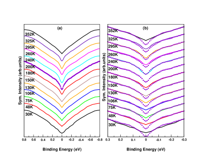
We have further analyzed the near EF spectral behavior across the PMM - FMM - AFMI transition. Panel (a) of Fig. 2 shows the high resolution near EF photoemission spectra from the Pr0.5Sr0.5MnO3 sample taken at a photon energy 50 eV. All the spectra at different temperatures are normalized at energies above eV from EF and then shifted along y-axis for clarity. The finer changes in the near EF region are depicted in panel (b) where the first-order derivative of the intensity is plotted against the binding energy. The FMM-AFMI transition, as expected, shows a smearing out of the Fermi edge. However, at temperatures below K (PMM phase) a dip at the EF starts to form gradually and becomes sharper through the FMM phase till the TN. This dip in the derivative spectra is a signature of a temperature dependent pseudogap existing in the PMM and FMM phases. The pseudogap is formed due to the transfer of spectral weight from the EF to higher binding energy positions with lowering of temperature. Our spectra show the energy scale over which a sharp dip in the FMM and the PMM phases occurs to be meV. This meV pseudogap-like feature is indicated by the solid line in the figure 2(b). Some indications of its temperature dependence are already obvious in the raw data and in the first derivative spectra. In order to confirm the pseudogap-like feature, we symmetrize the spectra and plot with a constant vertical shift as shown in Fig. 3(a). The numerical procedure removes any thermal broadening contribution arising from the Fermi function which are symmetric about the zero of the binding energy scale and gives the change in the density of states within 5KBT of EF. The analysis shows an interesting evolution with temperature, which leads us to identify a pseudogaplike structure at EF of meV. In Fig. 3(b), we have plotted the symmetrized spectra corresponding to each temperature together with that from the T K for a comparison. This figure shows that there are drastic changes in the electronic structure within 80 meV of EF and the pseudogaplike feature is more pronounced in the FMM and PMM phases. Thus the energy scale involved in the formation of pseudogap behavior ( meV) is comparable to the value of meV CO gap observed by Chainani et al. chainani . Across the FMM-AFMI transition, a finite intensity at the Fermi level is observed as we go down in temperature which suggests that the electronic structure of first few layers from the surface is different from that of the bulk. Our high resolution spectra show the existence of pseudogap in the PMM and FMM phases which is important for the metal-insulator transition and a gapless finite intensity at the EF in the CO-AFMI phase. As mentioned earlier, the crystal structure of the Pr0.5Sr0.5MnO3 is tetragonal in both the PMM and FMM phases. Further, both these states have an A-type orbital ordering where the Mn d orbitals are ordered and strongly hybridized with the O orbitals kajimoto . Finally with decreasing temperature from the PMM phase to the FMM phase this orbital ordering seems to be strengthened along with the magnetic ordering. This strengthening of the hybridization between the in-plane Mn d orbitals and the O orbitals can make the MnO6 octahedra elongated in the Z-axis. Such a distortion of the MnO6 octahedra may shift the out-of-plane Mn 3d orbitals where the eg electron is occupied, up in energy; like in the Q-modes of the Jahn Teller (JT) polaron millis1 . The spectral weight behavior and the consequent pseudogap in the PMM and FMM phases, found here, indicate the role of a strong electron - phonon interaction and formation of JT polarons.
IV CONCLUSION
Our study of the temperature dependent magnetic phase transitions in Pr0.5Sr0.5MnO3 compounds conclusively shows a pseudogap behavior in their near EF electronic spectrum over a large region of their phase diagram and a finite photoemission intensity at EF in the CO-AFMI state. The intensity at EF in the insulating phase is ascribed to the lack of CO in the first few layers from the surface. The formation of pseudogaps were discussed considering the strong electron - phonon interaction, consequent charge localizations and possible formation of JT polarons.
V Acknowledgments
P. P. is grateful to Carl Trygger Foundation for financial support.
References
- (1) V. B. Shenoy, T. Gupta, H. R. Krishnamurthy and T. V. Ramakrishnan, Phys. Rev. Lett. 98, 097201 (2007).
- (2) V. B. Shenoy, T. Gupta, H. R. Krishnamurthy and T. V. Ramakrishnan, Phys. Rev. B 80, 125121 (2009).
- (3) A. J. Millis, B. I. Shraiman, and R. Mueller, Phys. Rev. Lett. 77, 175 (1996).
- (4) A. J. Millis, R. Mueller, and B. I. Shraiman, Phys. Rev. B 54, 5405 (1996).
- (5) H. Aliaga, D. Magnoux, A. Moreo, D. Poilblanc, S. Yunoki, and E. Dagotto, Phys. Rev. B 68, 104405 (2003).
- (6) C. Zener, Phys. Rev. 82, 403 (1951).
- (7) P. W. Anderson, and H. Hasegawa, Phys. Rev. 100, 675 (1955).
- (8) K. H. Ahn and A. J. Millis, Phys. Rev. B 58, 3697 (1998).
- (9) A. Llobet, J. L. García-Muñoz, C. Frontera, and C. Ritter, Phys. Rev. B 60, R9889 (1999).
- (10) E. Z. Kurmaev, M. A. Korotin, V. R. Galakhov, L. D. Finkelstein, E. I. Zabolotzky, N. N. Efremova, N. I. Lobachevskaya, S. Stadler, D. L. Ederer, T. A. Callcott, L. Zhou, A. Moewes, S. Bartkowski, M. Neumann, J. Matsuno, T. Mizokawa, A. Fujimori and J. Mitchell, Phys. Rev. B 59, 12799 (1999).
- (11) A. Chainani, H. Kumigashira, T. Takahashi, Y. Tomioka, H. Kuwahara and Y. Tokura, Phys. Rev. B 56, R15513 (1997).
- (12) P. Pal, M. K. Dalai, R. Kundu, M. Chakraborty, B. R. Sekhar and C. Martin, Phys. Rev. B 76, 195120 (2007).
- (13) H. Kawano, R. Kajimoto, H. Yoshizawa, Y. Tomioka, H. Kuwahara and Y. Tokura, Phys. Rev. Lett. 78, 4253 (1997).
- (14) D. D. Sarma, N. Shanthi, S. R. Krishnakumar, T. Saitoh, T. Mizokawa, A. Sekiyama, K. Kobayashi, A. Fujimori, E. Weschke, R. Meier, G. Kaindl, Y. Takeda, and M. Takano, Phys. Rev. B 53, 6873 (1996).
- (15) M. K. Dalai, P. Pal, B. R. Sekhar, N. L. Saini, R. K. Singhal, K. B. Garg, B. Doyle, S. Nannarone, C. Martin, and F. Studer, Phys. Rev. B 74, 165119 (2006).
- (16) K. Ebata, H. Wadati, M. Takizawa, A. Fujimori, A. Chikamatsu, H. Kumigashira, M. Oshima, Y. Tomioka, and Y. Tokura, Phys. Rev. B 74, 064419, (2006).
- (17) P. Pal, M. K. Dalai, R. Kundu, B. R. Sekhar and C. Martin, Phys. Rev. B 77, 184405 (2008).
- (18) M. Chakraborty, P. Pal, and B. R. Sekhar, Solid State Commun. 145, 197, (2008).
- (19) H. Wadati, A. Chikamatsu, H. Kumigashira, A. Fujimori, M. Oshima, M. Lippmaa, M. Kawasaki, and H. Koinuma, Phys. Rev. B 79, 153106 (2009).
- (20) R. Kajimoto, H. Yoshizawa, Y. Tomioka and Y. Tokura, Phys. Rev. B 66, 180402(R) (2002).