Building micro-soccer-balls with evaporating colloidal fakir drops
Abstract
Evaporation-driven particle self-assembly can be used to generate three-dimensional microstructures. We present a new method to create these colloidal microstructures, in which we can control the amount of particles and their packing fraction. To this end, we evaporate colloidal dispersion droplets on a special type of superhydrophobic micro-structured surface, on which the droplet remains in Cassie-Baxter state during the entire evaporative process. The remainders of the droplet consist of a massive spherical cluster of the microspheres, with diameters ranging from a few tens up to several hundreds of microns. We present scaling arguments to show how the final particle packing fraction of these balls depends on the dynamics of the droplet evaporation.
pacs:
Evaporation-driven particle self-assembly is an ideal mechanism for constructing micro- and nanostructures at scales where direct manipulation is impossible. For example, in colloidal dispersion droplets with pinned contact lines, evaporation gives rise to the so-called coffee-stain effect Deegan et al. (1997): a capillary flow drags the particles towards the contact line to form a ring-shaped stain. Such a flow not only aggregates the particles, but is also able to organize them in crystalline phases Fan and Stebe (2004); Bigioni et al. (2006); Harris et al. (2007); Marín et al. (2011). Similar mechanisms such as the “convective assembly” Velikov et al. (2002); Meng et al. (2006) are currently successfully used to produce two-dimensional colloidal crystal films. To obtain three-dimensional clusters of micro-particles, colloidal dispersion droplets which are suspended in emulsions Velev et al. (2000); Manoharan et al. (2003); Dinsmore et al. (2002) or kept in Leidenfrost levitation Tsapis et al. (2005) are used. With these techniques, new colloidal structures arise from the geometrical constraints during the drying Lauga and Brenner (2004). This problem of organization of particles into a spherical topography dates back to the days of the first models of the atom and has been extensively studied by Bausch et al. Bausch et al. (2003); Lipowsky et al. (2005). The main drawback of these three-dimensional assembly techniques, however, is the lack of control on both the amount of particles and the particle arrangement in the remaining structures.
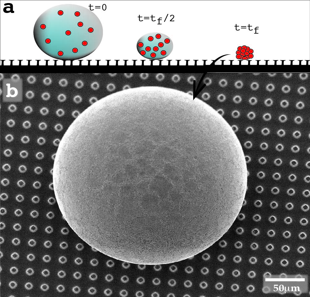
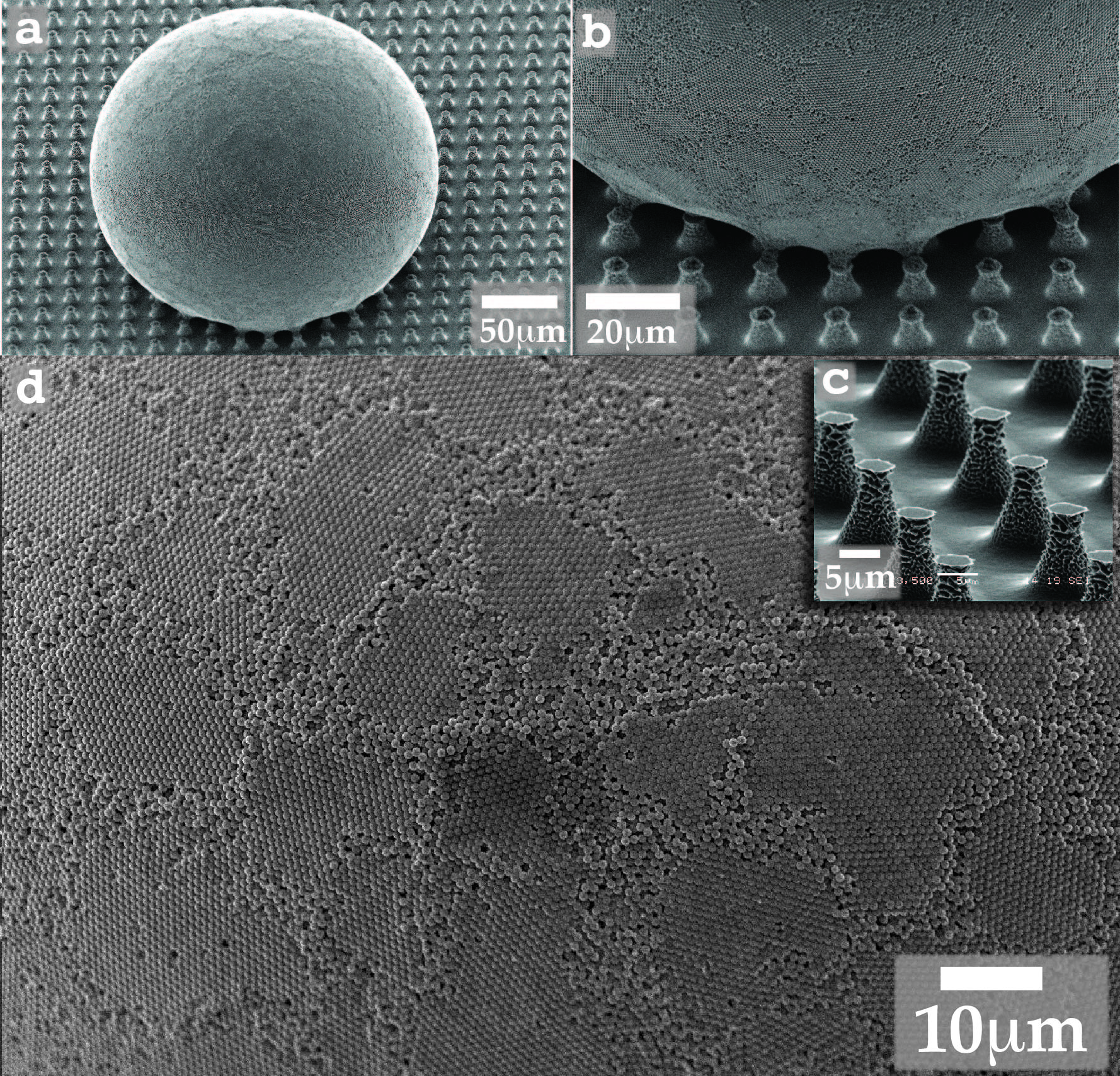
In this work, we devise a new, controlled way of generating on-demand self-assembled spherical micro-structures via droplet evaporation on a superhydrophobic surface (see figure 1). We present scaling arguments to predict the particle arrangement in the microstructures formed, based on the dynamics of the evaporation process. To generate the microstructures, we evaporate colloidal dispersion droplets on a special type of superhydrophobic substrates. In most of the cases, a liquid Cassie-Baxter state drop evaporating on a superhydrophobic surface will eventually suffer a wetting transition into a Wenzel state, i.e. it will get impaled into the structure and loose its spherical shape Reyssat et al. (2008); Tsai et al. (2010). Here, however, we use a surface that combines overhanging pillared structures Tuteja et al. (2008) with a hierarchical nano-structure (figure 2c). These surface properties impose a huge energy barrier for the wetting transition to occur, and therefore the droplet will remain almost floating over the structure in a Cassie-Baxter state during its entire life Susarrey-Arce et al. (2012). A typical result can be observed in figure 1: a water droplet containing polystyrene particles (concentration 0.08% weight and initial volume ) evaporates on the superhydrophobic surface at room temperature and 30% humidity. After a typical evaporation time of 45 minutes, the solvent is completely evaporated and only the colloids are left upon the substrate. Remarkably, the particles ( in this particular case) are not just lying scattered over the substrate but they have aggregated and form a spherical macro-cluster resting on top of the micro-pillars, which we call colloidal supraball. We do not observe shell formation and buckling during the evaporation of the droplets Tsapis et al. (2005): the supraballs we obtain are solid, and present a high mechanical resistance and stability. When looking closer at the surface of these particular colloidal supraballs as shown in figure 2d, one can identify crystalline flat patches which resemble the pentagonal patches present in a soccer ball.
To understand the final structure of these supraballs, it will turn out crucial to understand the dynamics of the droplet evaporation. The fact that we do not observe shell formation, suggests that the particles do not influence the droplet evaporation. To test this hypothesis, we compare the evaporation dynamics to that of a liquid drop that does not contain any particles. The evaporative mass loss from such a drop is typically governed by the diffusion of vapor molecules in the surrounding air Deegan et al. (1997); Popov (2005); Gelderblom et al. (2011). For diffusion-limited evaporation, the rate of volume change of the drop is given by
| (1) |
where is the drop radius, and , with the diffusion constant for vapor in air, the vapor concentration difference between drop surface and the surroundings and the liquid density Marín et al. (2011). One might have expected the evaporation rate from the drop surface to be proportional to the droplet surface area . However, the vapor concentration gradient is proportional to , and therefore the total evaporation rate is proportional to Eggers and Pismen (2010). If the droplet evaporates with a constant contact angle, we find that, since ,
| (2) |
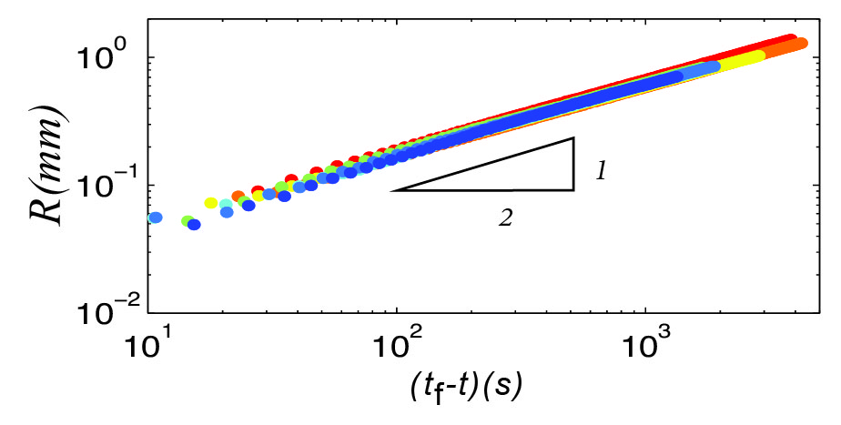
Here is the total droplet lifetime in case no particles are present, for which the drop radius reaches zero. In the present case the drop radius saturates at a finite radius, , at a time , corresponding to the moment where the particles become densely packed. In figure 3 we plot the colloidal droplet radius versus . Our experimental data for different number of particles are in very good agreement with the -power law. This confirms that the particles do not influence the evaporation process, until the final radius is reached. The scaling (1) implies that the speed with which the interface is moving inwards, is given by . Hence, the interface speed increases dramatically as the droplet shrinks and the maximum speed reached in the experiment will be determined by the final radius . As we will show further on, this increase in interface speed determines the particle packing inside the supraballs.
The final size of the ball depends on the number of particles inside the drop. This can be tuned by manipulating either the initial particle concentration or the droplet size. In our experiment, the ball size was in the range , with the particle radius. Clearly, the exact final size of the ball will not only depend on the amount of particles in the system but also on their packing fraction. We define the packing fraction as
| (3) |
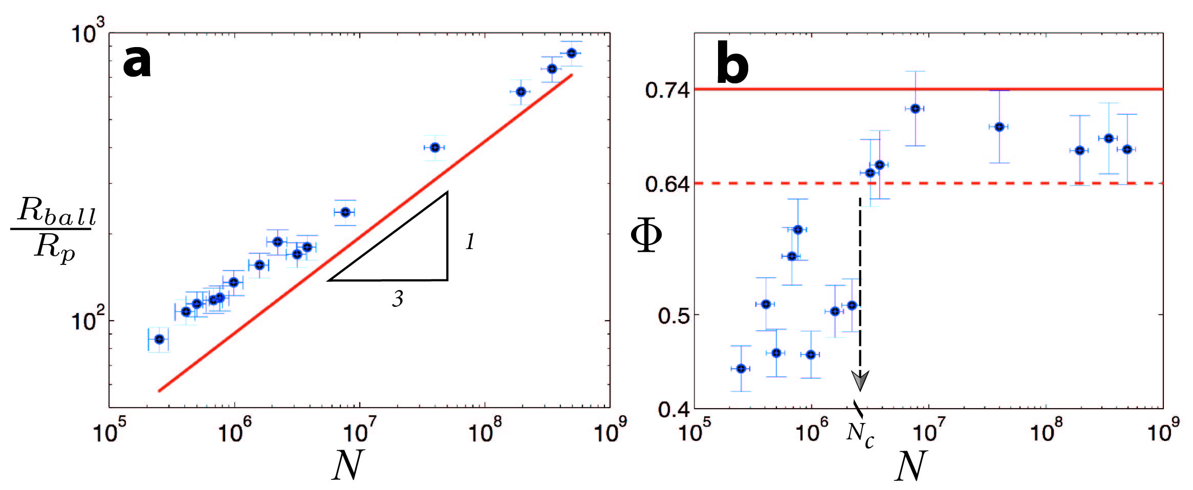
where is the total number of particles in the droplet. The final supraball radius is accurately determined from Scanning Electron Microscope (SEM) images. If the packing fractions were identical for all supraballs, one would expect that , as depicted in figure 4a 111All quantitative results shown in this paper have been performed with colloids of diameter (non-surface-modified fluorescent microspheres supplied by Thermo Scientific), but the same qualitative behavior has been observed for and . The colloidal solutions were always prepared with deionized water.. However, in figure 4b we see that the packing fraction strongly depends on the number of particles in the system.
As the number of particles increases, the packing fraction approaches that of a perfect Hexagonal Close Packing configuration, in which case one would find Kepler (1966), hence, we have an ordered particle packing inside the balls. On the other hand, the supraballs with a smaller amount of particles show remarkably low packing fractions, even below the Random Close Packing (RCP) limit () Weitz (2004), corresponding to a disordered particle arrangement. The balls which show packing fractions below the RCP limit contain several empty cavities. Remarkably, the final configuration reached, seems to depend on the number of particles in the system. In figure 4b we indicated the critical number of particles when the packing fraction reaches that of a RCP. For we get a loose, disordered particle packing in the supraball, whereas for we get a densely packed, ordered supraball.
What causes the transition from ordered to disordered packings, and what determines the critical number of particles? To explain this, we follow a similar approach as in Marín et al. Marín et al. (2011): we compare the time scale on which particles can arrange by diffusion to the hydrodynamic time scale for the particle transport by convection, given by the inward motion of the liquid-air interface. If the diffusion time is small compared to the hydrodynamic time, particles can arrange into an ordered packing. The diffusive time scale is , with the particle radius and the diffusivity of the particles in the liquid Marín et al. (2011). The hydrodynamic time-scale is . Here is the droplet radius, and is the typical inter-particle distance, which depends on the particle concentration as , as long as the solution is dilute (). We define the ratio of both time scales as:
| (4) |
where in the last step we used (1) to replace .
From (4) we observe that increases as the droplet radius becomes smaller during the evaporation (see figure 3), until the limit is reached. A cross-over between the time-scales is reached when the hydrodynamic time becomes equal to the diffusion time, hence when . If the cross-over is reached when , the amount of crystalline clusters is still very small. From this point in time onwards the interface speed is too high for the particles to further arrange in a crystalline way Marín et al. (2011). Instead, they are pushed together in a random arrangement, with a low packing fraction. If the cross-over is reached when , the particle packing is already dense and ordered, and we find a high packing fraction. For all droplets the evaporative mass loss, and hence the decrease in radius, is the same (see figure 3), hence, the moment when the particle packing becomes sufficiently dense for particles to arrange depends solely on the number of particles in the droplet. If is high (), this moment is reached relatively early, i.e. well before , and we get an ordered particle packing inside the supraballs. Using that and considering , we find from (4) the critical number of particles above which we obtain ordered supraballs
| (5) |
This result emphasizes that the transition is governed by two diffusion processes: the diffusion of vapor, determining the speed of evaporation, versus the diffusion of particles inside the drop. The ratio of diffusion constants selects the critical number of particles. In our experiment m2/s and m2/s, from which we find that . This is 2 to 3 orders of magnitudes larger than the experimentally observed . However, in the preceding analysis we have neglected all prefactors, and we emphasize that the result is strongly (to the third power) dependent on the experimental parameters included in , i.e. humidity, liquid density, diffusivity of vapor, and saturated vapor concentration.
To verify whether the final packing fraction indeed depends on the spacing between the particles the moment the cross-over time is reached, we go back to our experimental data. We define the time-dependent packing fraction as . As the droplet evaporates, this packing fraction will increase until it reaches its final value . At the cross-over, defined by , the droplets will have a packing fraction . If this is low, the amount of crystalline clusters is still very small. On the other hand, if is high, we expect crystalline clusters to have formed already. After the cross-over time, the interface moves too fast to allow for further ordering, and it just presses the ordered particle clusters closer together. In figure 5 we show that droplets with a high have a high : when we obtain a final packing fraction above the RCP limit.
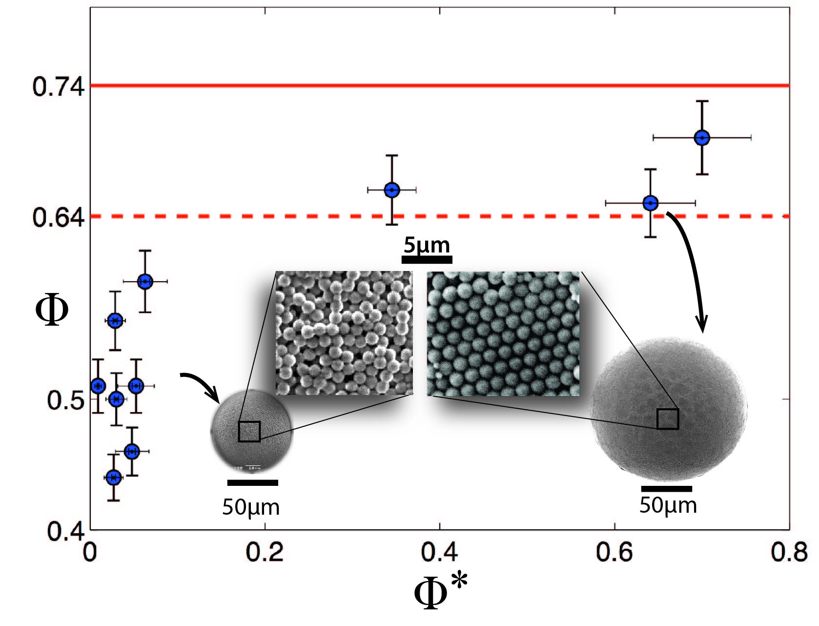
We cannot predict the critical theoretically. However, we can, retrospectively, use the experimental critical to compute . Using that at the cross-over, we obtain , which is in the same order of magnitude as our experimental results; see figure 4b.
The particle packing in the supraballs can not only be assessed by measuring the packing fraction, but it can also directly be seen in SEM images from the surface of the colloidal supraballs, as shown in figure 5. The size of the soccer-ball-like crystalline patches on the surface of the ordered supraballs depends on the ball size: bigger balls will show larger patches due to the reduced curvature at their surfaces. To explain the size of the crystalline domains, we hypothesize that a crystalline patch will bend radially no more than a particle size. Then, it follows by simple trigonometry that the size of a patch will be related to the ball radius and the particle size via: . This expression predicts a typical patch size of for a ball with and , which is in the right order of magnitude as one can observe in figure 2d.
In conclusion, in this Letter we devise a simple technique to create spherical colloidal supraballs relying only on droplet evaporation over a robust superhydrophobic surface. The supraballs show a highly ordered structure if the number of particles inside the drop is large enough to trigger early particle clustering. The critical number of particles required to obtain an ordered particle packing inside the balls depends on the parameters driving the droplet evaporation (through ) and the diffusivity of the particles. Hence, by controlling the humidity and ambient temperature the supraball packing fraction and hence size can be controlled. Massive fabrication of micro-compact-supraballs could easily be achieved by simply spraying a colloidal solution over the micro-structure in a controlled atmosphere. By tuning the wetting properties of the particles one could also be able to generate the well-known colloidosomes Dinsmore et al. (2002) using the same proposed technique.
Acknowledgements.
We gratefully acknowledge Vicenzo Vitelli, Martin van Hecke, Devaraj van der Meer, Chao Sun and many others for their useful comments and their support. The authors also acknowledge financial support by the NWO grant No. 700.10.408 and No. 700.58.041.References
- Deegan et al. (1997) R. D. Deegan, O. Bakajin, T. Dupont, G. Huber, S. Nagel, and T. Witten, Nature 389, 827 (1997).
- Fan and Stebe (2004) F. Fan and K. J. Stebe, Langmuir 20, 3062 (2004).
- Bigioni et al. (2006) T. P. Bigioni, X. M. Lin, T. T. Nguyen, E. I. Corwin, T. A. Witten, and H. M. Jaeger, Nature Materials 5, 265 (2006).
- Harris et al. (2007) D. J. Harris, H. Hu, J. Conrad, and J. Lewis, Phys. Rev. Lett. 98, 148301 (2007).
- Marín et al. (2011) A. G. Marín, H. Gelderblom, D. Lohse, and J. H. Snoeijer, Phys. Rev. Lett. 107 (2011).
- Velikov et al. (2002) K. P. Velikov, C. G. Christova, R. P. A. Dullens, and A. van Blaaderen, Science 296, 106 (2002).
- Meng et al. (2006) L. Meng, H. Wei, A. Nagel, J. Benjamin, L. Scriven, and D. Norris, Nano Letters 6, 2249 (2006).
- Velev et al. (2000) O. D. Velev, A. M. Lenhoff, and E. W. Kaler, Science 287 (2000).
- Manoharan et al. (2003) V. Manoharan, M. Elsesser, and D. Pine, Science 301, 483 (2003).
- Dinsmore et al. (2002) A. Dinsmore, M. Hsu, M. Nikolaides, M. Marquez, A. Bausch, and D. Weitz, Science 298, 1006 (2002).
- Tsapis et al. (2005) N. Tsapis, E. Dufresne, S. Sinha, C. Riera, J. Hutchinson, L. Mahadevan, and D. Weitz, Phys. Rev. Lett. 94 (2005).
- Lauga and Brenner (2004) E. Lauga and M. Brenner, Phys. Rev. Lett. 93, 238301 (2004).
- Bausch et al. (2003) A. Bausch, M. Bowick, A. Cacciuto, A. Dinsmore, M. Hsu, D. Nelson, M. Nikolaides, A. Travesset, and D. Weitz, Science 299, 1716 (2003).
- Lipowsky et al. (2005) P. Lipowsky, M. Bowick, J. Meinke, D. Nelson, and A. Bausch, Nature Materials 4, 407 (2005).
- Reyssat et al. (2008) M. Reyssat, J. Yeomans, and D. Quéré, Europhys. Lett. 81, 26006 (2008).
- Tsai et al. (2010) P. Tsai, R. Lammertink, M. Wessling, and D. Lohse, Physical Review Letters 104, 116102 (2010).
- Tuteja et al. (2008) A. Tuteja, W. Choi, J. Mabry, G. McKinley, and R. Cohen, Proceedings of the National Academy of Sciences 105, 18200 (2008).
- Susarrey-Arce et al. (2012) A. Susarrey-Arce, A. G. Marín, H. Nair, L. Lefferts, J. Gardeniers, D. Lohse, and A. van Houselt, Submitted to Soft Matter (2012).
- Popov (2005) Y. O. Popov, Phys. Rev. E 71, 036313 (2005).
- Gelderblom et al. (2011) H. Gelderblom, A. G. Marín, H. Nair, A. van Houselt, L. Lefferts, J. Snoeijer, and D. Lohse, Phys. Rev. E 83, 026306 (2011).
- Eggers and Pismen (2010) J. Eggers and L. M. Pismen, Phys. Fluids 22, 112101 (2010).
- Note (1) All quantitative results shown in this paper have been performed with colloids of diameter (non-surface-modified fluorescent microspheres supplied by Thermo Scientific), but the same qualitative behavior has been observed for and . The colloidal solutions were always prepared with deionized water.
- Kepler (1966) J. Kepler, Strena seu de nive sexangula, 1611 (English translation, Clarendon Press, Oxford, 1966).
- Weitz (2004) D. A. Weitz, Science 303, 968 (2004).