Photostimulated desorption of Xe from Au(001) surfaces via transient Xe- formation
Abstract
Photo-stimulated desorption (PSD) of Xe atoms from the Au(001) surface in thermal and nonthermal regimes was investigated by the time-of-flight measurement at photon energies of 6.4 and 2.3 eV. Xe was desorbed in a thermal way at high laser fluence, which was in good agreement with theoretical simulations. At a low laser fluence, on the other hand, desorption was induced only at a photon energy of 6.4 eV by a non-thermal one-photon process. We argue that the nonthermal PSD occurs via transient formation of Xe- on Au(001). The lifetime of Xe- is estimated to be 15 fs with a classical model calculation. Whereas the electron affinity of Xe is negative in the isolated state, it is stabilized by the metal proximity effect.
pacs:
68.43.Tj, 68.43.Vx, 79.20.La, 32.10.HqI Introduction
Photo-stimulated processes at solid surfaces have been a topic of extensive studies because they allow us to control adsorbates in either thermal or nonthermal ways.Ho1 ; Chuang ; Fukutani2003 Laser-induced thermal desorption (LITD) was investigated in detail for the systems of CO/Fe(110) (Ref. Wedler, ) and Xe/Cu,Hussla and has been successfully applied to the studies of surface diffusion combined with low-energy electron microscopyYim or with scanning tunneling microscopy.Schwalb Nonthermal photostimulated phenomena, on the other hand, provide us with pathways that are nonaccessible in a thermal process. The nonthermal photostimulated desorption (PSD) of rare gas atoms from metal surfaces has been investigated using photons of two energy regions: At 7 eV, the excitonic or ionic excitation of the mono and multi-layers of Ar and Kr induces desorption,Feulner1987 while infrared light at 1 eV causes the direct excitation of the vibrational mode in the physisorption well to a continuum state.Pearlstine
The non-thermal PSD of Xe/metal using 17 eV photons has been considered not to occur. Generally, the mechanism of nonthermal PSD using photons of 17 eV is understood in terms of formation of the transient negative ion (TNI)Richter and the Antoniewicz model,Antoniewicz where a substrate conduction electron is photoexcited to the adsorbate affinity level. The ground state Xe in the gas phase does not bind an electron stably,Buckman ; Nicolaides ; Bae ; Hird which has been confirmed both theoretically and experimentally with the exception of Ref. Haberland, . Xe atoms physisorb on a metal surface. Physisorption is assumed to occur with little influence on the electronic states. Hence, it has been anticipated that the PSD of Xe/metal via TNI is absent. Condensed Xe, however, has been reported to have modified electronic states compared with the isolated ones due to hybridization with the orbitals of neighboring atoms. It is known that it takes 0.5 eV to remove an excess electron from the bulk Xe,Schwentner1975 and also that the ground state XeN clusters with stably bind an electron,Haberland indicating that the electron affinity level of Xe is shifted downward or broadened depending on the phase of Xe. In this sense, adsorption of Xe onto metal surfaces may well result in a shift and/or broadening or even narrowing of its affinity level by hybridization of the unoccupied orbitals with the substrate electronic states, as is predicted by theoretical studies.Nordlander ; Silva
In the present paper, we report an experimental study of LITD and nonthermal PSD of Xe from a Au(001) surface at photon energies of 6.4 and 2.3 eV. At a high laser fluence, Xe desorption was thermally induced at both photon energies, which is in good agreement with theoretical calculations. At a low laser fluence, on the other hand, Xe desorption was induced nonthermally by 6.4 eV photons as a one-photon process, whereas little desorption was observed with 2.3 eV photons. We argue that the nonthermal PSD proceeds with a transient formation of Xe- as a result of the photoexcitation of substrate conduction electrons. A classical model calculation of Xe desorption reproduces both the experimentally observed TOF and nonthermal PSD cross section, assuming a value of the Xe- lifetime to be 15 fs.
II Experiment
A single-crystal disk of Au(001) was mounted on a cold head after being chemically and mechanically polished. The Au(001) surface was cleaned by several cycles of Ar+ ion sputtering at 0.5 keV and annealing at 700 K in an ultrahigh vacuum chamber ( Pa). The cleanliness of Au(001) was confirmed by observing the () reconstructed pattern with LEED (Ref. Vanhove, ) and no contamination in AES. A Xe monolayer was formed by dosing Xe gas of 3 L (1 L = Pa s) to the sample surface at 23 K (Ref. Dai, ) cooled by a closed-cycle He-compression type refrigerator. The ArF excimer laser (6.4 eV, 8 ns) and the second harmonics of Nd:YAG (yttrium aluminum garnet) laser (2.3 eV, 7 ns) pulses were guided onto the area of 1 mm2 on the sample surface with an incidence angle of 25∘ from the surface normal. The desorbing Xe atoms were detected by a quadrupole mass spectrometer (QMS) located in the surface normal direction with a flight distance of 10 cm. The output signal was amplified by a fast current amplifier and recorded with an oscilloscope synchronized with the laser pulse. The QMS was operated in a low-resolution mode, which enabled high-sensitivity detection of Xe atoms.
III Results
The time-of-flight (TOF) of desorbing Xe atoms upon laser irradiation was recorded at a wide range of laser pulse energy absorbed by the sample () for both 6.4 and 2.3 eV photons. was estimated by taking account of the reflectivity on Au (0.8 for 2.3 eV and 0.2 for 6.4 eV). Figure 1 shows typical TOF results. The data reveal a maximum at a TOF of 400 s with a tailing feature in the long TOF region. TOF was recorded with only one pulse for both 6.4 and 2.3 eV photons with 10 mJ/pulse cm2 [Figs. 1(a) and 1(b)]. With 10 mJ/pulse cm2, on the other hand, TOF was recorded by accumulating over 120 data [Figs. 1(c) and 1(d)] because the Xe desorption yield was small. Whereas a substantial desorption yield was observed with 6.4 eV photons as shown in Fig. 1(c), no significant signal was recorded with 2.3 eV photons as shown in Fig. 1(d). Solid curves in Fig. 1 are fits to the data with a sum of two Maxwell-Boltzmann (MB) velocity distributions described as , where and are mass of a Xe atom and the Boltzmann constant, respectively, and and the translational temperature are fitting parameters. In the analysis, the form is converted to the flux weighted form. In the following, we discuss only the fast component of the TOF.

Figures 2(a) and 2(b) show and the Xe desorption yield of the first component () plotted as a function of , respectively. We first focus on the region of mJ/pulse cm2. In this region, increases with increasing from about 200 K at 32 mJ/pulse cm2 and saturates at about 300 K, for both 6.4 and 2.3 eV photons. In Fig 2(b), shows a sharp increase at 32 mJ/pulse cm2. The behavior of and indicates that the Xe desorption is thermally activated with mJ/pulse cm2.Wedler ; Hussla For the quantitative analysis, we carried out numerical calculations of the surface temperature () during laser irradiation on the basis of the one-dimensional heat conduction equation.Hicks Subsequently, the time evolution of the Xe coverage and the Xe desorption rate (i.e., LITD) was deduced by employing the first-order desorption kinetics assuming the activation energy for desorption of Xe from Au(001) to be 240 meV.Mcelhiney By the calculations above, we obtained the surface temperature at maximum Xe desorption rate () and the maximum surface temperature () following laser pulse irradiation with .
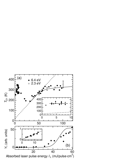
and deduced from the calculation are depicted as a function of in Fig. 2(a) as dashed and solid curves, respectively. obtained by the experimental results is in good agreement with , indicating that LITD of Xe is dominant with mJ/pulse cm2. deviates from with mJ/pulse cm2 because desorption occurs before the surface temperature reaches its maximum.Wedler The solid line in Fig. 2(b) shows a calculated result of the by LITD as a function of . The calculated sharply increases at 35 mJ/pulse m2 and saturates above 55 mJ/pulse cm2. This is in good agreement with the experimental data that the desorption yield exhibits a steep increase at 32 mJ/pulse cm2. This thresholdlike behavior is typical of LITD. However, the experimental data in Fig. 2(b) monotonously increases in contrast to the saturating behavior of the calculated curve, which may be caused by either spatial inhomogeneity of the desorption laser intensityKoehler or coverage dependence of the activation energy for desorption due to the attractive interactions between the adsorbates.Wedler
We turn next to the region of 24 mJ/pulse cm2, where desorption of Xe is observed only at 6.4 eV. As can be seen in Fig. 2(a), significantly deviates from the calculated result of in this region of . The inset in Fig. 2(a) shows a magnification of with mJ/pulse cm2. In this region, is independent of and constant at 30020 K which is much higher than (100 K). Since in the region of mJ/pulse cm2 the calculated result of LITD fails to account for the experimental data, other desorption mechanisms should be operative. Especially with mJ/pulse cm2, the LITD yield of Xe is negligible because is too low for the thermal activation of Xe desorption. Therefore, only non-thermal PSD of Xe atoms from Au(001) is operative in this region of . As shown in the inset of as a function of in Fig. 2(b), with 6.4 eV photons linearly increases with increasing , indicating that the observed nonthermal PSD is a one-photon process. The nonthermal PSD cross section was deduced to be 10-2110-22 cm2 by comparing the nonthermal PSD yield with the LITD yield of the Xe monolayer.
IV Discussion
We first discuss the initial excitation of the non-thermal PSD of Xe from Au(001) upon irradiation of 6.4 eV photons. As the initial excitation, we argue that the negative ion state of Xe is formed via the photoexcitation of the substrate electron.Richter Other excitation pathways can be excluded for the following reasons. The first-excitation energy of Xe from the ground state (55) to the metastable state (556) is 8.3 eV.Feulner1987 When Xe is condensed into a two-dimensional layer on a surface, the excitation energy might be modified, as denoted by the surface exciton. The value is, however, reported to be little modified in the monolayer adsorption regime,Schonhense suggesting that such excitation is unlikely to occur at 6.4 eV. The first ionization energy is 12.1 eV,Feulner1984 ; Moog which is also unreachable with 6.4 eV photons even though it is reduced due to the image charge effect by 2.9 eV.Schonhense Xe desorption from Ag nanoparticles (AgNP) or Si(001), on the other hand, is also reported to occur via surface-plasmon excitation of AgNP at 2.34.0 eV photonsWatanabe2007 and localized surface phonon excitation of Si(001) at 1.16.4 eV photons.Watanabe2000 Desorption via direct excitation from the bound state to a continuum state took place at a photon energy of lower than 1 eV.Pearlstine ; Rao All these desorption mechanisms can be ruled out because the nonthermal PSD of Xe was observed only at 6.4 eV and not at 2.3 eV photoirradiation.
The work function of the Au(001) surface is 5.0 eV, which is reduced by 0.5 eV with Xe adsorption. Therefore, the electronic states nearby the vacuum level are accessible with the hot electrons from the substrate band created by 6.4 eV photoexcitation, and not by 2.3 eV as schematically shown in Fig. 3(a). Although the electron affinity of Xe atoms in the gas phase is known to be negative,Buckman the following studies suggest stabilization of the affinity level due to Xe condensation. Bulk Xe has a conduction band minimum (CBM) at 0.5 eV below the vacuum level.Schwentner1975 ; Schwentner1973 Haberland et al. found that the ground-state XeN clusters are able to bind an electron stably with ,Haberland of which the electron affinity is calculated to be a few meV.Stampfli ; Martyna These studies suggest that interaction with neighboring atoms lowers the electron affinity level of Xe due to the mixing between the unoccupied orbitals. Furthermore, the image charge effect on metal surfaces shifts the electron affinity level downward by 1.0 eV.
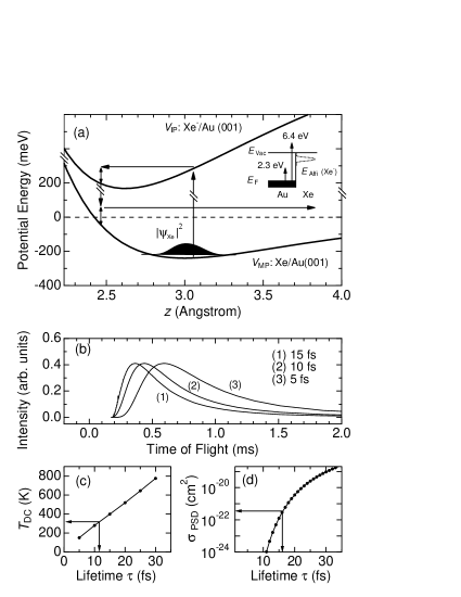
Assuming that the excitation intermediate is the negative ion state, a plausible desorption mechanism is the Antoniewicz model.Antoniewicz In the model, for the appreciable desorption to occur, the lifetime of the Xe- state is required to be long enough. We tentatively estimated the lifetime () of Xe- on Au(001) that reproduces the experimentally observed values of and . is a product of the photoionization cross section and desorption probability . We assume, as a first approximation, that the is as large as cm2.Moog TOF of desorbing Xe and are calculated on the basis of the Antoniewicz model and classical kinetics, as is depicted in Fig. 3(a). Initially, Xe atoms are trapped at the bottom of the physisorption well described by a Morse potential of the form , where , and represent the depth (240 meV),Mcelhiney the width (14 nm-1) and the position (3.0 Å),Silva respectively. The distribution of the initial position of Xe is accounted for as described in Ref. Moog, . Upon Xe- formation, the adiabatic potential of the Xe atom evolves into the form , where is the image charge potential and is an excitation energy. Due to the image charge attraction, the Xe atom is first attracted toward the surface, and is neutralized at a certain distance from the surface. The nuclear motion on is treated classically. If the Xe atom gains enough energy, it escapes the physisorption well leading to desorption. We assume that the neutralization rate of Xe- is described by independent of .
Figure 3(b) shows the TOF results of Xe calculated for lifetimes of 515 fs. Each TOF is well expressed by a single MB velocity distribution with a translational temperature . Figure 3(c) shows the obtained of the calculated TOF as a function of , where 13 fs reproduces the experimentally observed of 30020 K. Figure 3(d) shows the calculated result of as a function of , where fs reproduces the experimentally observed of 10-2110-22 cm2. It is worth emphasizing that the two experimental data of and are well reproduced by a common value of 15 fs based on the Antoniewicz model. The fact is indicative of the validity of the present model. Walkup et al. have shown that the classical adiabatic potential concerning the image charge potential is essentially correct, athough it is slightly different from the one obtained by a quantum-mechanical treatment. They have furthermore shown that the classical treatment of nuclear motion is valid as long as distribution of the initial Xe position is accounted for and that it qualitatively reproduces the kinetic-energy distribution.Walkup It is noted that the affinity level of Xe should lie below the vacuum level for the to be as long as 15 fs.
The obtained value of 15 fs corresponds to the linewidth of 70120 meV for the Xe- state. Padowitz et al. found that in using the two-photon photoemission spectroscopy, the image charge state on clean Ag(111) is shifted by Xe adsorption due to the coupling with the Xe orbitals.Padowitz ; Merry The linewidth obtained in the present study is similar to the value of 2550 meV observed for the image charge states () on Xe/Ag(111) at 0.60.1 eV below the vacuum level. Hence, we suggest the image charge state of Xe/Au(001) is resonanced with the affinity level of Xe, which causes the PSD. We note that a smaller estimation of 10-18 cm2 in the model calculation results in a linewidth of 35 meV.
As already mentioned above, two possible mechanisms of Xe- stabilization are hybridization of unoccupied orbitals and the image charge effect. Since unoccupied orbitals have an extended feature compared with occupied orbitals, unoccupied states could be appreciably hybridized with substrate states even in a weakly bound physisorption well. In addition to these two factors, we discuss another possible reason for the Xe- formation on a metal surface. In the gas phase, contrary to the ground state Xe (55), metastable Xe∗ (556) binds an electron to form a transient Xe- (556) with a large cross section (10-16 cm2).Blagoev In the gas phase, the 556 state is located at 8.3 eV above the ground state. A recent density functional studySilva has shown that the Xe adsorption on a metal surface results in a partial depletion of the occupied Xe 5 state and a partial occupation of the previously unoccupied Xe 6 and 5 states. This indicates mixing of the 556 state upon adsorption on a metal surface, which may contribute to the stabilization of the Xe- state.
Lastly, we comment on the result of an earlier study on the nonthermal PSD of Xe from Ru(001) surfaces.Feulner1987 In the study, no significant desorption was observed from Xe mono and multilayers following 730 eV photoirradiations, whereas desorption from Ar mono- and multilayers and Kr multilayers were observed. As discussed in the present paper, Xe- is expected to be formed following the photoirradiations of 6 eV, and subsequently Xe desorption is expected to occur. Although the desorption cross section is not mentioned in Ref. Feulner1987, , we suspect of 10-22 cm2 was too small for the signal to be detected in their experimental condition. Arakawa et al. reported that the absolute yield of the PSD from solid Ar following 1250 eV photons is as large as 0.1 atoms/photon,Arakawa2 indicating that the cross section of the Xe PSD via the Xe- formation observed in the present study is several orders of magnitude smaller than those of Ar via exciton excitation.
V Conclusion
In conclusion, we have investigated the PSD of Xe on Au(001) at photon energies of 2.3 and 6.4 eV. With decreasing pump laser fluence, the desorption was found to undergo transition from thermal to non-thermal regimes. The non-thermal PSD of Xe occurred only at 6.4 eV as a one-photon process, and the desorption proceeds via the Antoniewicz model with transient negative ion formation. On the basis of the model calculation, the lifetime of Xe- is estimated to be 15 fs. These results strongly suggest that the affinity level of Xe is substantially stabilized by the metal proximity effect.
Acknowledgements.
This work was supported by Grant-in Aid for Scientific Research (A) of Japan Society for the Promotion of Science (JSPS) and by the Sasakawa Scientific Research Grant from the Japan Science Society. A. I. acknowledges support from a Research Assistant of the Global Centre of Excellence for Physical Science Frontier of Tokyo University, Japan.References
- (1) H.-L. Dai and W. Ho, Laser Spectroscopy and Photo-chemistry on Metal Surfaces Part I and Part II (World Scientific, Singapore, 1995).
- (2) T. J. Chuang, Surf. Sci. Rep. 3, 1 (1983).
- (3) K. Fukutani, K. Yoshida, M. Wilde, W. A. Dino, M. Matsumoto, and T. Okano, Phys. Rev. Lett. 90, 1972 (2003).
- (4) G. Wedler and H. Ruhmann, Surf. Sci. 121, 464 (1982).
- (5) I. Hussla, H. Coufal, F. Trager, and T. J. Chuang, Can. J. Phys. 64, 1070 (1986).
- (6) C. M. Yim, K. L. Man, X. Xiao, and M. S. Altman, Phys. Rev. B 78, 155439 (2008).
- (7) C. H. Schwalb, M. Lawrenz, M. Dürr, and U. Höfer, Phys. Rev. B 75, 085439 (2007).
- (8) P. Feulner, T. Muller, A. Puschmann, and D. Menzel, Phys. Rev. Lett. 59, 791 (1987).
- (9) K. A. Pearlstine and G. M. Mcclelland, Surf. Sci. 134, 389 (1983).
- (10) L. J. Richter and R. R. Cavanagh, Prog. Surf. Sci. 39, 155 (1992).
- (11) P. R. Antoniewicz, Phys. Rev. B 21, 3811 (1980).
- (12) S. J. Buckman and C. W. Clark, Rev. Mod. Phys. 66, 539 (1994).
- (13) C. A. Nicolaides and G. Aspromallis, Phys. Rev. A 44, 2217 (1991).
- (14) Y. K. Bae, J. R. Peterson, A. S. Schlachter, and J. W. Stearns, Phys. Rev. Lett. 54, 789 (1985).
- (15) B. Hird and S. P. Ali, Can. J. Phys. 57, 867 (1979).
- (16) H. Haberland, T. Kolar, and T. Reiners, Phys. Rev. Lett. 63, 1219 (1989).
- (17) N. Schwentner, F. J. Himpsel, V. Saile, M. Skibowski, W. Steinmann, and E. E. Koch, Phys. Rev. Lett. 34, 528 (1975).
- (18) P. Nordlander, Phys. Rev. B 46, 2584 (1992).
- (19) J. L. F. D. Silva, C. Stampfl, and M. Scheffler, Phys. Rev. Lett. 90, 066104 (2003).
- (20) M. A. Van Hove, R. J. Koestner, P. C. Stair, J. P. Biberian, L. L. Kesmodel, I. Bartos, and G. A. Somorjai, Surf. Sci. 103, 218 (1981).
- (21) P. Dai, T. Angot, S. N. Ehrlich, S. K. Wang, and H. Taub, Phys. Rev. Lett. 72, 685 (1994).
- (22) J. M. Hicks, L. E. Urbach, E. W. Plummer, and H.-L. Dai, Phys. Rev. Lett. 61, 2588 (1988).
- (23) G. Mcelhiney and J. Pritchard, Surf. Sci. 60, 397 (1976).
- (24) B. G. Koehler and S. M. George, Surf. Sci. 248, 158 (1991).
- (25) G. Schönhense, A. Eyers, and U. Heinzmann, Phys. Rev. Lett. 56, 512 (1986).
- (26) P. Feulner, D. Menzel, H. J. Kreuzer, and Z. W. Gortel, Phys. Rev. Lett. 53, 671 (1984).
- (27) E. R. Moog, J. Unguris, and M. B. Webb, Surf. Sci. 134, 849 (1983).
- (28) K. Watanabe, K. H. Kim, D. Menzel, and H.-J. Freund, Phys. Rev. Lett. 99, 225501 (2007).
- (29) K. Watanabe, H. Kato, and Y. Matsumoto, Surf. Sci. 446, L134 (2000).
- (30) R. M. Rao, R. J. Beuhler, and M. G. White, J. Chem. Phys. 109, 8016 (1998).
- (31) N. Schwentner, M. Skibowski, and W. Steinmann, Phys. Rev. B 8, 2965 (1973).
- (32) P. Stampfli and K. H. Bennemann, Phys. Rev. A 38, 4431 (1988).
- (33) G. J. Martyna and B. J. Berne, J. Chem. Phys. 88, 4516 (1988).
- (34) R. E. Walkup, P. Avouris, N. D. Lang, and R. Kawai, Phys. Rev. Lett. 63, 1972 (1989).
- (35) D. F. Padowitz, W. R. Merry, R. E. Jordan, and C. B. Harris, Phys. Rev. Lett. 69, 3583 (1992).
- (36) W. R. Merry, R. E. Jordan, D. F. Padowitz, and C. B. Harris, Surf. Sci. 295, 393 (1993).
- (37) A. Blagoev, I. Ivanov, T. Mishonov, and T. Popov, J. Phys. B 17, L647 (1984).
- (38) I. Arakawa, T. Adachi, T. Hirayama, and M. Sakurai, Low Temp. Phys. 29, 259 (2003).