On the feasibility and utility of exploiting real time database search to improve adaptive peak selection
Abstract
Rationale: In a shotgun proteomics experiment with data-dependent acquisition, real-time analysis of a precursor scan results in selection of a handful of peaks for subsequent isolation, fragmentation and secondary scanning. This peak selection protocol typically focuses on the most abundant peaks in the precursor scan, while attempting to avoid re-sampling the same m/z values in rapid succession. The protocol does not, however, incorporate analysis of previous fragmentation scans into the peak selection procedure.
Methods: In this work, we investigate the feasibility and utility of incorporating analysis of previous fragmentation scans into the peak selection protocol. We demonstrate that real-time identification of fragmentation spectra is feasible in principle, and we investigate, via simulations, several strategies to make use of the resulting peptide identifications during peak selection.
Results: Our simulations fail to provide evidence that peptide identifications can provide a large improvement in the total number of peptides identified by a shotgun proteomics experiment.
Conclusions: These results are significant because they point out the feasibility of using peptide identifications during peak selection, and because our experiments may provide a starting point for others working in this direction.
Running head: Real time database search to improve adaptive peak selection
Introduction
In broad terms, the goal of many tandem mass spectrometry experiments is to identify, from a given complex mixture, as many distinct peptides as possible in an efficient fashion. Accordingly, many aspects of a typical shotgun proteomics workflow are tuned to optimize the number of high quality spectra derived from distinct peptides. For example, the liquid chromatography phase provides the mass spectometer, at each time point, with a sample of greatly reduced complexity relative to the initial sample, thereby reducing the probability of observing hard-to-identify chimeric spectra, derived from a population of two or more peptide species. Similarly, the flow of sample off the liquid chromatography column is calibrated so that each MS1 scan over intact peptides is followed by sufficient MS2 scans to characterize a large proportion of the identifiable peptide species.
In this work, we focus on a particular component of this workflow, namely, the selection of peaks within an observed MS1 spectrum for subsequent isolation and characterization via MS2 scans. With each MS1 scan, the spectrometer’s on-board computer must select a subset of peaks to isolate, fragment, and measure in an MS2 scan. This task is challenging because, in each duty cycle, hundreds of MS1 peaks may be reasonable candidates for MS2 analysis, but the available time typically permits only a small number (5–10) to be analyzed. Ideally, a peak within an MS1 spectrum indicates the presence of a homogeneous population of peptides, and the peak height can be used as a proxy for peptide abundance. Existing protocols therefore use this MS1 information to select a set of windows for subsequent fragmentation and MS2 characterization.
Today, the most common peak selection strategy is based upon the notion of an exclusion list, which is a dynamically maintained list of m/z values that are excluded from being selected for fragmentation [1]. The standard exclusion list protocol works as follows. A list of maximum fixed size values (in the range of 50–500, depending on the instrument) is maintained in the on-board computer’s memory and is initialized to be empty. With each MS1 scan, the highest-intensity peaks are examined in order. Any peak already on the exclusion list is ignored. Any peak not on the exclusion list is selected for fragmentation and is placed on the exclusion list. This process continues until the start of the next duty cycle. Once a peak is placed on the exclusion list, it remains there for a user-specifiable period of time, typically around 30 seconds.
The main goal of the exclusion list protocol is to maximize the number of distinct peptides identified, which it does using a two-part approach. First, the protocol aims to gather spectra principally from abundant peptides, which are likely to be of higher quality and hence easier to identify. Second, the exclusion list attempts to avoid re-sampling the same peptide species twice, thereby ensuring the selection of a diverse set of peptide species. Taken together, these goals are intended to lead to a broad sampling across abundant peptides in the sample.
A variety of methods have been proposed for improving upon the standard exclusion list by performing analyses interleaved between repeated rounds of MS experiments. Thus, peptides identified in an initial round are excluded from analysis in subsequent rounds [2, 3, 4, 5] or precursor peaks identified in an MS analysis are used to select peaks for analysis in a subsequent MS/MS experiment [6, 7, 8, 9, 10]. Both types of approach yield a larger number of total identifications overall.
In contrast, we consider the possibility of augmenting the peak selection protocol with information about whether a selected MS1 peak was successfully identified in the current experiment. Such an approach is feasible because we have recently demonstrated that spectrum identification via database search can be carried out extremely rapidly [11]—at a rate of 1550 spectra/s for a fully tryptic digestion of proteins from a complex eukaryote. This high-speed search procedure raises the possibility of performing real-time spectrum identification and exploiting the resulting identifications in the context of a peak selection protocol.
In this work, we first establish the technical feasibility of real-time spectrum identification. We then explore the hypothesis that the information provided by real-time identification can be successfully exploited by an adaptive peak selection protocol to yield better peptide identifications over the course of a shotgun proteomics experiment. We refer to such a protocol as ID-informed adaptive peak selection. However, prior to implementing real-time identification software and actually coupling it with an on-board peak selection protocol, we first chose to simulate a variety of ID-informed adaptive peak selection schemes. Unfortunately, these simulations did not strongly support our initial hypothesis: among many algorithms that we tried, only a few led to an increase in the estimated number of distinct peptides identified, and the increases were generally quite modest.
This article can therefore be read in several ways: as a statement of work in progress, as a cautionary tale, or as a stimulant for other researchers working in this area. On the one hand, we are quite certain that real-time spectrum identification is technically feasible. On the other hand, we cannot at this time provide strong evidence for the utility of such an approach.
Methods
Data
In our simulation, we pooled MS2 data from a previously described collection of 11 replicate experiments [10]. Briefly, C. elegans were grown on enriched peptone plates seeded with the OP50 strain of E. coli at 20°C. After lysing and centrifugation, the lysate was denatured using 0.1% RapiGest SF (Waters Corporation, Milford, MA) in 50 mM ammonium bicarbonate pH 7.8. The resulting C. elegans digest (4 µg) was loaded from the autosampler onto a 75-m capillary column placed in line with a Waters NanoAcquity HPLC and autosampler. Peptide elution was performed using two buffer solutions, as described previously. Tandem mass spectra were acquired using either traditional data-dependent acquisition with dynamic exclusion turned on or PAnDA. In PAnDA, MS/MS scans are performed on ions selected from an m/z inclusion-exclusion list computed to prefer ions, which, based on prior analysis of the LC-MS on the same sample, were expected to be abundant peptides that had not been selected in earlier replicate experiments. In both cases, a single high resolution mass spectrum was acquired at 60,000 resolution (at m/z 400) in the Orbitrap mass analyzer in parallel with 5 low resolution MS/MS spectra acquired in the LTQ.
| Experiment | Spectra | IDs |
|---|---|---|
| e10 | ||
| e10PAnDA | ||
| e11 | ||
| e11PAnDA | ||
| e12 | ||
| e12PAnDA | ||
| e14 | ||
| e14PAnDA | ||
| e15 | ||
| e15PAnDA | ||
| e16 |
Searching MS2 data
Each experiment was searched using Tide [11] against a forward and a reversed protein database consisting of the predicted open reading frames from C. elegans and common contaminants (Wormpep v160, 27,499 proteins). Tide was run with the following parameters: (1) fully-tryptic digestion of database proteins; (2) peptide lengths between and residues; (3) peptide masses between and Da; (4) static cysteine modification of Da. (carboxamidomethylation); (5) mass tolerance of Da.; (6) monoisotopic precursor. The Tide results were further processed using q-ranker [12] with the default parameters. Peptide-spectrum matches were accepted as “correct” using a -value threshold of . For the simulation results shown in Figure 5–6, a peptide was considered to be identified if at least one PSM mapped to it. Table 1 provides a summary of each data set, including the number of spectra and the number of PSMs at .
Aligning MS1 data
To pool the MS2 data required first aligning the MS1 data from experiment e10 with respect to each of the 10 experiments. This alignment was done by considering, for a given pair of experiments, all peptides that were succesfully identified (using a -value threshold of ) exactly once in each of the two experiments. The pairs of retention times were then used in a regression minimizing perpendicular least squares. The resulting regression line allowed mapping an observed retention time in a given experiment to its corresponding retention time in experiment e10.
Results
Requirements for real-time peptide identification
We begin by establishing the technical feasibility of real-time spectrum identification. Before doing so, however, we introduce a few concepts that are necessary to understand the discussion. Any database search solution to spectrum identification is based on the idea of enumerating, for each observed spectrum, all of the candidate peptides whose intact mass is close to the inferred mass associated with the spectrum. Here, “close to” is measured in Daltons, and is a parameter set by the user. In the SEQUEST algorithm, which we focus on below, each candidate peptide generates a corresponding theoretical spectrum. A score function, called XCorr, computes the quality of the match between an observed spectrum and a theoretical spectrum. Note that, although the original SEQUEST algorithm used a two-pass scoring scheme involving a prelimimary score (Sp) and XCorr, more recent implementations omit the Sp scoring [13]. SEQUEST thus returns, for each spectrum, the candidate peptide whose theoretical spectrum matched with the highest XCorr value.
To achieve real-time peptide identification, we need a system that can do three things, of which a typical peptide database search program can do only the first: (1) Perform a database search of reasonable complexity, (2) achieve high bandwidth on a single computer, and (3) achieve low latency between spectrum acquisition and identification.
To achieve reasonable search complexity for a typical shotgun proteomics experiment, we want to be able to search a database of proteins from a single target organism, plus contaminants, with some reasonable settings, such as a semi-tryptic digest and a sufficiently wide precursor tolerance window. Reasonable database size, digest, and tolerance window are significant contributors to search time. All modern peptide search software, can perform reasonably complex searches.
| Semi-tryptic | Fully tryptic | |||
|---|---|---|---|---|
| S. cerevisiae | 536 | 1198 | 2738 | 3244 |
| C. elegans | 77.2 | 413 | 1554 | 1980 |
For the second item in the list above, we have recently described Tide [11], which is a reimplementation of the SEQUEST algorithm [14] that achieves extremely high bandwidth on a single computer. Table 2 shows Tide’s throughput, measured in spectra per second on a single thread of a single CPU, for eight different experimental settings: using a protein database derived from S. cerevisiae or C. elegans, generating peptides using a fully tryptic or semi-tryptic digestion, and selecting candidate peptides from the database using a large (3.0 Da) or small (0.25 Da) precursor window. Even for the most demanding setting (C. elegans, semi-tryptic, 3.0 Da), Tide’s throughput is approximately an order of magnitude higher than the 5–10 spectra/s that would be required for a real-time pipeline.
However, the current implementation of Tide does not achieve the third aim listed above. Instead, Tide assumes that all spectra to be searched are available at the start of computation. This assumption, while reasonable in the context of most proteomics laboratory pipelines, fails in the context of real-time peptide identification.
To achieve ID-informed adaptive peak selection, we need to identify spectra in time to make on-the-fly decisions on which peaks to select for fragmentation. We therefore need a low-latency peptide identification method, meaning that spectra are identified within a very short time (say, a few seconds) after the spectrum has been acquired. This requirement is distinct from that of high bandwidth, though the two requirements are related and can be addressed using many of the same approaches.
Adapting Tide for real-time computation
Tide begins processing by sorting the input spectra by neutral mass. A low-latency version of Tide would not be able to access all spectra at the start of processing, and hence cannot this initial sort. Some alternative is required, and we discuss this next.
The initial sort of spectra, coupled with a pre-sorted database of peptides, achieves two important objectives: avoiding redundant computation by computing a theoretical spectrum for the same peptide in the context of identifying two different spectra and maintaining a small memory footprint over the course of computation. The first objective is critical for speed, and some alternative implementation is required to achieve the same level of performance. However, the second objective—maintaining a small memory footprint—is of intermediate importance: there needs to be sufficient memory available to complete the computation, but there is no need to conserve beyond that point.
A simple substitute strategy for the initial sort is to preload all theoretical spectra in memory in advance of collecting the first spectrum. Before any spectrum is processed, the index of peptides is loaded from disk and the accompanying data structures are all stored in memory. This preloading approach has the potential for enormous impact on the program’s memory usage, but it also leaves the total running time (including the preloading step) almost exactly the same as for the current Tide. This approach maintains the guarantee that each theoretical spectrum will be computed at most once, at program startup. Thereafter, the theoretical spectra will not have to be recomputed at all. All the functions of Tide are performed under the preloading approach, but in a different order than for Tide. Except for the larger memory footprint, the resource usage does not change.
We now address the question of whether enough memory would be available to perform a reasonable peptide identification experiment under the preloading strategy. We first measure empirically how much memory would be consumed. The current implementation of Tide interleaves the preprocessing of the observed spectra, the computation of the theoretical spectra, and computing the XCorr. The interleaving allows us to free memory used by the theoretical spectrum computation once the relevant candidate peptides are no longer needed. If we do not free the corresponding memory, then we can measure how much memory would be used by the alternative approach described.

Figure 1 shows the total disk usage and cumulative memory allocated (ignoring reuse) by Tide when run in each of four modes. We see that the worm dataset, which needs the larger database, requires more memory than yeast under comparable settings. We also see that semi-tryptic digestion is considerably more memory-intensive than fully-enzymatic digestion. However, even in the most intensive case, only 6GB of memory is allocated in total. These results show that on a modern workstation, there is sufficient memory not only for a fully tryptic search, but even for a substantially larger semi-tryptic search, even without substantial changes to data structures.
We have not implemented a low-latency version of Tide according to the method described here. We opted first to perform simulations to ascertain the feasibility of ID-informed adaptive peak selection. Had we seen more positive results from testing, then a low-latency implementation of Tide would have been the next step.
Simulation protocol
Before attempting to implement ID-informed adaptive peak selection in the context of a real shotgun proteomics experiment, we wanted to prove the concept first. To this end, we simulated the action of a spectrometer able to select peaks adaptively. In the simulation, the spectrometer has access to real-time peptide identifications as they would be performed by a real-time Tide implementation and is able accordingly to select peaks for fragmentation.
 |
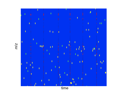 |
In a perfect simulation, an MS2 scan would be available for any MS1 peak that a peak selection algorithm might choose. To approximate this setting in our simulation, we pooled MS2 data from a previously described collection of 11 repicate experiments [10] (see Methods). Pooling the data required first aligning the replicate experiments by retention time. One experiment, called e10, was used as the basis for MS1 data that the simulator would give to the candidate peak picker. Then the MS2 data was taken from each of the other 10 experiments and compared with the MS2 data of e10. Figure 2(A) shows the computed retention-time alignment between e10 and another replicate, e12, as an example. Such an alignment allows us to map any identified MS1 peak onto the baseline experiment e10. Figure 2(B) shows the result graphically: MS1 data from experiment e10 is interleaved with MS/MS data from all replicates. The hope is that if a candidate peak picker requests an MS/MS scan of a particular peak and it wasn’t selected in the actual run of e10, then one of the other experiments may have taken a scan of the same peak at about the same time.
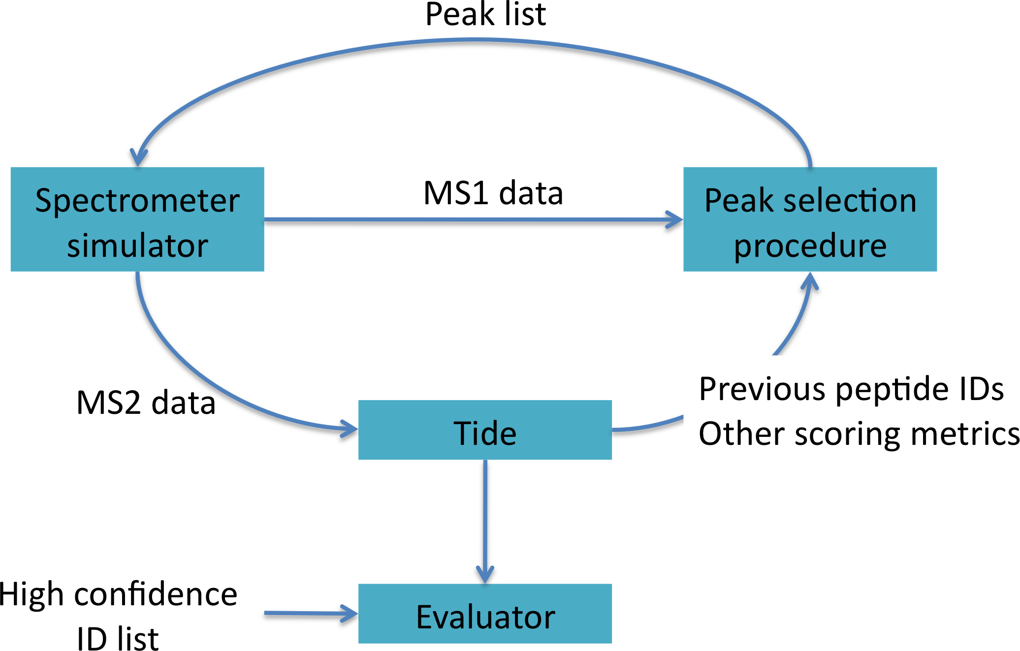
Given this data, the simulator works in conjunction with a given experimental peak-selection procedure, Tide and an evaluator module, as shown in Figure 3. We implemented in Python a framework for simulation according to the diagram. The spectrometer simulator, the Tide results module, and the evaluator were implemented in one Python program which was allowed to run with any separately-supplied peak picker procedure. This approach enabled us to run simulations under varying parameter settings.
After completion of one simulation run, we are primarily interested in the total number of unique peptides identified over the course of the entire spectrometry experiment. However, because of the limits inherent in simulation—in particular, the simulator’s inability to return every MS2 spectrum requested by the peak picker—we could not use this simple metric to make reasonable comparisons against the basic exclusion list, for which complete corresponding MS2 data is available. We therefore selected a two-dimensional metric: the number of unique peptides identified versus the number of spectrum requests for which an MS2 scan was available.
Performance of the baseline method
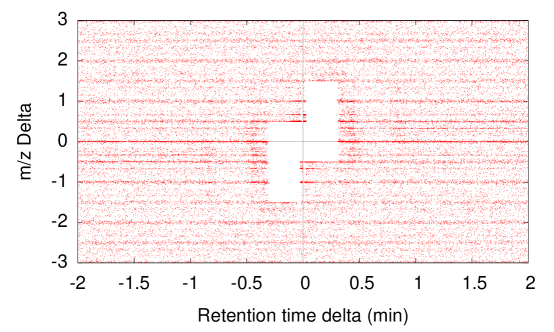
To establish a baseline for our simulation, we began by attempting to replicate, as closely as possible, the behavior of the actual exclusion list that was used to generate our experimental data. The basic settings included an exclusion duration of seconds and an exclusion window of relative to any excluded peak. That is, when a candidate peak is considered for fragmentation, it is checked for two criteria against every other peak previously chosen: if the candidate occurs within 18 seconds of a previously chosen peak and also falls within the exclusion window of that peak by , then the candidate may not be picked. A picture of these settings can be drawn by plotting the differences between all pairs of selected peaks within e10, by precursor and retention time (Figure 4). Such a picture is symmetric by construction, and the blank region highlights the excluded differences. This picture shows that our settings for the exclusion window and exclusion duration are correct.
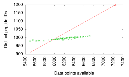
The results of this initial simulation are shown in Figure 5. In the figure, each point corresponds to a different setting of simulation parameters, where the parameters define the tolerance of the simulator for returning MS2 data whose retention time or precursor differed from that requested. The most striking observation to be drawn from Figure 5 is that the simulated exclusion list yields far fewer MS2 spectra and, correspondingly, fewer distinct peptide identifications than the real experiment (shown as a point in the upper right corner of the plot). Even if we select the simulation parameters that yield the best results ( tolerance of Da/charge and retention time tolerance of minutes), the simulator only successfully returns MS2 spectra, which is fewer spectra than were actually produced in experiment e10. On the other hand, the rate of successful identifications is sometimes higher and sometimes lower than in the real experiment, as shown by the relative locations of the green simulation points and the red diagonal line.
We hypothesize that the tendency to return fewer MS2 spectra in the simulation arises for two reasons. First, we observed that, in some cases, the on-board computer selected peaks that are not taken from the top few peaks by intensity. Although some of the skipped-over peaks could be explained by their presence on the exclusion list, not all of the skipped peaks could be “explained away” in this fashion. We therefore suspect that the actual peak selection algorithm includes some features of which we were not aware. Second, we observed that the exclusion list is surprisingly unstable, in that slight perturbations in the choice of peaks early in the experiment lead to far-reaching differences in the choice of peaks later on. This phenomenon appears to be the result of a feedback effect between the peak selector and the exclusion list over the course of a run: a change in peak selection leads to a different exclusion list, which leads to a subsequent change in peak selection, and so on. The second effect thus magnifies the impact of small differences in peak selections early in the experiment. Overall, the mismatch between reality and simulation underscores the importance of establishing a simulation baseline against which to compare the results of alternative peak selection procedures.
Variable-Duration Exclusion List
 |
 |
 |
 |
 |
 |
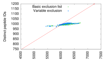
The hypothesis underlying our first peak-picking method is that the basic exclusion list duration is not optimal for acquiring a diversity of peptides, once the peptides are knowable. The 18-second exclusion list duration represents a mean duration time, but performance could be improved if peaks corresponding to quickly-eluting peptides came off the list quickly, whereas peaks corresponding to slowly-eluting peptides should remain on the list for a longer period of time. Based on this intuition, we reasoned that if we know the identity of the selected peaks, then a repeated identification is a good indication that the peptide is still eluting. In that case, a longer than normal exclusion list duration should apply for that peak.
To implement this strategy, we gather peaks in order of intensity, just as for the basic exclusion list, but we modify the decision to include or exclude them. We allow a peak to be requeried after the normal exclusion time. However, if a peak is requeried and is identified as the same peptide as previously, the peak subsequently remains on the exclusion list for three times longer.
The results from the variable duration exclusion list procedure are shown in the top two panels of Figure 6. We can see that for a majority of simulation settings (most individual plotted points), the variable exclusion list identifies slightly more distinct peptide than the simulated basic exclusion list. However, the difference in performance is extremely modest: an average increase of 1.9 distinct peptides out of an average total of 997 for the basic exclusion list.
Weibull-score Triage
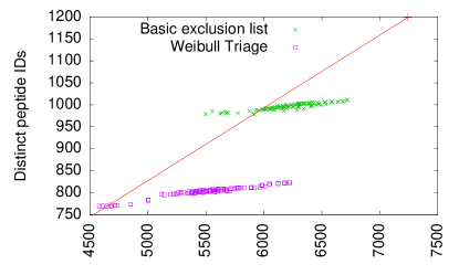
The second peak-picking method that we investigated is based on the hypothesis that if a peak is not confidently identified, then it may be beneficial query the same peak a second time. The method subdivides spectra into three groups: (1) those with high scores that do not need to be re-queried, (2) those with very low scores for which a re-query is not likely to lead to a successful identification, and (3) spectra with intermediate scores that should be re-queried. This hypothesis sets up a natural triage as follows. If a peak is identified with high confidence or low confidence, then it should be placed on the exclusion list according to the standard protocol. Alternatively, if the peak is identified with middling confidence, then we allow it to be requeried after a much shorter interval.
We used a previously described confidence estimation metric, based on fitting a Weibull curve to the tail of the scoring distribution over the candidate peptides [15]. We employed this score on the theory that, if this method worked well, we could introduce the Weibull-based confidence estimator into Tide while keeping the performance high enough to meet the throughput and latency requirements for real-time peptide identification.
We then applied the following specific protocol:
-
1.
If a selected peak was identified with a -value of according to the Weibull curve fit, then it was placed on the exclusion list normally.
-
2.
If a selected peak failed to be identified confidently (-value ), then it was also placed on the exclusion list normally.
-
3.
If a selected peak was identified with intermediate confidence (-value between and ), then it was placed on the exclusion list only briefly and was allowed to be requeried after 5 seconds.
This approach performed noticeably poorly relative to the basic exclusion list—about 19% fewer identifications than the basic exclusion list, averaged across a variety of tolerances, albeit with 11% fewer available simulation peaks. Detailed results are shown in the middle two panels of Figure 6. We investigated other triage thresholds and obtained similarly poor results (data not shown).
Charge state exclusion
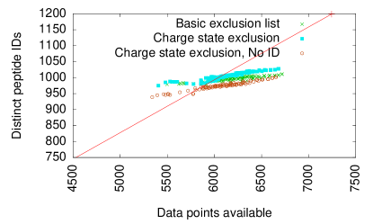
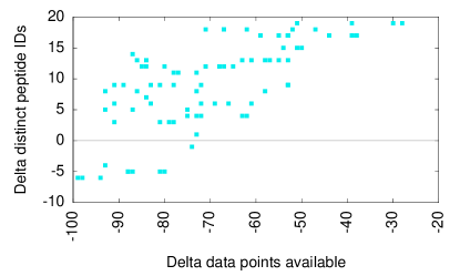
The third peak-picking method we investigated is based on the fact that the same peptide may elute roughly simultaneously at two different charge states. The basic peak selection protocol may then end up choosing both such peaks. No effort is made to prefer one over the other, because it may be easier to identify the peptide at one charge state or the other, and the spectrometer does not know a priori which is preferable. However, we hypothesize that if a peak is identified with high confidence at one charge state, then we can reasonably exclude the corresponding peaks at the other charge states as well, because they are likely to be the same peptide species. If a peak is not identified with high confidence, then it may be treated as before.
The results, shown in the bottom two panels of Figure 6, are somewhat more promising than for the other peak picking methods we examined, but remain modest: an average of 10 more unique peptide identifications (1%) over the simulated basic exclusion list. To be sure of the effect, we also show the results (shown in red crosses) of simply excluding peaks at sister charge states on a first-come-first-served basis, without regard to whether the identification at the first attempted charge state was made confidently. These results are worse than the basic exclusion list and show that real-time identification was critical in gaining the additional identifications seen in the charge state exclusion method.
Discussion
Overall, our results point to one positive conclusion and one negative conclusion. On the one hand, we have demonstrated that real-time assignment of peptides to fragmentation spectra is feasible, in principle. Much larger searches, such as non-enzymatic digest or searches allowing multiple modifications per peptide, would require more memory and more time than the above strategy provides. Hence, there certainly remain limits to what can be achieved in a real-time context, but our analysis brings closer together the notions of high-throughput and of real-time peptide identification.
On the other hand, our simulations have failed to provide evidence that real-time identifications yield significant value in the context of selecting MS1 peaks for fragmentation. In particular, we could not demonstrate that any of the three peak-selection strategies would yield more than a 1% increase in unique peptide identifications relative to the standard exclusion list.
Among the peak-selection strategies that we tried, the Weibull-triage method fared poorly, while the other two methods showed very slight improvements over the basic exclusion list, with the charge-state exclusion being the most promising. Thus, it seems that the better-performing approaches rely on aggressively excluding peaks rather than relaxing the exclusion criteria. Rather than requerying peaks that failed to be identified previously, we are better off excluding those peaks that have any likelihood of being a duplicate. Our interpretation is that this is because the generally low identification rate of peptides is a stronger effect than that of noise in the spectrum.
We were stymied in getting more positive peak selection results by at least three effects. The first was the insufficiency of data for a good simulation: many requested peaks did not have corresponding scans. We attempted to address this problem by aggregating over replicate experiments, but clearly, aggregating over a larger set of experiments would lead to a better simulation. We also applied a two-dimensional success metric that attempts to correct for missing simulation data.
The second effect was that results were more sensitive to uninteresting parameters, such as simulation tolerances, than interesting ones. To address this problem, we allowed the simulation parameters to vary, and we aggregated over all of those results.
The third effect was that the actual peak-picking strategy employed by the spectrometer is apparently more complex than a straightforward exclusion list. In principle, we could attempt to reverse engineer the mechanism of the on-board peak selector; however, the instability of the exclusion list argues against this approach. Experimental peak selectors will necessarily deviate from the on-board peak selector’s choices, so the subsequent effects of exclusion list instability are unavoidable in evaluating alternative peak selectors. It therefore makes sense in evaluation to use a baseline that is subject to the same instability.
References
- [1] B. Kohli, J. Eng, R. Nitsch, and U. Konietzko. An alternative sampling algorithm for use in liquid chromatography/tandem mass spectrometry experiments. Rapid Communications in Mass Spectrometry, 19(589–596), 2005.
- [2] H.-S. Chen, T. Rejtar, V. Andreev, E. Moskovets, and B. L. Karger. Enhanced characterization of complex proteomic samples using LC-MALDI MS/MS: Exclusion of redundant peptides from MS/MS analysis in replicate runs. Analytical Chemistry, 77(7816–7825), 2005.
- [3] N. Wang and L. Li. Exploring the precursor ion exclusion feature of liquid chromatography-electrospray ionization quadrupole time-of-flight mass spectrometry for improving protein identification in shotgun proteome analysis. Analytical Chemistry, 80(4696–4710), 2008.
- [4] S. Bendall, C. Hughes, J. Campbell, M. Stewart, P. Pittock, S. Liu, E. Bonneil, P. Thibault, M. Bhatia, and G. Lajoie. An enhanced mass spectrometry approach reveals human embryonic stem cell growth factors in culture. Molecular Cell Proteomics, 8(3):421–432, 2009.
- [5] A. Scherl, P. Francois, V. Converset, M. Bento, J. A. Burgess, J.-C. Sanchez, D. F. Hochstrasser, J. Schrenzel, and G. L. Corthals. Nonredundant mass spectrometry: A strategy to integrate mass spectrometry acquisition and analysis. Proteomics, 4:917–927, 2004.
- [6] O. Rinner, L. Mueller, M. Hubálek, M. Müller, M. Gstaiger, and R. Aebersold. An integrated mass spectrometric and computational framework for the analysis of protein interaction networks. Nature Biotechnology, 25:345–352, 2007.
- [7] P. Picotti, R. Aebersold, and B. Domon. The implications of proteolytic background for shotgun proteomics. Molecular and Cellular Proteomics, 6:1589–1598, 2007.
- [8] A. Schmidt, N. Gehlenborg, B. Bodenmiller, L. Mueller, D. Campbell, M. Mueller, R. Aebersold, and B. Domon. An integrated, directed mass spectrometric approach for in-depth characterization of complex peptide mixtures. Molecular and Cellular Proteomics, 7:2138–2150, 2008.
- [9] A. Zerck, E. Nordhoff, A. Resemann, E. Mirgorodskaya, D. Suckau, K. Reinert, H. Lehrach, and J. Gobom. An iterative strategy for precursor ion selection for LC-MS/MS based shotgun proteomics. Journal of Proteome Research, 8:3239–3251, 2009.
- [10] M. R. Hoopmann, G. E. Merrihew, P. D. von Haller, and M. J. MacCoss. Post analysis data acquisition for the iterative MS/MS sampling of proteomics mixtures. Journal of Proteome Research, 8(4):1870–1875, 2009.
- [11] B. Diament and W. S. Noble. Faster sequest searching for peptide identification from tandem mass spectra. Journal of Proteome Research, 10(9):3871–3879, 2011. PMC3166376.
- [12] M. Spivak, J. Weston, L. Bottou, L. Käll, and W. S. Noble. Improvements to the Percolator algorithm for peptide identification from shotgun proteomics data sets. Journal of Proteome Research, 8(7):3737–3745, 2009.
- [13] J. K. Eng, B. Fischer, J. Grossman, and M. J. MacCoss. A fast SEQUEST cross correlation algorithm. Journal of Proteome Research, 7(10):4598–4602, 2008.
- [14] J. K. Eng, A. L. McCormack, and J. R. Yates, III. An approach to correlate tandem mass spectral data of peptides with amino acid sequences in a protein database. Journal of the American Society for Mass Spectrometry, 5:976–989, 1994.
- [15] A. A. Klammer, C. Y. Park, and W. S. Noble. Statistical calibration of the sequest XCorr function. Journal of Proteome Research, 8(4):2106–2113, 2009.