Plasmonic enhanced lasers of nano-colloidal fluids
Abstract
ABSTRACT:
Localized surface plasmon resonances have recently attracted considerable attention due to their ability to
dramatically enhance near-field optical intensities and boost nanoscale light-matter interactions.
Here we demonstrate unambiguously that polyhedral silver nanoparticles can be tailored to promote enhanced coherent emission from organic dye
dispersions in a cavity. In particular, we observe that nanoparticles supporting resonances close to the absorption peak
of the dye yield to more efficient lasing, up to one order of magnitude, which is also evidenced by the narrowing of the
emission spectral line down to 3 nm. Moreover we observe emission saturation and energy dependent spectral shifts associated with Kerr effects,
which demonstrate the general relevance of non-resonant nonlinearities in plasmonic enhanced laser emissions.
Works in this area will contribute to the design of novel kind of laser sources providing ultra-short pulsed operation and
structural tunability.
Understanding the fundamental light-matter interactions at a scale much smaller than the wavelength is pivotal to any effort to reliably engineer laser emission.
This relies on controlling frequency and bandwidth Landau and Lifshitz (1960), to ultimately design ultra-short laser pulses.
Localized surface plasmons (LSP) can be optically excited and are associated with sustained oscillations of free electrons localized
at the metal/dielectric interface. Barnes et al. (2003); Zayats and Smolyaninov (2003) LSP oscillations are coherent and accumulate energy from the external excitation.
These LSP can be tuned with the size and the shape of metallic structures which can be fabricated through a wide range of approaches
including lithography and wet-chemistry.Forestiere et al. (2012); Volpe et al. (2009); Di Falco et al. (2010); Rodríguez-Fernández
et al. (2009); Scholl et al. (2012); Link and El-Sayed (1999); Jain et al. (2006) Such nanometer scale sources of intense coherent electromagnetic fields can enhance
inherently weak physical processes, such as fluorescence and Raman scattering of single molecules with potential applications ranging from
biomedicine to lithography, and from microscopy to information technology.Maier et al. (2003); Anker et al. (2003); Pile (2011); Berini and De Leon (2012); Ploschner et al. (2012); Lindquist et al. (2012); Oulton (2012); Yin et al. (2012); Kabashin et al. (2009); Liu et al. (2011a); Di Falco et al. (2011)
Another important potential of LSPs lies in triggering coherent light emission amplified by plasmonic resonances.
Coupling between a dye and the LSP can occur when the photoluminescence
spectrum of the semiconductor overlaps with the LSP resonance. Beyond a specific pump threshold, a resonant energy transfer between
the dye and the LSP can support lasing Gather (2012). Examples of such systems are Spasers made of metal nano-particles
embedded in a gain medium, Noginov et al. (2009); Stockman (2008); De Luca et al. (2011); Zheludev et al. (2008); Stockman (2011); Liu et al. (2011b) plasmonic lasers and nano-cavities, Oulton et al. (2009); Russell et al. (2012) random lasers with metallic scatterers. Popov et al. (2006); Dice et al. (2005)
Despite this cumulative effort in the field, the experimental demonstration of Spasers available in the literature are still very limited.
There is thus an opportunity to gain insight into the dynamics of surface plasmons supported lasing even in very simple systems.
This in turn offers the possibility to investigate more complex lasing regimes, for example at pump energies that trigger higher order nonlinear effects.
Here we report on the lasing from systems based on polyhedral silver nanoparticles (nPs), with plasmonic response determined by
their shape and polydispersity. We show that a liquid dye solution in a parallelepiped cell acts as a laser once the metallic nPs with tailored plasmonic resonance are added:
the system is hence a standard Fabry-Perot laser cavity, made by the facets of the cell and the gain is the dye solution, whose fluorescence
is enhanced by the plasmonic resonances Noginov et al. (2006). The lasing efficiency is strongly dependent on the resonance wavelength:
the closer the resonance peak to the dye absorption maximum, the more efficient the process. Lasing is clearly demonstrated by the
strong increase of the emitted radiation intensity and by the spectral narrowing through pump threshold. Moreover, we show that the
lasing process evidenced by Fabry-Perot resonances of the cavity, gain saturation and non-resonant nonlinearities indicate that
coherent emission can be observed in very dilute transparent samples, in contrast with random lasing, Wiersma et al. (1995); Leonetti et al. (2011) relying on the
multiple scattering of light involving several particles in a volume.
Of interest for both fundamental and applied sciences and applications, the synthesis of nPs has been associated with intense activities dedicated to further control the size and the shape of nanometer size colloids, soluble in a wide range of solvents. Metal nPs are subject to a wide range of investigations including optoelectronic devices, Ahn et al. (2009); Ribierre et al. (2011) sensing, Maheshwari and Saraf (2006); Talapin et al. (2010) catalysis, and plasmonics Govorov and Carmeli (2007); Righini et al. (2009); Tao et al. (2007) to name but a few. In this work, the silver nP syntheses were carried out under ambient conditions according to modified protocols reported in the literature. Silver nitrate (AgNO3, 99.0% ACS grade), trisodium citrate (C6H5O7Na3.2H2O, 99% ACS grade), sodium borohydride (NaBH4, 99% ReagentPlus), polyvinylpyrrolidone (PVP, M 55 kD), L-Arginine (C6N4H14O2, 98% reagent grade) were purchased from Sigma-Aldrich and used without further purification. For a typical preparation of spherical silver nPs, Yang et al. (2005) silver nitrate solution (200 L, 100 mM) was diluted in DI-water, the citrate solution (160 L, 400 mM) was then added to act as a stabilizer for the nPs. Under vigorous stirring, a sodium borohydride solution (300 L, 150 mM) was added drop-wise, initially leading to a black solution quickly turning into a yellow-brown solution. If stirring is stopped before the ageing is completed then part of the nPs falls out of solution. To address this effect and to obtain very stable colloidal solutions, the syntheses were left under stirring to age for at least 24 hrs. The total volume of the nPs solution was set to 20 mL but can easily be scaled up. Photoreduction has become a popular pathway to produce nPs of controlled shape including prism, disc and decahedral nPs. Jin et al. (2001, 2003); Xue et al. (2007, 2008); Zhang et al. (2010); Maillard et al. (2003); Wu et al. (2008); Bastys et al. (2006); Callegari et al. (2003); Kim et al. (2002); Niidome et al. (2003) Starting from small spherical nPs, varied light exposure conditions allow the preparation of nPs with tunable decahedron and prism populations. Pietrobon and Kitaev (2008) In a typical synthesis, citrate solution (127 L, 250 mM), PVP (19 L, 50 mM), L-arginine (16 L, 20 mM) and silver nitrate solution (25 L, 50 mM) are successively mixed into DI-water. Sodium borohydride solution (102 L, 100 mM) is swiftly injected leading a yellow pale solution which was exposed to light sources. The exposure conditions varied from 24 h to 2 weeks, and from monochromatic to white lights. Extraction by ultra-centrifugation was used to purify the as-prepared nPs solutions. Transmission electron microscopy (TEM) images were recorded using a Gatan CCD camera on a JEOL JEM-2011 electron microscope operating at 200 kV. Rhodamine B and the nPs aqueous solutions were mixed with the final dye concentration set to 1 mM for all the samples. A fraction of the resulting solutions were transferred in parallelepiped quartz cuvettes cells with a 1 mm optical path, with the water solution acting as gain medium once the system is pumped. A Coherent Surelite Q-switched frequency doubled Nd:YAG laser at wavelength =532 nm (repetition rate 10 Hz, pulse duration 6 ns and with 8 mm beam waist) was used for optical pumping. For each sample, the laser was focused down to a spot of 1 mm lateral dimension. The emitted radiation was collected from the input face, after focalization into an optical fiber connected to a Horiba MicroHR spectrograph equipped with Symphony electrically cooled CCD array detector, with gratings density of 600 mm-1 and 1800 mm-1, for low and high resolution spectra respectively. The exposure time was set to s and each spectrum results from the average of 10 pump shots.
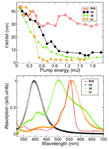
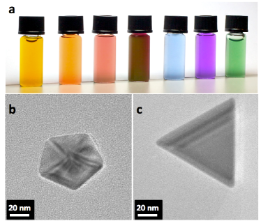
Figure 1a presents a selection of the samples used in this work. The different colors correspond to nPs with varied polyhedral morphologies, obtained by tuning the synthetic parameters. The transmission electron microscopy (TEM) images of two examples are shown in Figure 1b-c, for decahedral and prismatic shapes, respectively. The detailed population diagrams are presented in the Supporting Information, for the lasing experiments we focused mainly on the most representative samples SA, SB and SC.
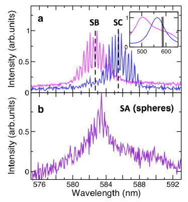
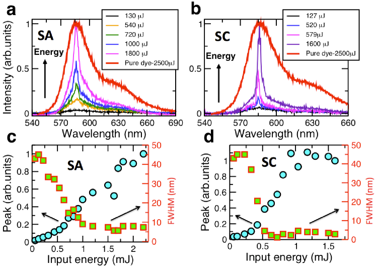
Figure 2 shows the measured laser emission from these samples: by increasing the input pump energy the fluorescence
spectrum of Rhodamine-B displays a pronounced peak that progressively becomes sharper and more intense,
as reported in the panel a and b for samples SA with ellipsoidal nPs and SC with anisotropic and polyhedral nPs, respectively.
In the same graphs the pure dye emission at much higher energy (2.5 mJ) is depicted showing that without nPs no lasing occurred (until being exposed to at least 4.5 mJ, not shown).
This evidences clearly that lasing is provoked by the enhancement of dye fluorescence by the LSP resonances of the metallic colloids.
Figure 2c-d presents the spectral width and the relative intensity of the emission as a function of the pump energy,
unveiling the transition to a coherent emission.
These results show that different samples have different lasing efficiency as evidenced by the spectral narrowing and by the saturation
of the emission in SC, corresponding to a trend of the peak intensity deviating from a straight line as shown in Figure 2d.
To understand the reason of such dependency we report in Figure 3a the linewidth vs. pump energy for
samples SA (spheres), SB, SC and pure Rhodamine solution, with a line-width of 3 nm for the polyhedrals and 10 nm for the spheres.
In contrast, the control pure Rhodamine solution has a line width around 30 nm, in the same range of pump energy.
Figure 3b shows the normalized absorption spectra of the plasmonic samples together and the pure dye.
The resonance located around 400 nm is characteristic of spherical silver nPs and presents little variation with the size of the nPs. Hao and Schatz (2004)
However, lower energy resonances are associated with silver nPs of a varied aspect ratios and dimensions. Mock et al. (2002)
The data demonstrate that samples with plasmonic resonance wavelength close to the dye absorption peak are more efficient lasers as the emission line is narrower.
The different observed efficiency cannot be ascribed to the nPs concentration, being the concentration of the samples: [SA] = 17 nM, [SB] = 250 nM and [SC] = 14 nM.
The low concentrations also evidence that the reported lasers are not random lasers, because the systems are totally transparent and there is no multiple light scattering.
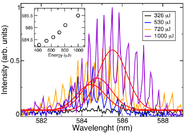
To gain further insight into the spectral emission we show in Figure 4a and Figure 4b the high resolution emission spectra of
the samples respectively with polyhedral nPs (SB and SC) and with spherical nPs (SA), for pump energy of 1 mJ.
The results require further analysis. We associate the emission spectra with the lasing modes of the Fabry-Perot cavity (the glass cuvette),
hosting an active medium whose effective gain is enhanced by the metallic nPs. This claim is supported by the following evidences:
i) The first observation is that, independently on the sample, the position of the maximum of the emission spectra approximately coincides with the emission peak of the Rhodamine ( 580 nm).
The absolute position shifts with the sample under consideration, likely due to a slightly different effective overlap between the absorption spectrum of the nPs and that of the dye.
The inset in Figure 4a further highlights both these aspects.
ii) The spacing of the spectral features is 0.5 nm for all the samples with polyhedral particles considered.
This value has the same order of magnitude of the expected spacing (2nL) = 0.13 nm of a
Fabry-Perot cavity for normal incidence, with n = 1.33 (Water), L = 1mm and = 532 nm. The difference between the experimental
value and the model are most likely due to the resolution of our experimental setup (0.3 nm) and to a different effective index of the active medium.
iii) The fringes are only visible for the samples with polyhedral nPs at this pump energy level, as evidenced in Figure 4b and from the
high resolution emission spectra of pure RhodamineB-water solution reported in the Supplementary Information.
This result reinforces the experimental findings of Figure 3,
where nPs with absorption peak close to that of the dye yield to more efficient lasing. Control experiments with water alone confirm the lack of lasing.
A calibration experiment with Rhodamine only aqueous solutions (no nP present, see Supplementary Information) shows that lasing occurs at a threshold of 4.5 mJ.
Assuming as lasing threshold the point of abrupt change of the slope of the emission peak intensity vs. pump energy in
Figure 2c-d, we can extrapolate the effective gain of the nP-dye solution.
For example, for SC we experimentally obtain approximately a ten times increase in the effective gain, with respect to the dye alone, in the range of cm-1
(assuming that a 0.01 mM Rhodamine B solution in diethylene-glycol at 0.5 nJ/m2 has a gain of 100 cm-1). Leonetti and Lopez (2012)
This estimate assumes that the emission is the super-positioned output of many nanoparticles excited simultaneously.
Thus variations in particle size and shape are expected to blur out any regular multi-mode emission spectrum.
The last aspect to analyze is the emission dynamics in the high pump energy range. From Figure 2 it emerges that exceptionally, the lasing intensity only saturates for SC,
which is the sample with the highest effective gain. This is a strong indication that the concentration of electromagnetic
energy associated with the plasmonic resonances could result in optical Kerr effects characterized by a spectral shift towards longer wavelength, with increasing energy.
To verify this hypothesis we recorded several high-resolution spectra of the emission for sample SC, for increasing pump energy, as shown in Figure 5.
The experimental results illustrate how increasing the input energy induces a red-shift of the emission shown in the inset of Figure 5.
This is consistent with our hypothesis and demonstrates the general relevance of non-resonant nonlinearities in plasmonic enhanced laser emissions.
It should be noted that the pulsed regime excludes that the shift could be due to thermal effects.
In the present experiments, as the resonances shift with increasing energy, their position with respect to the maximum (absorption) gain changes and this negative feedback results into a limited amplification.
Remarkably, Figure 5 also shows that the fine spectral resonances emerge nonlinearly from a spontaneous emission background,
as they get more pronounced and visible by increasing pump.
This is another evidence of the connection between the fine Fabry-Perot lasing modes and the laser efficiency, both boosted by the presence of resonant nPs.
In conclusion we have unveiled novel aspects of the rich dynamics of plasmonic enhanced dye lasers, and specifically the pronounced non-linear regimes that can be achieved. Highlighting extremely strong light-matter interactions, these novel features include gain saturation due to Kerr effect. These results pave the way to the exploration of novel fundamental physical phenomena relative to nonlinear regimes at a spatial scale much smaller than the wavelength, as well as to innovative applications such as novel kind of laser sources providing ultra-short pulsed operation and structural tunability, realization of ultrafast microprocessors working at THz clock speed, supercontinuum generation, ultrasensing, ultradense and ultrafast information storage.
I Associated Content
I.1 Supporting Information
Figure Supplementary1 reports population distribution of samples SA, SB and SC. Figure Supplementary2 shows high resolution emission spectra of the aqueous Rhodamine-B solution used as gain material and the recorded peak intensity vs. pump energy.
II AUTHOR INFORMATION
II.1 Corresponding Authors
neda.ghofraniha[AT]roma1.infn.it
pascal.andre[AT]st-andrews.ac.uk
III Acknowledgements
The research leading to these results has received funding from the European Research Council under the European Community’s Seventh Framework Program (FP7/2007-2013)/ERC grant agreement n.201766, project Light and Complexity and from the Italian Ministry of Education, University and Research under the Basic Research Investigation Fund (FIRB/2008) program/ CINECA grant code RBFR08M3P4. We thank MD Deen Islam for the technical support in the laboratory. P.A. would like acknowledge support of the Royal Society (RG080158) for the nP growth reactor design and fabrication, and the Scottish University Physics Alliance (SUPA) as well as the Canon Foundation in Europe to support his Advanced Research Fellowship at the University of St Andrews (UK) and his visiting scientist position at the RIKEN (Japan), respectively. A.D.F. is supported by an EPSRC Career Acceleration Fellowship (EP/I004602/1).
References
- Landau and Lifshitz (1960) L. D. Landau and E. M. Lifshitz, Electrodynamics of continuous media (Pergamon Press, Oxford, 1960).
- Barnes et al. (2003) W. L. Barnes, A. Dereux, and T. W. Ebbesen, Nature 424, 824 (2003).
- Zayats and Smolyaninov (2003) A. V. Zayats and I. I. Smolyaninov, J. Opt. A: Pure Appl. Opt. 5, S16 (2003).
- Forestiere et al. (2012) C. Forestiere, A. J. Pasquale, A. Capretti, G. Miano, A. Tamburrino, S. Y. Lee, B. M. Reinhard, and L. Dal Negro, Nano Lett. p. 120307134031002 (2012).
- Volpe et al. (2009) G. Volpe, S. Cherukulappurath, R. Juanola Parramon, G. Molina-Terriza, and R. Quidant, Nano Lett. 9, 3608 (2009).
- Di Falco et al. (2010) A. Di Falco, M. Ploschner, and T. F. Krauss, New Journal Of Physics 12, 113006 (2010).
- Rodríguez-Fernández et al. (2009) J. Rodríguez-Fernández, C. Novo, V. Myroshnychenko, A. M. Funston, A. Saánchez-Iglesias, I. Pastoriza-Santos, J. Pérez-Juste, F. J. García de Abajo, L. M. Liz-Marzán, and P. Mulvaney, J. Phys. Chem. C 113, 18623 (2009).
- Scholl et al. (2012) J. Scholl, A. Koh, and J. Dionne, Nature 483, 421 (2012).
- Link and El-Sayed (1999) S. Link and M. A. El-Sayed, J. Phys. Chem. B 103, 4212 (1999).
- Jain et al. (2006) P. K. Jain, K. S. Lee, I. H. El-Sayed, and M. A. El-Sayed, J. Phys. Chem. B 110, 7238 (2006).
- Maier et al. (2003) S. Maier, M. Brongersma, P. Kik, S. Meltzer, A. Requicha, B. Koel, and H. Atwater, Adv. Mater. 15, 562 (2003), ISSN 1521-4095.
- Anker et al. (2003) J. N. Anker, W. P. Hall, O. Lyandres, N. C. Shah, J. Zhao, and R. P. Van Duyne, Nat. Mater. 7, 442 (2003).
- Pile (2011) D. Pile, Nat. Photonics 5, 517 (2011).
- Berini and De Leon (2012) P. Berini and I. De Leon, Nat. Photonics 6, 16 (2012).
- Ploschner et al. (2012) M. Ploschner, T. Čižmár, M. Mazilu, A. Di Falco, and K. Dholakia, Nano Lett. 12, 1923 (2012).
- Lindquist et al. (2012) N. C. Lindquist, P. Nagpal, K. M. McPeak, D. J. Norris, and S.-H. Oh, Rep. Prog. Phys. 75, 036501 (2012).
- Oulton (2012) R. F. Oulton, Materials Today 15, 26 (2012).
- Yin et al. (2012) Y. Yin, T. Qiu, J. Li, and P. K. Chu, Nano Energy 1, 25 (2012).
- Kabashin et al. (2009) A. V. Kabashin, P. Evans, S. Pastkovsky, W. Hendren, G. A. Wurtz, R. Atkinson, R. Pollard, V. A. Podolskiy, and A. V. Zayats, Nat. Mater. 8, 867 (2009).
- Liu et al. (2011a) N. Liu, M. L. Tang, M. Hentschel, H. Giessen, and A. P. Alivisatos, Nat. Mater. 10, 631 (2011a).
- Di Falco et al. (2011) A. Di Falco, Y. Zhao, and A. Alú, Appl. Phys. Lett. 99, 163110 (2011).
- Gather (2012) M. C. Gather, Nat. Photonics 6, 708 (2012).
- Noginov et al. (2009) M. A. Noginov, G. Zhu, A. M. Belgrave, R. Bakker, V. M. Shalaev, E. E. Narimanov, S. Stout, E. Herz, T. Suteewong, and U. Wiesner, Nature 460, 1110 (2009).
- Stockman (2008) M. I. Stockman, Nat. Photonics 2, 327 (2008).
- De Luca et al. (2011) A. De Luca, M. P. Grzelczak, I. Pastoriza-Santos, L. M. Liz-Marzán, M. La Deda, M. Striccoli, and G. Strangi, Nano Lett. 5, 5823 (2011).
- Zheludev et al. (2008) N. I. Zheludev, S. L. Prosvirnin, N. Papasimakis, and V. A. Fedotov, Nat. Photonics 2, 351 (2008).
- Stockman (2011) M. I. Stockman, Phys. Rev. Lett. 106, 156802 (2011).
- Liu et al. (2011b) S.-Y. Liu, J. Li, F. Zhou, L. Gan, and Z.-Y. Li, Opt. Lett. 36, 1296 (2011b).
- Oulton et al. (2009) R. F. Oulton, V. J. Sorger, T. Zentgraf, R.-M. Ma, C. Gladden, L. Dai, G. Bartal, and X. Zhang, Nature 461, 629 (2009).
- Russell et al. (2012) K. J. Russell, T.-L. Liu, S. Cui, and E. L. Hu, Nat. Photonics 6, 459 (2012).
- Popov et al. (2006) O. Popov, A. Zilbershtein, and D. Davidov, Appl. Phys. Lett. 89, 191116 (2006).
- Dice et al. (2005) G. Dice, S. Mujumdar, and A. Elezzabi, Appl. Phys. Lett. 86, 131105 (2005).
- Noginov et al. (2006) M. A. Noginov, G. Zhu, M. Bahoura, J. Adegoke, C. Small, B. A. Ritzo, V. P. Drachev, and V. M. Shalaev, Appl. Phys. B 86, 455 (2006).
- Wiersma et al. (1995) D. S. Wiersma, M. P. Vanalbada, and A. Lagendijk, Nature 373, 203 (1995).
- Leonetti et al. (2011) M. Leonetti, C. Conti, and C. Lopez, Nat. Photonics 5, 615 (2011).
- Ahn et al. (2009) B. Y. Ahn, E. B. Duoss, M. J. Motala, X. Guo, S. I. Park, Y. Xiong, J. Yoon, R. G. Nuzzo, J. A. Rogers, and J. A. Lewis, Science 323, 1590 (2009).
- Ribierre et al. (2011) J. C. Ribierre, T. Aoyama, T. Muto, and P. André, Org. Electron. 12, 1800 (2011).
- Maheshwari and Saraf (2006) V. Maheshwari and R. F. Saraf, Science 312, 1501 (2006).
- Talapin et al. (2010) D. V. Talapin, J. S. Lee, M. V. Kovalenko, and E. V. Shevchenko, Chem. Rev. 110, 389 (2010).
- Govorov and Carmeli (2007) A. O. Govorov and I. Carmeli, Nano Lett. 7, 620 (2007).
- Righini et al. (2009) M. Righini, P. Ghenuche, S. Cherukulappurath, V. Myroshnychenko, F. J. Garcia de Abajo, and R. Quidant, Nano Lett. 9, 3387 (2009).
- Tao et al. (2007) A. Tao, P. Sinsermsuksakul, and P. Yang, Nat. Nanotechnol. 2, 435 (2007).
- Yang et al. (2005) J. Yang, J. Y. Lee, and H. P. Too, J. Phys. Chem. B 109, 19208 (2005).
- Jin et al. (2001) R. Jin, Y. Cao, , E. Hao, C. A. Mirkin, G. C. Schatz, and J. G. Zheng, Science 294, 1901 (2001).
- Jin et al. (2003) R. Jin, C. Cao, , E. Hao, G. S. Métraux, G. C. Schatz, and C. A. Mirkin, Nature 425, 487 (2003).
- Xue et al. (2007) C. Xue, J. E. Millstone, S. Li, and C. A. Mirkin, Angew. Chem. 119, 8588 (2007).
- Xue et al. (2008) C. Xue, G. S. Métraux, J. E. Millstone, and C. A. Mirkin, J. Am. Chem. Soc. 130, 8337 (2008).
- Zhang et al. (2010) J. Zhang, M. R. Langille, and C. A. Mirkin, J. Am. Chem. Soc. 132, 12502 (2010).
- Maillard et al. (2003) M. Maillard, P. Huang, and L. Brus, Nano Lett. 3, 1611 (2003).
- Wu et al. (2008) X. Wu, P. L. Redmond, H. Liu, Y. Chen, M. Steigerwald, and L. Brus, J. Am. Chem. Soc. 130, 9500 (2008).
- Bastys et al. (2006) V. Bastys, I. Pastoriza-Santos, B. Rodríguez-Gonález, R. Vaisnoras, and L. Liz-Marzán, Adv. Funct. Mater. 16, 766 (2006).
- Callegari et al. (2003) A. Callegari, D. Tonti, and M. Chergui, Nano Lett. (2003).
- Kim et al. (2002) F. Kim, J. H. Song, and P. Yang, J. Am. Chem. Soc. 124, 14316 (2002).
- Niidome et al. (2003) Y. Niidome, K. Nishioka, H. Kawasaki, and S. Yamada, Chem. Commun. p. 2376 (2003).
- Pietrobon and Kitaev (2008) B. Pietrobon and V. Kitaev, Chem. Mater. 20, 5186 (2008).
- Hao and Schatz (2004) E. Hao and G. C. Schatz, J. Chem. Phys. 120, 357 (2004).
- Mock et al. (2002) J. Mock, M. Barbic, D. Smith, D. Schultz, and S. Schultz, J. Chem. Phys. 116, 6755 (2002).
- Leonetti and Lopez (2012) M. Leonetti and C. Lopez, Appl.Phys. Lett. 101, 251120 (2012).