Probing of optical near-fields by electron rescattering on the 1 nm scale
Abstract
We present a new method of measuring optical near-fields within of a metal surface, based on rescattering of photoemitted electrons. With this method, we precisely measure the field enhancement factor for tungsten and gold nanotips as a function of tip radius. The agreement with Maxwell simulations is very good. Further simulations yield a field enhancement map for all materials, which shows that optical near-fields at nanotips are governed by a geometric effect under most conditions, while plasmon resonances play only a minor role. Last, we consider the implications of our results on quantum mechanical effects near the surface of nanostructures and discuss features of quantum plasmonics.
The excitation of enhanced optical near-fields at nanostructures allows the localization of electromagnetic energy on the nanoscale Novotny and Hecht (2006); Stockman (2011). At nanotips, this effect has enabled a variety of applications, most prominent amongst them are scanning near-field optical microscopy (SNOM) Wessel (1985); Inouye and Kawata (1994); Kawata et al. (2009); Schnell et al. (2011); Hartschuh (2008), which has reached a resolving power of Raschke et al. (2005), and tip-enhanced Raman spectroscopy (TERS) Wessel (1985); Stöckle et al. (2000). Because of the intrinsic nanometric length scale, measuring and simulating the tips’ near-field has proven hard and led to considerably diverging results (see Refs. Novotny and Hecht (2006); Hartschuh (2008) for overviews). Here we demonstrate a nanometric field sensor based on electron rescattering, a phenomenon well known from attosecond science Corkum and Krausz (2007). It allows measurement of optical near-fields, integrating over only right at the structure surface, close to the length scale where quantum mechanical effects become relevant Zuloaga et al. (2010); Marinica et al. (2012); Ciracì et al. (2012); Wachter et al. (2012); Teperik et al. (2013). Hence, this method measures near-fields on a scale that is currently inaccessible to other techniques (such as SNOM or plasmonic methods in electron microscopy García de Abajo (2010); Koh et al. (2011); Willets (2012)), and reaches down to the minimum length scale where one can meaningfully speak about a classical field enhancement factor. In the future, the method will allow tomographic reconstruction of the optical near-field and potentially the sensing of fields in more complex geometries such as bow-tie or split-ring antennas.
In general, three effects contribute to the enhancement of optical electric fields at structures that are smaller than the driving wavelength Martin et al. (2001); Hartschuh (2008); Zhang et al. (2009); Martin and Girard (1997). The first effect is geometric in nature, similar to the electrostatic lightning rod effect: the discontinuity of the electric field at the material boundary and the corresponding accumulation of surface charges lead to an enhanced near-field at any sharp protrusion or edge. This effect causes singularities in the electric field at ideal edges of perfect conductors. For real materials at optical frequencies, the electric field is not as strongly enhanced and remains finite Van Bladel (1996). The second effect occurs at structures whose size is an odd multiple of half the driving wavelength: optical antenna resonances can be observed there. The third effect concerns only plasmonic materials like gold and silver, where an enhanced electric field can arise due to a localized surface plasmon resonance. Antenna and plasmon resonances depend critically on the shape and material of the structure in question and occur only for specific wavelengths. In contrast, geometric effects are inherently broadband and result in a monotonically increasing field enhancement for increasing sharpness of the structure and for increasing discontinuity in the dielectric constant at the boundary. In spite of their different nature and properties, all three effects can be modeled in the framework of Maxwell’s equations with linear optical materials. However, field enhancement calculations remain challenging because they crucially depend on the shape of the illuminated object, while analytic solutions of Maxwell’s equations are known only for a few special cases like spheres and infinite cylinders. Accurate field enhancement measurements are equally challenging because of the nanometric length scale and the often unknown exact shape of the structure.
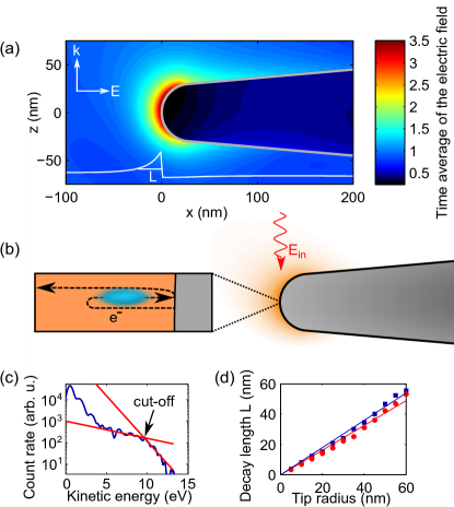
In this letter, we present experimental measurements with a new technique, the results of which we compare to numerical simulations of optical field enhancement at nanometric metal tips. Illuminating such a tip with light polarized parallel to the tip axis leads to the excitation of an enhanced near-field, which is spatially confined in all directions on the length scale of the tip radius Martin et al. (2001); Zhang et al. (2009); Novotny and Hecht (2006) (see Fig. 1(a)). The near-field drives a localized source of electrons at the tip apex Hommelhoff et al. (2006a, b); Ropers et al. (2007). Such photoemission experiments have found applications in a variety of different contexts aside from nanotips Petek and Ogawa (1997); Aeschlimann et al. (2007); Dombi et al. (2013). Very recently, it has been observed that part of the electrons can be driven back to the parent tip within a single cycle of the optical field. There, the electrons can scatter elastically and gain more energy in the optical field Krüger et al. (2011); Yalunin et al. (2011); Herink et al. (2012); Wachter et al. (2012). This process, well known from atomic physics Corkum (1993); Corkum and Krausz (2007); Paulus et al. (1994), has been called rescattering and leads to pronounced spectral features that are sensitive to the local electric field. Here we exploit the rescattering effect to probe the near-field in the immediate vicinity of the tip surface, as illustrated in Fig. 1(b).
Our experiment consists of an almost atomically smooth metal tip with a radius of curvature to . Its apex lies in the focal spot of few-cycle laser pulses derived from a Ti:sapphire oscillator (wavelength , repetition rate , pulse duration ). The setup is described in more detail in Ref. Schenk et al. (2010). While this laser system reaches intensities of up to in the focus, we do not observe any influence of possible optical non-linearities on the field enhancement factor, and all our results (e.g., the linear dependence of the rescattering cut-off on laser intensity Wachter et al. (2012)) are consistent with a linear model of the metal’s optical response.
Optical field enhancement enables us to observe electron rescattering at moderate pulse energies of less than . We measure the energy distribution of the electrons emitted in the forward direction using a retarding field spectrometer. The recorded spectra yield information about the dynamics of the emitted electrons. A typical spectrum is shown in Fig. 1(c). At small energies, such spectra display an exponential decrease in count rate, followed by a plateau towards larger energies. The latter is an indication of electron rescattering Paulus et al. (1994); Becker et al. (2002); Krüger et al. (2011, 2012); Wachter et al. (2012). This process has found utmost attention as it is at the core of attosecond science Corkum and Krausz (2007).
It has been shown that rescattering is highly sensitive to the peak electric field strength via the ponderomotive potential the electron experiences in the light field Paulus et al. (1994); Becker et al. (2002); Krüger et al. (2012): Here, is the laser wavelength, and are the electron’s charge and mass, and is the speed of light. The cut-off kinetic energy (see Fig. 1(c)) after rescattering is given by , where denotes the tip’s work function Busuladžić et al. (2006). Measuring hence yields FES .
Series of spectra for both tungsten and gold tips Eisele et al. (2011) with various tip radii yield the dependence of the field enhancement factor on tip radius and material. We extract the cut-off position of the rescattering plateau and deduce, via the above expressions, the peak electric field . We stress that , the field acting on the electron, is the enhanced field present at the tip’s surface. We thus obtain the field enhancement factor , with the laser field deduced from intensity measurements.
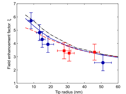
Figure 2 shows the field enhancement factor as a function of the tip radius . For tungsten tips, grows by around a factor of with decreasing , from at to at . For gold nanotips with radii between and , we obtain field enhancement factors between and . We have been unable to produce sharper gold tips with a well-controlled surface. Tip radii are determined in situ with the ring counting method in field ion microscopy or, for radii , using a scanning electron microscope FES .
We compare our results to fully independent simulations of field enhancement at tungsten, gold, and silver tips. They were performed using Lumerical (7.0.1), a commercial Maxwell solver implementing the finite-difference time-domain (FDTD) algorithm Taflove and Hagness (2005). From each simulation, we extract the field enhancement factor by fitting a quadratic decay to the near-field at the moment of greatest enhancement and extrapolating the result to the tip surface. This and other measures are essential to obtain meaningful results, as the finite mesh of the FDTD solver, together with the different length scales involved, makes this problem a tricky one. Further details and simulation results will be published elsewhere. The results for tungsten, gold, and silver are shown in Fig. 2. Experimental and simulation results agree well within the error bars. Note that this agreement is obtained without any free parameters. Both experiment and simulation show that increases smoothly for sharper tips, an indication of field enhancement due to a geometric effect.
Comparing our results to literature values of , we find good agreement for tungsten tips (experiment Neacsu et al. (2005); Yanagisawa et al. (2010), theory Martin et al. (2001); Yanagisawa et al. (2010)), while previous results for gold tips are inconsistent (experiment Neacsu et al. (2005); Ropers et al. (2007), theory Martin et al. (2001); Bouhelier et al. (2003)) with some authors reporting much higher enhancement Bouhelier et al. (2003); Neacsu et al. (2005); Ropers et al. (2007). A possible explanation for this disagreement is that the near-field at plasmonic materials like gold is exceptionally sensitive to the geometry of the tip (the opening angle in particular Martin et al. (2001); Zhang et al. (2009)) and its surface condition. This is supported by the large variance in enhancement factors at gold tips reported in Ref. Neacsu et al. (2005). Note also that far higher field enhancement factors are observed for tips in close vicinity () to surfaces Yang et al. (2009).
In our experiments with gold tips, we do not observe a large variance of field enhancement factors even though not all the tips had the ideal conical shape assumed in the simulations. A possible reason for this is that, before any measurement, we use field ion microscopy in conjunction with field evaporation to clean the tip surface and to ensure that the tip is almost ideally spherical in the vicinity of the apex FES . This is likely not the case in many other experiments. Evidently, more research is needed to fully understand the behavior of tips made of plasmonic materials. Such an investigation is beyond the scope of this letter. In the following analysis, we only consider perfectly smooth, conical tips (see Fig. 1(a)) with small opening angles between and .
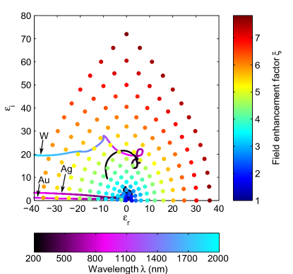
It appears, at first glance, surprising that the field enhancement factor of such different materials as tungsten and gold is so similar (see Fig. 2), considering that gold supports the excitation of surface plasmons Novotny and Hecht (2006); Stockman (2011). We therefore analyze the dependence of the field enhancement factor on tip material in a series of simulations where we vary the complex dielectric constant of the tip. This allows us to give a field enhancement map for all materials with , shown in Fig. 3. The results demonstrate that field enhancement occurs for any material with , even for pure dielectrics. Furthermore, the enhancement factor increases monotonically with the absolute value of the dielectric constant , confirming that we observe field enhancement due to a geometric effect. We also note a slightly larger enhancement for than for , which corresponds to an -dependent phase shift () of the near-field with respect to the driving field: the field enhancement factor is proportional to the maximum of the total electric field, which is reduced if the driving field and the near-field are out of phase. The -dependent simulations reveal why the field enhancement factor of tungsten, gold, and silver tips is similar: they have a similar value of at .
In order to obtain higher enhancement factors, materials with larger values of are required. For example, we find for aluminum tips ( at , beyond the range of our simulations in Fig. 3). Alternatively, can be increased by using longer wavelengths, because both the tip sharpness relative to the wavelength and the absolute dielectric constant of many materials increase for longer wavelengths. We expect considerably higher field enhancement at sharp metal tips for mid- and far-infrared or terahertz radiation. An enhancement factor has already been reported in SNOM experiments with terahertz radiation Huber et al. (2008).
In contrast to the increase with , there is one point in Fig. 3 close to that shows a significantly higher enhancement than the points surrounding it. This can be interpreted as a localized plasmon resonance, similar to what is known from nanospheres Martin et al. (2001). It can be observed with a wavelength of at silver tips Zhang et al. (2009) or at gold tips.
In the analysis of our experimental results, we have neglected the spatial variation of the near-field on the rescattered (field-probing) electrons’ path, assuming instead a constant electric field. This is justified as the decay length of the near-field (see Fig. 1(d)) is much longer than the maximum extension of the electron’s path : For our parameters, both classical Krüger et al. (2011, 2012) and quantum mechanical Wachter et al. (2012); Yalunin et al. (2013) simulations indicate that the electrons’ path extends approximately from the surface before rescattering. On this scale, the sharpest tips we investigate show a near-field variation of . Including this spatial variation into classical calculations of rescattering changes the enhancement factor by only, even for the sharpest tip in our experiments. This is less than the measurement uncertainty. For longer wavelengths or higher field strengths, the maximum extension increases. In this case, the effect may be more significant and can even suppress rescattering completely Herink et al. (2012).
One intriguing application of our method is the investigation of quantum effects in nanoplasmonics, a new field that has recently been named quantum plasmonics. Self-consistent quantum mechanical calculations of small nanoparticles (radius of curvature ) illuminated by laser pulses show that the excited surface charge density, the root cause of the optical near-field, extends over several ångströms beyond the surface Zuloaga et al. (2010). This “electron spill-out” reduces the strength of the near-field by up to . For small nanoparticles, it was shown that these effects are noticeable only at a distance of from the surface, while the near-field retains its classical shape for larger distances. As fully quantum mechanical calculations of larger nanoparticles remain difficult (although large steps are being made in this direction Teperik et al. (2013)), it is unclear if the length scale of nonclassical behavior depends on the size of the nanoparticle. The authors of Ref. Zuloaga et al. (2010) suspect to increase for larger nanoparticles.
While a fully integrated quantum calculation of both field enhancement and electron rescattering is beyond the scope of this letter, we will discuss the effects of quantum plasmonics on rescattering qualitatively. They depend on three length scales: the extent of nonclassical field reduction , the near-field’s decay length , and the rescattered electron’s maximum extension . If as in Ref. Zuloaga et al. (2010), the maximum of the near-field is significantly reduced, which implies a corresponding reduction of the cut-off energy. Extremely sharp nanostructures () will be required to reach this regime if does not scale with structure size. As discussed earlier, rescattering may be suppressed in this case, depending on the relation of and Herink et al. (2012).
If , only a small fraction of the near-field’s extent is reduced in strength so that the maximum of the near-field is almost unchanged. In this case, quantum effects are only noticeable if , because the rescattered electron would not be sensitive to the reduced field strength otherwise. The parameters in our experiments are (including a non-zero tunneling distance Busuladžić et al. (2006); Krüger et al. (2012); Hickstein et al. (2012)) and , so quantum effects should only be visible if becomes larger than for larger nanostructures. The agreement between experimental results and classical theory seems to suggest that . Hence, Q does not seem to scale with structure size, as hypothesized in Ref. Zuloaga et al. (2010). However, there is still the possibility of quantum plasmonic effects on a larger scale within the error bars of our results. An increase of for larger tips might explain the steeper decrease of for larger radii we observe in the experiment as compared to the simulation (see Fig. 2).
In conclusion, we demonstrate a new method of probing optical near-fields within distance from the surface of a nanoscale metal tip. The method is based on rescattering of electrons driven by short laser pulses. The length scale on which the near-field is measured reaches down to dimensions that are of utmost interest in the emerging field of quantum plasmonics. Experimental results for the field enhancement factors of tungsten and gold tips agree well with Maxwell simulations. Based on these results, we give a field enhancement map for a wide range of materials. Furthermore, the simulations reveal that geometric effects are the predominant mechanism of optical field enhancement at nanotips in most cases. Exceptions exist close to plasmon resonances. In the future, a tomographic reconstruction of the near-field, likely in three dimensions, will be possible by measuring the cut-off energy of the rescattered electrons while varying the laser power or wavelength.
Acknowledgements.
We would like to thank Peter Nordlander, Markus Raschke, and Hirofumi Yanagisawa for insightful discussions as well as Philipp Altpeter, Jakob Hammer, and Sebastian Stapfner for assistance with scanning electron microscope imaging.References
- Novotny and Hecht (2006) L. Novotny and B. Hecht, Principles of Nano-Optics (Cambridge University Press, 2006).
- Stockman (2011) M. I. Stockman, Optics Expr. 19, 22029 (2011).
- Wessel (1985) J. Wessel, J. Opt. Soc. Am. B 2, 1538 (1985).
- Inouye and Kawata (1994) Y. Inouye and S. Kawata, Optics Lett. 19, 159 (1994).
- Kawata et al. (2009) S. Kawata, Y. Inouye, and P. Verma, Nature Photon. 3, 388 (2009).
- Schnell et al. (2011) M. Schnell, P. Alonso-Gonzalez, L. Arzubiaga, F. Casanova, L. Hueso, A. Chuvilin, and R. Hillenbrand, Nature Photon. 5, 283 (2011).
- Hartschuh (2008) A. Hartschuh, Angewandte Chemie International Edition 47, 8178 (2008).
- Raschke et al. (2005) M. B. Raschke, L. Molina, T. Elsaesser, D. H. Kim, W. Knoll, and K. Hinrichs, ChemPhysChem 6, 2197 (2005).
- Stöckle et al. (2000) R. M. Stöckle, Y. D. Suh, V. Deckert, and R. Zenobi, Chem. Phys. Lett. 318, 131 (2000).
- Corkum and Krausz (2007) P. B. Corkum and F. Krausz, Nature Phys. 3, 381 (2007).
- Zuloaga et al. (2010) J. Zuloaga, E. Prodan, and P. Nordlander, ACS Nano 4, 5269 (2010).
- Marinica et al. (2012) D. Marinica, A. Kazansky, P. Nordlander, J. Aizpurua, and A. G. Borisov, Nano Letters 12, 1333 (2012).
- Ciracì et al. (2012) C. Ciracì, R. T. Hill, J. J. Mock, Y. Urzhumov, A. I. Fernández-Domínguez, S. A. Maier, J. B. Pendry, A. Chilkoti, and D. R. Smith, Science 337, 1072 (2012).
- Wachter et al. (2012) G. Wachter, C. Lemell, J. Burgdörfer, M. Schenk, M. Krüger, and P. Hommelhoff, Phys. Rev. B 86, 035402 (2012).
- Teperik et al. (2013) T. V. Teperik, P. Nordlander, J. Aizpurua, and A. G. Borisov, Phys. Rev. Lett. 110, 263901 (2013).
- García de Abajo (2010) F. J. García de Abajo, Rev. Mod. Phys. 82, 209 (2010).
- Koh et al. (2011) A. L. Koh, A. I. Fernández-Domínguez, D. W. McComb, S. A. Maier, and J. K. W. Yang, Nano Letters 11, 1323 (2011).
- Willets (2012) K. A. Willets, Progress in Surface Science 87, 209 (2012), ISSN 0079-6816.
- Yanagisawa et al. (2010) H. Yanagisawa, C. Hafner, P. Doná, M. Klöckner, D. Leuenberger, T. Greber, J. Osterwalder, and M. Hengsberger, Phys. Rev. B 81, 115429 (2010).
- Martin et al. (2001) Y. C. Martin, H. F. Hamann, and H. K. Wickramasinghe, J. Appl. Phys. 89, 5774 (2001).
- Zhang et al. (2009) W. Zhang, X. Cui, and O. J. F. Martin, Journal of Raman Spectroscopy 40, 1338 (2009).
- Martin and Girard (1997) O. J. F. Martin and C. Girard, Applied Physics Letters 70, 705 (1997).
- Van Bladel (1996) J. Van Bladel, Singular Electromagnetic Fields and Sources, IEEE Press Series on Electromagnetic Wave Theory (Wiley, 1996).
- Hommelhoff et al. (2006a) P. Hommelhoff, Y. Sortais, A. Aghajani-Talesh, and M. A. Kasevich, Phys. Rev. Lett. 96, 077401 (2006a).
- Hommelhoff et al. (2006b) P. Hommelhoff, C. Kealhofer, and M. A. Kasevich, Phys. Rev. Lett. 97, 247402 (2006b).
- Ropers et al. (2007) C. Ropers, D. R. Solli, C. P. Schulz, C. Lienau, and T. Elsaesser, Phys. Rev. Lett. 98, 043907 (2007).
- Petek and Ogawa (1997) H. Petek and S. Ogawa, Prog. Surf. Sci. 56, 239 (1997).
- Aeschlimann et al. (2007) M. Aeschlimann, M. Bauer, D. Bayer, T. Brixner, F. J. de Abajo, W. Pfeiffer, M. Rohmer, C. Spindler, and F. Steeb, Nature 446, 301 (2007).
- Dombi et al. (2013) P. Dombi, A. Hörl, P. Rácz, I. Márton, A. Trügler, J. R. Krenn, and U. Hohenester, Nano Letters 13, 674 (2013).
- Krüger et al. (2011) M. Krüger, M. Schenk, and P. Hommelhoff, Nature 475, 78 (2011).
- Yalunin et al. (2011) S. V. Yalunin, M. Gulde, and C. Ropers, Phys. Rev. B 84, 195426 (2011).
- Herink et al. (2012) G. Herink, D. R. Solli, M. Gulde, and C. Ropers, Nature 483, 190 (2012).
- Corkum (1993) P. B. Corkum, Phys. Rev. Lett. 71, 1994 (1993).
- Paulus et al. (1994) G. G. Paulus, W. Nicklich, H. Xu, P. Lambropoulos, and H. Walther, Phys. Rev. Lett. 72, 2851 (1994).
- Schenk et al. (2010) M. Schenk, M. Krüger, and P. Hommelhoff, Phys. Rev. Lett. 105, 257601 (2010).
- Becker et al. (2002) W. Becker, F. Grasbon, R. Kopold, D. Milošević, G. Paulus, and H. Walther, in Advances in Atomic, Molecular, and Optical Physics (Academic Press, 2002), vol. 48, pp. 35–98.
- Krüger et al. (2012) M. Krüger, M. Schenk, M. Förster, and P. Hommelhoff, Journal of Physics B: Atomic, Molecular and Optical Physics 45, 074006 (2012).
- Busuladžić et al. (2006) M. Busuladžić, A. Gazibegović-Busuladžić, and D. B. Milošević, Laser Phys. 16, 289 (2006).
- (39) See supplemental material below for details.
- Eisele et al. (2011) M. Eisele, M. Krüger, M. Schenk, A. Ziegler, and P. Hommelhoff, Rev. Sci. Instr. 82, 026101 (2011).
- Lide (2004) D. Lide, CRC handbook of chemistry and physics: a ready-reference book of chemical and physical data, CRC Handbook of Chemistry and Physics, 85th Ed (CRC Press, 2004).
- Taflove and Hagness (2005) A. Taflove and S. Hagness, Computational Electrodynamics: the Finite-Difference Time-Domain Method (Artech House, 2005).
- Neacsu et al. (2005) C. C. Neacsu, G. A. Reider, and M. B. Raschke, Phys. Rev. B 71, 201402 (2005).
- Bouhelier et al. (2003) A. Bouhelier, M. Beversluis, A. Hartschuh, and L. Novotny, Phys. Rev. Lett. 90, 013903 (2003).
- Yang et al. (2009) Z. Yang, J. Aizpurua, and H. Xu, Journal of Raman Spectroscopy 40, 1343 (2009), ISSN 1097-4555.
- Huber et al. (2008) A. J. Huber, F. Keilmann, J. Wittborn, J. Aizpurua, and R. Hillenbrand, Nano Letters 8, 3766 (2008).
- Haynes and Lide (2011) W. Haynes and D. Lide, CRC Handbook of Chemistry and Physics, 92nd Edition (CRC Press, 2011).
- Yalunin et al. (2013) S. V. Yalunin, G. Herink, D. R. Solli, M. Krüger, P. Hommelhoff, M. Diehn, A. Munk, and C. Ropers, Annalen der Physik 525, L12 (2013).
- Hickstein et al. (2012) D. D. Hickstein, P. Ranitovic, S. Witte, X.-M. Tong, Y. Huismans, P. Arpin, X. Zhou, K. E. Keister, C. W. Hogle, B. Zhang, et al., Phys. Rev. Lett. 109, 073004 (2012).