Spontaneous symmetry breaking in active droplets provides a generic route to motility
Abstract
We explore a generic mechanism whereby a droplet of active matter acquires motility by the spontaneous breakdown of a discrete symmetry. The model we study offers a simple representation of a “cell extract” comprising, e.g., a droplet of actomyosin solution. (Such extracts are used experimentally to model the cytoskeleton.) Actomyosin is an active gel whose polarity describes the mean sense of alignment of actin fibres. In the absence of polymerization and depolymerization processes (‘treadmilling’), the gel’s dynamics arises solely from the contractile motion of myosin motors; this should be unchanged when polarity is inverted. Our results suggest that motility can arise in the absence of treadmilling, by spontaneous symmetry breaking (SSB) of polarity inversion symmetry. Adapting our model to wall-bound cells in two dimensions, we find that as wall friction is reduced, treadmilling-induced motility falls but SSB-mediated motility rises. The latter might therefore be crucial in three dimensions where frictional forces are likely to be modest. At a supra-cellular level, the same generic mechanism can impart motility to aggregates of non-motile but active bacteria; we show that SSB in this (extensile) case leads generically to rotational as well as translational motion.
Living cells can move themselves around in a variety of different conditions and environments, and they exploit a range of strategies and mechanisms to do so. Uncovering the generic pathways to cell motility remains central to many important processes ranging from wound healing and tissue development Weijer1 to immunological response and diseases such as cancer Poincloux . The best characterized case is that of a crawling cell on a planar 2D substrate or wall. Here motility is generally attributed to cytoskeletal actin filaments which polymerize at one end () and depolymerize at the other () in a process called treadmilling. So long as the system has nonzero polarity (where is a unit tangent oriented from to and angle brackets denote an average over filaments), treadmilling leads to macroscopic motion. This exploits a Brownian ratchet mechanism in which forward fluctuations of the cell perimeter are locked in by polmerization Ratchet . However this mechanism requires a relatively solid anchor-point from which to propel the cell. For cells crawling on a 2D surface, this is provided by focal adhesions and other integrin rich structures Theriot-Kondev ; Theriot-Mogilner .
In vivo, cells often move in a three-dimensional environment such as an extracellular matrix and/or a tissue of cells Poincloux ; Evan-Ram ; Hawkins . Especially in quasi-spherical motile cells, both integrin-rich structures and mechanical anchoring are less in evidence, and the mechanisms of motility in such 3D environments remain unclear Friedl . A recent study on tumour cells moving inside an elastic gel suggests that an important role in 3D locomotion may be played not by polymerization but by myosin contractility. This can lead to collective internal flows of actin that may ultimately propel the cell forward Hawkins . The contractile effect arises by a motor spanning two fibres causing an inward force pair (Fig. 1A) Liverpool . This creates an active stress, usually modelled as with the concentration of active material, Greek suffices denote Cartesian directions, and is an activity parameter ( for contractile systems). This raises an important issue of principle: how exactly does a tensorial active stress result in a vectorial propulsion velocity?

Several studies use minimal models to address the fundamental physics of how activity imparts cell motility. Experimental progress has focused on “cell extracts” Carlier ; Bausch : unregulated bags of cytoskeletal filaments (actin) and molecular motors (myosin), enclosed by a membrane. However, most of this work focuses on 2D crawling via the treadmill-ratchet mechanism described above. (3D systems are harder to study, and selective inhibition of the treadmill dynamics is biochemically difficult Gerisch .) On the modelling side, generic theories have been proposed to make contact with the cell-extract data, again focusing mainly on 2D crawling and the treadmill-ratchet mechanism treadmilling ; Wolgemuth ; Ziebert . While the influence of myosin contractility on cell shape during locomotion has been addressed Wolgemuth ; Ziebert , in 2D this has not so far been thought sufficient by itself to lead to motility.
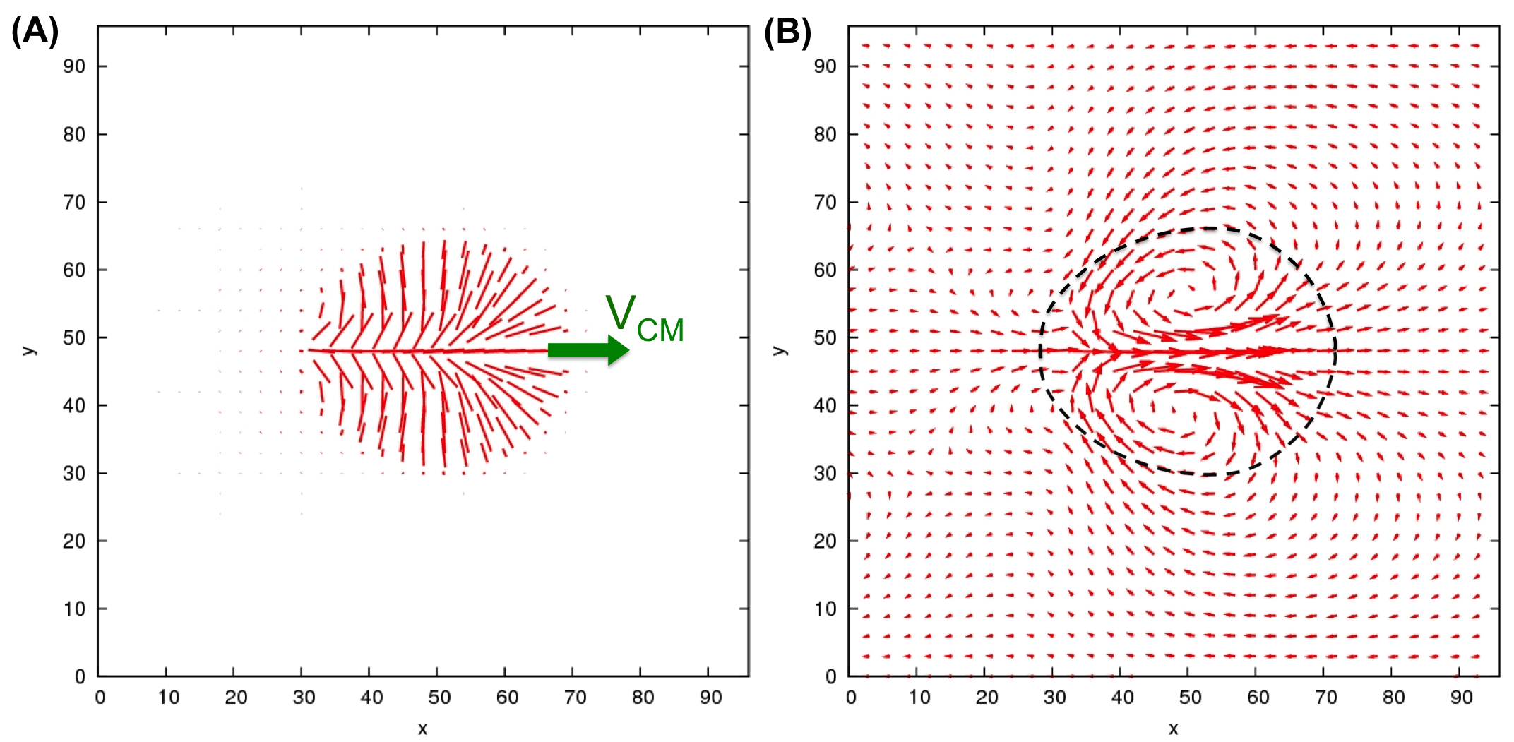
Here we provide a detailed computational study of the effects of active stresses in a minimal 2D model of an actomyosin cell extract. To confirm that our proposed motility mechanism remains pertinent in 3D, we additionally perform selective (computationally intensive) simulations in that case. Our simulation model comprises a droplet of an active fluid or gel Kruse ; Tjhung , confined by interfacial tension , and surrounded by a Newtonian host fluid. Our model equations are based on established continuum precepts and outlined in Methods and Materials. Myosin contractility is represented by an active stress as detailed above. This term is invariant under global polarity inversion as are, with treadmilling absent, the full equations of motion (for further discussions of their symmetries, refer to Appendix). We show then that when the activity parameter exceeds a given threshold, an initially circular or spherical droplet spontaneously breaks that inversion symmetry, leading to an elastic splay of the polarity field and to motion along . This spontaneous symmetry breaking (SSB) manifests itself as a supercritical Hopf bifurcation, which can alternatively be viewed as a continuous nonequilibrium phase transition. (The threshold value depend on both , and an effective elastic constant penalising distortions of the ordered polar state.) In 2D, the resulting motile droplets have crescent-like shapes similar to some crawling cells Theriot , whereas in 3D our model predicts both spherical and concave shapes. Representing wall friction by a depth-averaged drag term in 2D, we find this slows down the SSB-mediated motion, but does not stop it altogether unless a critical drag is exceeded.
We then introduce the treadmilling effect, which we represent by a self-advection parameter . This breaks the global symmetry, but it does so directly, not spontaneously. (Note that in our model polarity is present even without treadmilling: see Appendix A for a discussion of the relevant physics.) This is the speed at which, relative to the local suspending fluid (of velocity ), each filament is self-propelled along its own tangent. Even though asymmetric polymerization does not lead directly to mass transport, for actomyosin the resulting mass flux should capture, in a highly simplified manner, the preferential growth of filaments by addition of monomers at one end and loss at the other; see Fig. 1C. This simplified description is possible because we exclude the bath of monomers from the local mass density of active material. (We assume that on average, the monomers nonetheless keep up with the moving gel.) In general, self-advection leads to density gradients, which in turn cause hydrodynamic flows in the direction opposite to . This backflow severely limits the effectiveness of self-advection in creating motility, almost cancelling it for a droplet in free space as we discuss below. Our 2D study of the effect of wall friction shows however that high enough friction, by reducing towards zero, restores treadmilling-induced motility with a speed that approaches .
Although our main focus is on actomyosin cell extracts, our framework provides a broader generic approach to droplet motility. We can thus investigate what happens when we reverse the sign of the active stresses to consider the extensile case, Baskaran ; SoftMatter . The primary experimental relevance of this case is to suspensions of bacteria, which push fluid out along their major axes and draw it in around the equator; see Fig. 1A. In this context, is the bacterial swim speed; although this is nonzero for motile species, one may create virtually non-motile mutants (called “shakers”) which still create extensile active stresses, e.g. by excessively increasing the tumbling rate tumblers . A droplet of such organisms can be created either by inducing an attraction between them (as our model effectively assumes) jana or perhaps by confining them in an emulsion droplet. To attain nonzero one further requires these particles to have net polar order (as opposed to a nematic state, for which there is orientational order, but equal numbers of particles with tangent locally (see Appendix)). Setting aside the possible difficulties in meeting all those requirements experimentally, we predict that such “shaker” bacterial droplets could again break the symmetry and start moving spontaneously. Intriguingly, the predicted trajectories in this extensile case are more complicated than those of the contractile model. This is because, in extensile droplets, the SSB-mediated velocity forcing is in a direction perpendicular, rather than parallel, to the polarization vector . Restoring nonzero to describe the case of motile bacteria, the composition of these two motions leads to circular or spiralling trajectories of the droplet as a whole.
I Results
We first present results for the 2D contractile case, then briefly describe our findings in 3D, and finally give some further 2D results on extensile droplets. We initialized our simulation runs with a circular (or spherical) droplet within which the concentration of active material is taken to be a constant (), with outside. The polarization field within the droplet is initially uniform along the horizontal () axis and varies with concentration as magnitude with a threshold value for polarization onset. We choose so that is nonzero inside the droplet and zero (isotropic) outside, so that we assume the actomyosin network inside our active droplets is initially polarised. The system is then evolved via the equations of motion as specified in Materials and Methods (discussed further in Appendix A), with chosen values of the activity parameter and the self-advection (treadmilling) parameter . On a relatively short timescale both the internal concentration and the polarisation field relax towards the equilibrium values and (while still and externally) with some interfacial tension , set by minimization of our chosen free energy. (Our choice creates no anchoring of at the surface so, in the absence of symmetry breaking, the polarization remains uniform; see Fig. 2A left. The effect of a soft anchoring is discussed in the Appendix A.) Having made one such relaxed droplet, the dependence of its behavior on and/or was systematically explored by incrementing those quantities and waiting for steady state, before incrementing again. Because we are primarily interested in trends and symmetry breaking phenomena, rather than quantitative predictions of where these will occur for specific materials, we report all results below in the natural units for lattice Boltzmann simulations (LBU); the connection between these and physical units is discussed in Appendix D.
I.1 Contractile stress can create motility via SSB
Although contractile motor stress and actin treadmilling are generally both present in motile cells, it is illuminating to study these two mechanisms separately. We first consider a droplet with no external drag term (no wall friction) and no treadmilling term (), and vary the activity parameter . For low activity , below some critical value , the droplet polarisation field remains aligned uniformly along its initial direction and the droplet remains stationary. However it becomes slightly elongated in the direction perpendicular to the polarisation vector as a result of the competition between the contractile stress and the interfacial tension (see Fig. 2A middle). In this regime, the contractile stress set up a quadrupolar fluid flow around the droplet (Fig. 2B left), so that the whole droplet behaves as a large contractile element (compare Fig. 1A right). However it does not translate: there is no motility, and the droplet is a “shaker” rather than a “mover” Ramaswamy .

As we increase beyond , the uniform polarization field becomes unstable with respect to a splay deformation. This happens because the contractile stress is large enough to overcome the resistance to deformation mediated by the elastic constant . The splay creates a state in which neighboring vectors either fan outwards () or inwards (). The first is shown in Fig. 2A right; the second is found by first taking its mirror image and then reversing . This choice is made at random, spontaneously breaking the global polarity inversion symmetry. As soon as this happens, the droplet starts to move along the direction set by . This motion is attributable to the formation of a pair of flow vortices inside the droplet (Fig. 2B right). Such spontaneous propulsion is somewhat reminiscent of the self-electrophoretic motion of a vesicle with active membrane pumps in an ionic solution Mitchell ; Lammert .
We note that in bulk active contractile fluids, the state of uniform is also generically unstable to splay fluctuations in 1D Baskaran ; Simha ; Giomi-Marchetti , which then lead to the onset of spontaneous flow. At one level, the SSB-induced motility transition described here can be viewed as a manifestation of that bulk instability, albeit with two variations. First, spontaneous flows are present on both sides of our transition: as discussed above there is a quadrupolar flow field already in the non-moving state. Second, in bulk the critical activity level is nonzero only in finite systems, for which the transition is discontinuous, unlike ours (see below and Fig. 2C), and the resulting velocity field much more complicated Tjhung ; SoftMatter .
To illustrate our symmetry breaking motility mechanism more clearly, we plot the magnitude of the centre of mass velocity of the droplet as a function of activity in Fig. 2C. This bifurcation diagram shows a continuous non-equilibrium transition from a stationary and uniform state to a moving and splayed state. Moreover, to within numerical accuracy the observations are consistent with a supercritical Hopf bifurcation, for which . This mean-field like exponent is perhaps unsurprising as there is no noise in our simulations. Accordingly it might change in the presence of activity-generated noise harvardSGR , depending on whether the bifurcation remains low-dimensional or acquires a many-body critical character.
I.2 SSB-induced motility is diminished by friction
The results above are for a droplet in 2D surrounded by Newtonian fluid. To better describe experiments involving cell-crawling on a substrate, we now consider an additional frictional force between the solid wall and the cell. To do this within our 2D continuum model, we introduce an additional force density to the momentum balance equation (see Materials and Methods and Appendix). Here is an effective coefficient of friction which depend on whether we have non-slip or partial-slip boundary conditions on the substrate and also on the thickness of the cell. It may represent conventional friction and/or a coarse-grained model of focal adhesions and other localized mechanical contacts. The presence of this friction will significantly quench the hydrodynamic flow . Since our SSB-induced motility requires a hydrodynamic vortex flow inside the droplet, the frictional force can greatly reduce the droplet’s velocity, bringing it to rest beyond a critical level , at which point the symmetry is restored; the value of depends on activity and other model parameters. This is seen in a plot of against in Fig. 3.

I.3 Self-advective motility is enhanced by friction
We now consider the case when there is only treadmilling (modeled as self-advection ) but no contractile stress (). As discussed previously, the droplet will move along the polarization vector with speed . Since there is no spontaneous symmetry breaking involved, translational motion occurs for any nonzero , in contrast to the threshold behavior seen for contractile SSB-motility. The most interesting aspect is the role of the friction parameter . Again we plot the velocity of the droplet as a function of in Fig. 3. In contrast to the previous case, motility is significantly enhanced by the presence of friction. Indeed, in in the limit , we have and (see Fig. 3 inset).
The intersection of the two plots of droplet velocity versus friction (found respectively by switching off activity or self-advection) defines a characteristic friction scale . For contractile stresses dominate cell motility, while for , self-advection is dominant.
In most experiments on 2D crawling of cells/cell extracts Theriot-Mogilner , the involvement of focal adhesions suggests that the high friction (treadmilling dominated) limit generically prevails. On the other hand, some recent experiments Yam directly identify spontaneous symmetry breaking of the actomyosin network as the initiator of polarized cell motility in keratocytes. Our work emphasizes that spontaneous breaking of global polarity inversion symmetry arises from contractile motor activity, not from treadmilling. It is therefore arguable that the role of motor activity in 2D motility has so far been underestimated. We note however that an equivalent discrete symmetry breaking would create motility if a pure treadmilling state of zero , but finite nematic order (), spontaneously acquires polarity locally (see Appendix).

I.4 3D droplets show a window of SSB-induced motility
In the context of experiments on 3D tumour cells, it has been argued that motility is driven primarily by contractile stress Poincloux ; Hawkins , suggesting that the low-friction limit of our model prevails here. This accords with the much diminished part played by focal adhesions in 3D Friedl . For the 3D case we therefore neglect the friction term, and run selected simulations to confirm that the SSB route to motility remains operative.
Fig. 4 shows steady state polarization fields inside a 3D contractile droplet with increasing values of the activity magnitude from A to D. As expected, the first steady state encountered is a symmetric but deformed immotile droplet (Fig. 4A). The active stress contracts the droplet along resulting in a lenticular shape. As we increase beyond a critical , splay instability spontaneously breaks symmetry and causes the droplet to move along just as in the 2D case. The droplet shape is concave and (as in the lenticular case) both it and the flow field resemble that created by rotating the 2D droplet about the axis (Fig. 4B). The resulting hydrodynamic flow (Fig. 4E) therefore corresponds to a toroidal vortex ring. It would be interesting to see how these predictions compare with intracellular actin and fluid flow maps that might in future be measured for cells moving in 3D environments, for instance those studied in Ref. Poincloux .
Interestingly, as is increased further, the droplet becomes increasingly spherical (Fig. 4C), and finally symmetry is restored, creating an immotile spherical droplet with a ‘hedgehog’ defect (of topological charge 1 as dictated by the polar ordering Chaikin-Lubensky ) at the centre (Fig. 4D).
I.5 Extensile SSB creates transverse or circular motility
Our final results are for 2D extensile droplets (Fig. 1A). Continuum descriptions of uniform extensile active fluids are widely used to describe dense bacterial suspensions Baskaran ; Ramaswamy ; Suzanne . These results may therefore be relevant to bacterial droplets formed by aggregation in the presence of attractive forces jana , or possibly by confinement of bacteria within a droplet emulsion. Again, for fixed thermodynamic parameters controlling the elasticity and interfacial tension of the confined material, one can vary the activity parameter (now positive) and the self-advection parameter which corresponds to the swimming speed of individual bacteria and is nonzero for “movers” but zero for “shakers” Baskaran ; Simha .
Fig. 5A shows steady state configurations of a 2D purely extensile () droplet at different ranges of activity. For the droplet remains stationary but again elongates symmetrically, this time along the direction of the polarization field . The extensile stress creates the quadrupolar flow field expected of a large, extensile shaker. For beyond the critical value , the droplet again becomes unstable, but now with respect to bend deformation as opposed to splay. This gives rise to a horizontal vortex pair inside the droplet as opposed to the vertical one in the contractile case (Fig. 2B right). This flow field causes the droplet to move in a direction set by the sense of bending as (which can be upwards or downwards according to Fig. 5A). As can be seen from figure Fig. 5A this stationary-to-motile transition can still be characterised as a continuous SSB transition, however, the droplet speed attains a maximum at intermediate before falling to zero again. Beyond a second critical activity threshold ( in Fig. 5A), the polarisation pattern oscillates continuously while remaining symmetric at all times and consequently, the droplet again becomes non-motile.
Turning finally to the case of nonzero self-advection () we find that this, combined with the extensile motility, can give rise to an intriguing spiralling motion (see Fig. 5B). This arises because the SSB-induced motility is at right angles to the polarization (Fig. 5B right). On the other hand, self-advection everywhere transports material along locally; since the polarization field is spontaneously curved, this by itself would give a circular droplet orbit. Compounding these two motions typically leads to outward spiral trajectories as shown in Fig. 5B. This outcome contrasts with the contractile case where both the SSB-induced and the self-advective motion (the latter averaged over the droplet configuration) point either together or oppositely along the direction and only straight line motion can result.

II Discussion and Conclusions
Our simulations of contractile droplets can be viewed as a simple in silico analogue of the in vitro cell-extract studies that have been used to dissect the biophysical ingredients of motility. Our simulation work powerfully complements these studies, by allowing us to isolate the role of contractile (motor) activity in cell locomotion: this is very difficult in the laboratory, where current strategies for inhibiting polymerization dynamics (treadmilling) can severely impair other key subcellular processes Gerisch .
Our approach likewise complements those of previous theories Wolgemuth ; Ziebert ; Doubrovinski which have concentrated on treadmilling as the main driver of motility. We have shown in 2D that contractile stresses alone can not only shape the rear of a crawling cell (Fig. 2), but also create motility itself, provided that the motor forces are large enough to create an asymmetric circulatory flow as in Fig. 2B. Interestingly, keratocyte cells crawling on glass seemingly do exploit myosin activity to set up an intracellular actin flow in the rear of a cell which ultimately polarises it and makes it motile Yam . Nonetheless, our study of the the effects of a frictional term, which promotes the motility created by treadmilling but inhibits that caused by contractile stress, lends support to the view that cell crawling on a wall is usually dominated by the treadmilling.
In contrast, in 3D cell motility, recent work suggests that treadmilling plays at most a minor role Poincloux ; Hawkins . In the 3D case, therefore, our work describes a simple and compelling mechanism for how spontaneous translational motion can in principle arise solely by the action of a contractile stress. This requires spontaneous symmetry breaking, mediated in our case by splay deformation in response to that stress. Our 2D and 3D simulations go beyond the 2D theory of Ref. Hawkins by addressing the dynamics of the polarization field. We do however make some important simplifications: our droplets are confined only by interfacial tension not by an elastic membrane; we treat treadmilling as a simple self-advection; and we do not address any direct transition between nematic and polar order, despite assuming polarity inversion symmetry at thermodynamic level. Improving the model in these respects will require a more detailed microscopic derivation which we shall leave to future work. To test whether our model indeed captures the biophysics of 3D cell motility, it would be exciting to visualize experimentally the detailed cytoskeletal organization and flow fields, e.g. for cells moving through matrigel Poincloux ; Hawkins .
Our generic framework is not limited to contractile actomyosin networks. Indeed we have discussed the case of extensile droplets, possibly relevant to aggregates or emulsions of active but immotile bacteria; here translational motility arises by spontaneous symmetry breaking only at intermediate activity, and is mediated by bend rather than splay deformation. The addition of self-advection along the bent polarization field then leads in addition to rotational motion. We note that rotational and translational motility of small bacterial aggregates was recently observed, but attributed to a somewhat different mechanism where symmetry is broken by frozen-in statistical fluctuations rather than SSB jana .
Finally, our hydrodynamic equations of motion, or close variants of these, might in some cases be applicable to concentrated eukaryotic cell masses such as tissue Ranft . Within a tissue each cell exerts forces on its neighbors which at the lowest order continuum level creates a certain density of force dipoles Ranft ; the velocity field then describes the slow migration of cells inside the tissue. It is intriguing to note that the large-scale tissue flow in animal cells during gastrulation may break the symmetry to form vortices, similarly to our active droplets. This is the case of the so-called “polonaise movements” which are observed in the developing chick embryo Weijer1 , and which are important to form the correct supercellular structure. In this context a “polarisation” field is sometimes used to describe the orientation of individual cells Vasiev . The relation, if any, between the onset of this vortex flow and that seen in our droplets remains to be explored.
III Materials and Methods
We briefly outline here the hydrodynamic model used in this work (more details are in Appendix). We consider a fluid, comprising a mixture of active material and solvent, with constant total mass density . The hydrodynamic variables whose dynamics we monitor are: (i) the concentration of active material , (ii) the fluid velocity (with incompressibility requiring ), and (iii) the polarization field as defined previously.
Although an active droplet is a nonequilibrium system, we introduce the following free energy functional to describe its equilibrium physics in the passive limit of zero activity:
Here is a phenomenological free energy amplitude, determines the droplet interfacial tension, and is an effective elastic constant. This choice of leads to a continuous isotropic-to-polar transition at . To confine the active material into a droplet, we choose: and set . This creates two free energy minima corresponding to a phase of pure passive solvent (external to the droplet, and ) and a polar active phase (inside the droplet, and ).
Treating the active material as locally conserved, the time evolution of the concentration field can then be written as a convective-diffusion equation:
| (2) |
where is a thermodynamic mobility parameter and is the self-advection parameter.
The dynamics of the polarisation field follows an “active nematic” evolution SoftMatter , given by
| (3) |
where and are the symmetric and anti-symmetric parts of the velocity gradient tensor . is the rotational viscosity and is related to the geometry of the active particles Kruse .
Force balance is ensured through the Navier-Stokes equation, where is the isotropic pressure, is the friction force per unit volume and is the total stress in the fluid which includes viscous, elastic/Ericksen, interfacial and “active” stresses (see Appendix for details and for a discussion of the effects of additional terms, allowed by symmetry, in the equations of motion). The active stress is Ramaswamy where for contractile activity and for extensile.
To solve these equations in 2D and 3D, we performed hybrid lattice Boltzmann simulations, as done previously for other active flows Tjhung ; SoftMatter .
Acknowledgements.
We thank R. Voituriez for very useful discussions. ET thanks SUPA for a Prize Studentship and MEC holds a Royal Society Research Professorship.References
- (1) Dormann D., Weijer C.J. (2006) Imaging of cell migration. EMBO J. 25:3480-93.
- (2) Poincloux R., Collin O., Lizarraga F., Romao M., Delbray M., Piel M., Chavrier P. (2011) Contractility of the cell rear drives invasion of breast tumor cells in 3D Matrigel. Proc. Natl. Acad. Sci. USA 108:1943-1948.
- (3) Peskin C. S., Odell G. M., Oster G. F. (1993) Cellular motions and thermal fluctuations - the Brownian ratchet. Biophys. J. 65:316-324.
- (4) Phillips R., Kondev J., Theriot J. A. (2008) Physical biology of the cells, (Garland Science).
- (5) Barnhart E. L., Lee K. C., Keren K., Mogilner A., Theriot J. A. (2011) An adhesion-dependent switch between mechanisms that determine motile cell shape. PLoS Biology 9(5):e1001059.
- (6) Evan-Ram S., Yamada K. M. (2005) Cell migration in 3D matrix. Curr. Opin. Cell Biol. 17:524-532.
- (7) Hawkins R. J., Poincloux R., Benichou O., Piel M., Voituriez R. (2011) Spontaneous contractility-mediated cortical flows generates cell migration in three-dimensional environments. Biophys. J. 101:1041-1045.
- (8) Friedl P., Wolf K. (2003) Tumour-cell invasion and migration: diversity and escape mechanism. Nat. Rev. Cancer 3:362-374.
- (9) Liverpool T. B., Marchetti M. C. (2006) Rheology of active filament solutions. Phys. Rev. Lett. 97:268101.
- (10) Loisel T., Boujemaa R., Pantaloni D., Carlier M.-F. (1999) Reconstitution of actin-based movement using pure proteins. Nature 401:613-616.
- (11) Köhler S., Schaller V., Bausch A. R. (2011) Structure formation in active networks. Nature Mat. 10:462–468.
- (12) Gerisch G., Bretschneider T., Muller-Taubenberger A., Simmeth E., Ecke M. et al. (2004) Mobile actin clusters and traveling waves in cells recovering from actin depolymerization. Proc. Natl. Acad. Sci. USA 87:2493-3503.
- (13) Sambeth R., Baumgaertner A. (2001) Autocatalytic polymerization generates persistent random walk of crawling cells. Phys. Rev. Lett. 86:5196-5199.
- (14) Wolgemuth C. W., Stajic J., Mogilner A. (2011) Redundant mechanisms for stable cell locomotion revealed by minimal models. Biophys. J. 101:545-553.
- (15) Ziebert F., Swaminathan S., Aranson, I. S. (2011) Model for self-polarisation and motility of keratocyte fragments. J. Roy. Soc. Interface, doi:10.1098.
- (16) Kruse K., Joanny J. F., Julicher F., Prost J., Sekimoto K. (2004) Asters,Vortices, and Rotating Spirals in Active Gels of Polar Filaments. Phys. Rev. Lett. 92:078101.
- (17) Tjhung E., Cates M. E., Marenduzzo D. (2011) Nonequilibrium steady states in polar active fluids. Soft Matter 7:7453-7464.
- (18) Keren K., Pincus Z. Allen G. M., Barnhart E. L., Marriott G., Mogilner A., Theriot J. A. (2008) Mechanism of shape determination in motile cells. Nature 453:475-480.
- (19) Baskaran A,. Marchetti M.C. (2009) Statistical mechanics and hydrodynamics of bacterial suspensions. Proc. Natl. Acad. Sci. USA 106:15567-15572.
- (20) Cates M. E., Henrich O., Marenduzzo D., Stratford K. (2009) Lattice Boltzmann simulations of liquid crystalline fluids: active gels and blue phases. Soft Matter 5:3791-3800.
- (21) Parkinson J. S., Parker S. R., Talbert P. B, Houts S. E. (1983) Interactions between chemotaxis genes and flagellar genes in Escherichia coli. J. Bacteriol. 155:265–274.
- (22) Schwarz-Linek J., Valeriani C., Cates M. E., Cacciuto A., Marenduzzo D., Morozov A. N., Poon W. C. K. (2012) Phase separation and rotor self-assembly in active particle suspensions. Proc. Natl. Acad. Sci. USA 109:4052-4057.
- (23) Hatwalne Y., Ramaswamy S., Rao M., Simha R. A. (2007) Rheology of active-particle suspensions. Phys. Rev. Lett. 92:118101.
- (24) Mitchell P. (1972), Self-electrophoretic locomotion in microorganisms – bacterial flagella as giant ionophores. FEBS Letters 28:1.
- (25) Lammert P. E., Prost J., Bruinsma R. (1996) Ion drive for vesicles and cells. J. Theor. Biol. 178:387-391.
- (26) Simha R. A., Ramaswamy S. (2002) Hydrodynamic fluctuations and instabilities in ordered suspensions of self-propelled particles. Phys. Rev. Lett. 89:058101.
- (27) Giomi L., Marchetti M. C. (2012) Polar patterns in active fluids. Soft Matter 8:129-139.
- (28) Stamenovic, D., Rosenblatt, N., Montoya-Zavala, M., Matthews, B.D., Hu, S. et al. (2007) Rheological Behavior of living cells is timescale-dependent. Biophys. J. 93:L39-L41.
- (29) Yam P. T., Wilson C. A., Ji L., Hebert B., Barnhart E. L. et al. (2007) Actin-myosin network reorganization breaks symmetry at the cell rear to spontaneously initiate polarized cell motility. J. Cell. Biol. 178: 1207-1221.
- (30) Chaikin P. M., Lubensky T. C. (2000) Principles of condensed matter physics, (Cambridge University Press).
- (31) Cates M. E., Fielding S. M., Marenduzzo D., Orlandini E., Yeomans J. M. (2008) Shearing active gels close to the isotropic-nematic transition. Phys. Rev. Lett. 101:068102.
- (32) Doubrovinski K., Kruse K. (2011) Cell motility resulting from spontaneous polymerization waves. Phys. Rev. Lett. 107:258103.
- (33) Ranft J., Basan M., Elgeti J., Joanny J.-F., Prost J., Julicher F. (2010) Fluidization of tissues by cell division and apoptosis. Proc. Natl. Acad. Sci. USA 107:20863-20868.
- (34) Vasiev B., Balter A., Chaplain M., Glazier J. A., Erijer C. J. (2010) Modeling gastrulation in the chick embryo: formation of the primitive streak. PLoS One 5:e10571.
- (35) de Gennes P.-G., Prost. J. (1993) The physics of liquid crystals, (Clarendon Press, Oxford).
- (36) Frank F. C. (1958) On the theory of liquid crystals. Discussions Faraday Soc. 25:19.
- (37) Ericksen J. L. (1961) Conservation laws for liquid crystals. Trans. Soc. Rheol. 5:23.
- (38) Edwards S. A., Yeomans J. M. (2009) Spontaneous flow states in active nematics: a unified picture EPL 85:18008.
- (39) deGroot S. R., Mazur P. (2011) Non-Equilibrium Thermodynamics, (Dover Publications).
- (40) Small J. V., Herzog M., Anderson K. (2008) Actin filament organization in the fish keratocyte lamellipodium. J. Cell Biol. 129:1275-1286.
- (41) Kung W., Marchetti M. C., Saunders K. (2006) Hydrodynamics of polar liquid crystals. Phys. Rev. E 73:031708.
- (42) Bertin E., Droz M., Gregoire G. (2006) Boltzmann and hydrodynamic descriptions for self-propelling particles. Phys. Rev. E 74:022101.
- (43) Marchetti M. C., Liverpool T. B. (2007) Hydrodynamics and rheology of active polar filaments, in Cell Motility, ed. P. Lenz, Springer-Verlag, NY.
- (44) Voituriez R., Joanny J. F., Prost J. (2005) Spontaneous flow transition in active polar gels. EPL 70(3):404-410.
- (45) Cates M. E., Stratford K., Adhikari R., Stansell P., Desplat J.-C., Pagonabarraga I., Wagner A. J. (2004) Simulating colloid hydrodynamics with lattice Boltzmann methods. J. Phys. Cond. Mat. 16:S3903-S3915.
- (46) Rubinstein B., Fournier M. F., Jacobson K., Verkhovsky A., Mogilner A. (2009) Actin-myosin viscoelastic flow in the keratocyte lamellipod. Biophys. 97:1853-1863.
Appendix A Hydrodynamic description
A.1 Choice of order parameters
We model actin filaments in the cytoskeleton, or bacteria in suspension, as rod-shaped active polar particles. In the hydrodynamic (continuum) limit, the dynamics of these dense suspensions of active polar particles can be described by a few continuum variables.
Our chosen hydrodynamic variables are: the concentration of the active particles ; the average velocity field of both the active particles and the solvent; and finally the polarisation field which is defined as a mesoscopic average orientation of the polar particles: (with the average taken over molecular orientations ; see main text). The total mass density is assumed to be constant throughout (so the fluid is incompressible).
Importantly, in this paper we assume to be nonzero, even in the absence of self-advection (). We believe this to be the correct description for actomyosin gels in a cytoskeletal context: that is, in any mesoscopic region containing aligned actin fibres, we assume an excess of one orientation over the other that scales as , not as (as would arise for random orientations) PGG . Moreover, relaxation of in the absence of activity requires slow reversal of filament directions; therefore the local polarization will not suddenly disappear if all activity is abruptly switched off. The resulting presence of a quasi-static polarization is important in principle, because it restricts the nature of the allowed orientational defects in the system to those of integer topological charge Chaikin-Lubensky .
An alternative theory would suppose that without self-advection the system is generically in a nematic state, which has but a nonzero tensor order parameter . This describes a different situation in which half-integer defects become possible (at least when ). A brief discussion of the onset of spontaneous motility in a nematic model is given in Appendix B. Meanwhile we emphasize that orientation of actin, as distinct from self-advection of any kind, is what the polarization field represents in our model.
Note that in some parts of the liquid crystal literature Frank ; Ericksen ; Yeomans , the term “nematic” is used for a system whose mathematical description involves a nonzero vectorial order parameter (say ), but whose governing equations respect the global symmetry
| (4) |
(as will arise for in the equations to be developed below). We believe this terminology in our specific problem is best avoided: it gives the impression that the spontaneous breakdown of this global symmetry is equivalent to a genuine transition from nematic () to polar (). But mathematically there is no such equivalence. Indeed, the nematic-to-polar transition is from a state in which half-integer defects are allowed, to one in which they are forbidden. In contrast, spontaneous breakdown of the symmetry Eq. 4 represents a transition between two states that both forbid half-integer defects. Put differently, the global symmetry in Eq. 4 is not equivalent to a local symmetry, in which one can freely reverse the orientation of all rods within any arbitrary subvolume. That local symmetry is the defining feature of the nematic state. It is fully built into any description based on the tensor order parameter , but the local symmetry is hidden if one replaces this with a director field (a unit vector whose orientation is defined by the major principal axis of ). For instance, a fictitious discontinuity (domain wall) between two slabs with (say) correctly has zero energy cost in the tensorial description but acquires a spurious, and indeed infinite, square gradient energy in the director field approach. The corresponding domain wall would, in contrast, be physically real in a system with polar order.
A.2 Hydrodynamic equations
The time-evolution of hydrodynamic variables in active systems can either be derived analytically Baskaran or phenomenologically Ramaswamy – we will follow the latter avenue in this work.
In deriving phenomenologically the hydrodynamic equations, we first introduce the free energy functional:
In the passive limit the system tends to minimise this free energy. Note that we have assumed the single elastic constant () approximation. Furthermore, within our theory in the passive limit there is a second order transition from isotropic () to polar () phase at critical concentration . Other phenomenological parameters are , which controls the isotropic-to-polar transition, and , which in conjunction with determines the interfacial tension.
To enable droplet formation, we choose:
| (6) |
and set . This creates two free energy minima corresponding to a phase of pure passive solvent (external to the droplet, and ) and a polar active phase (inside the droplet, and ). Then and are found by minimizing in a state of uniform and . The resulting surface tension will depend on , , and the elastic constant . In the special case of , the expression for and are:
| (7) | |||||
| (8) |
where is a unit vector.
Finally, the last term in Eq. A.2, , represents a soft anchoring of to the droplet interface (so that will tend to point outwards at the droplet perimeter for ). For simplicity this was set to zero in the main text. Note that for given and , determines a dimensionless ratio where are the interfacial tensions with polarity directed along the outward or inward normal.
A nonzero value of (and hence of ) is allowed by symmetry and indeed in a passive system of simple amphiphilic molecules one would expect value of order unity. (The interfacial tension is quite different for amphiphiles correctly oriented at an interface than for ones pointing the wrong way.) However, for an actomyosin droplet, much smaller values of appear likely: indeed we see no obvious mechanism to suggest a strong preference of either the positive or negative end of an actin fibre for the droplet surface. Accordingly in the main text we set , so that the free energy is invariant under the global symmetry transformation of Eq. 4. (This can still be broken by .) However, in Fig. 6 we show the effect of adding a small term on the spontaneous symmetry breaking transition that leads to motility in the absence of self-advection (). Here, we choose which roughly corresponds to . The effect resembles that of applying a weak external field to a ferromagnet undergoing an Ising-type phase transition. Just as in that case, we can expect a detailed understanding of the zero-field () case to offer fruitful mechanistic insights, even if in reality a small field is generically present.
Since the total number of active particles is conserved, the time evolution of the concentration field can now be written as a convective-diffusion equation:
| (9) |
where is the mobility of the active particles, related to the diffusion constant by and is the self-advection or the speed of the active particles relative to the bulk fluid. In the main text, we stated that (there denoted ) is related to the velocity of actin treadmilling (which is, in turn, proportional to the rate of actin polymerisation at the positive end of the filament). However, a contribution to may also arise due to other active processes in actomyosin systems such as when the motor proteins preferentially pull the actin filaments towards ther positive ends more favourably. Note also that the term is the chemical potential derived from the free energy in Eq. A.2.
The dynamics of the polarisation field is borrowed from polar liquid crystal theory, and can be written as (see Marchetti for a more thorough derivation and justification)
| (10) |
where and are the symmetric and anti-symmetric parts of the velocity gradient tensor . is the rotational viscosity and is a shape factor related to the geometry of the active particles: for rod-like particles and for oblate particles. Here we take positive as seems appropriate for filamentary contractile matter such as actin networks. The parameter also determines whether the particles are shear-aligning (for ) or shear-tumbling (for ). In this paper we assume . Note that Eq. 10 contains in principle a second self-advection parameter that, for an arbitrary model of activity, need not equal . However, it seems highly plausible that (at least for a treadmilling mechanism) both concentration and polarity should advect at the same rate; in this work we therefore set . Note in addition that an even more complete approach would allow two further terms, and to appear in Eq. 10 extra_A . Inclusion of these terms could lead to a significant rounding of the transition along the lines discussed above for the term.
Force balance in our system is enforced through the incompressible Navier-Stokes equation,
| (11) | |||||
| (12) |
where is the isotropic pressure and is the friction force, already discussed in the main text. Here is the total hydrodynamic stress which includes the active stress. There are four contributions to the hydrodynamic stress:
| (13) |
The first one is the viscous/dissipative stress which can be written as: where is the shear viscosity and the greek indices indicate cartesian coordinates. Next there is the elastic/Ericksen stress borrowed from the liquid crystal dynamics:
| (14) |
The interfacial stress between the active phase and the passive phase is
| (15) |
similar to that of binary fluid deGroot-Mazur , with defined to be the free energy density. The active stress can be derived by summing the contributions from each force dipole and coarse-graining Ramaswamy which results in:
| (16) |
where is the activity parameter which is positive for contractile particles (pullers) and negative for extensile particles (pushers). Note that active stress contributions proportional to are also allowed by symmetry in principle extra_B . These higher order terms are plausibly relevant to bacterial swimmers which may have a high degree of asymmetry between the particle’s head and tail. However, for actomyosin solutions (comprising elongated fibres of uniform width, with the activity provided by dilute motor proteins crawling along those fibres) any such terms are likely to be small and we omit them from our model. The magnitude of the activity is proportional to the strength of the force dipoles. The active stress is fundamental in our theory, as it is this term which drives the system out of equilibrium.

Appendix B Alternative models
As noted previously, in the absence of self-advection () and with no anchoring term (), the equations above are invariant under the global symmetry of Eq. 4. This is spontaneously broken at large enough activity to create a new route to motility, as explored in the main text.
As already explained, when defects are allowed for, the breaking of a global symmetry (which leads to motility in our case) is mathematically distinct from a nematic-to-polar transition. Therefore one possible but inequivalent route to motility, already mentioned in the main text, is where a truly nematic system () becomes polar (). If this happens in a system where treadmilling (say) of individual rods is already present at the molecular level, then this transition should cause the onset of macroscopic self-advection with speed . This describes a mechanistically different way in which spontaneous symmetry breaking can lead to motility; however no motility would ever arise, by this route, in the absence of self-advection ().
A way in which spontaneous motility can arise even with , which remains mathematically distinct from ours but which is much more closely related to it, is when a truly nematic active droplet (), spontaneously breaks spatial symmetry to create a splay or bend deformation. For a uniaxial nematic, with the director field; so long as defects are not involved, the dynamics in this case should be extremely similar to a polar system with Chaikin-Lubensky . For instance, Fig. 7 shows one example of a spontaneously moving state in a droplet of an active nematic. Here, the splay instability due to contractile stress creates a vortex flow inside the droplet which is indeed similar to the polar droplet case discussed in the main text. The droplet shape is somewhat different, but not dissimilar to the ones found for the polar droplet at somewhat higher activity values than shown in Fig. 2(B) of the main text, whose 3D analog is seen in Fig. 4(C).
On the other hand, as shown already in Fig. 4(D) of the main text, defects can and do arise in our active droplet system. If, as we claimed above, nonzero is the generic situation for actomyosin gels even under quasi-static conditions and with Anderson , then our description based on the dynamics of is preferable on principle to one based on .
Appendix C Simple scaling analysis
In the absence of self-advection, we observe a stationary-to-motile transition at some critical activity which is accompanied by spontaneous symmetry breaking of the global symmetry in Eq. 4. As described in the main text, below the critical activity , the droplet remains stationary while the polarisation field is uniform inside the droplet. In addition, the droplet is also elongated due the competition between the active stress and the interfacial tension. In general the shortest radius of the elongated droplet is a function of the activity and surface tension , or .
At large enough activity, above the critical value , the droplet becomes motile. This transition may be understood as driven by spontaneous elastic deformations (bending or splay) – therefore the critical value can be estimated by equating the active and elastic stresses, as follows
| (17) |
Eqs. (14) and (16) lead to and , hence to the following scaling law:
| (18) |
which represents a mean-field estimate of the location of the critical point. Notice that the surface tension will also depend on in general. This scaling law also has the same form as that of 1D spontaneous flow transition Voituriez . To test this scaling law, we may plot the droplet speed as a function of a dimensionless quantity:
| (19) |
near the critical point for different values of elastic constant and initial radius of the droplet . These plots are given in Fig. 8. We can see from the figure that all the four curves corresponding to different parameters have almost the same critical point at , thereby validating our approximate scaling analysis.

Appendix D Lattice Boltzmann units
To establish an approximate correspondence between the natural simulation units (lattice Boltzmann units) and those of typical cell extract experiments, we choose the length-scale, time-scale and force-scale to be: , , and respectively. In lattice Boltzmann/simulation units these three scales are all defined to be equal to one exactly: that is, the lattice parameter is taken as the unit of length and the timestep the unit of time. The variables and parameters used in our model are given in the Table 1 with their respective values in simulation and physical units. These parameters have been chosen to be roughly in the same order of magnitude to the ones quoted in Rubinstein ; Theriot-Mogilner . Note that, in line with standard practice in Lattice Boltzmann simulations, we chose the fluid mass density to be much larger than the actual mass density of a real solvent (water) codef . This is acceptable so long as the role of inertial (characterized by the Reynolds number) remains small; the procedure speeds up the computations by several orders of magnitude. In this case the choice of the force/density scale above gives the Reynolds number of about which is small enough for the flow to be laminar though much larger than the actual experimental value which is of the order of .
Table. 1. Typical values of the physical quantities used in the simulations. This choice of parameters are made to be consistent to other physical estimates in Rubinstein ; Theriot-Mogilner . Model variables and parameters Simulation units Physical units Effective shear viscosity, Effective elastic constant, Shape factor, dimensionless Self-advection/polymerisation speed, Average concentration of actin-myosin pairs, Critical concentration, Effective diffusion constant, Rotational viscosity, Friction coefficient (if focal adhesion is present), Activity,