Mechanically driven growth of quasi-two dimensional microbial colonies
Abstract
We study colonies of non-motile, rod-shaped bacteria growing on solid substrates. In our model, bacteria interact purely mechanically, by pushing each other away as they grow, and consume a diffusing nutrient. We show that mechanical interactions control the velocity and shape of the advancing front, which leads to features that cannot be captured by established Fisher-Kolmogorov models. In particular, we find that the velocity depends on the elastic modulus of bacteria or their stickiness to the surface. Interestingly, we predict that the radius of an incompressible, strictly two-dimensional colony cannot grow linearly in time. Importantly, mechanical interactions can also account for the nonequilibrium transition between circular and branching colonies, often observed in the lab.
pacs:
87.18.Hf, 87.18.Fx, 87.10.-eActive matter, which constantly takes energy from its environment in order to do work Ramaswamy (2010), has recently attracted much interest. Particular examples are collections of cells such as tissues and suspensions of swimming bacteria Sokolov et al. (2007); Simha and Ramaswamy (2002); Basan et al. (2011), and microbial colonies, in which activity is caused by growth, death and migration of cells. The combination of these three factors has been shown to lead to a variety of interesting and universal patterns Ben-Jacob et al. (1994); Cates et al. (2010); Kawasaki et al. (1997); Bonachela et al. (2011). For example, bacteria such as B. subtilis or E. coli grown on Petri dishes form patterns ranging from circular, through Eden-like Eden (1961), to diffusion-limited aggregation-like patterns Fujikawa and Matsushita (1989). Such patterns have been traditionally modelled using a system of diffusive Fisher-Kolmogorov equations Ben-Jacob et al. (2000); Murray (2003) which combine migration (diffusion of bacteria), bacterial growth, and nutrient diffusion. This approach, however, does not accurately represent the growth on surfaces on the microscopic level, where expansion is caused by cells pushing each other out of the way as they grow, rather than by migration.
In this paper, we study the role of mechanical interactions between cells in the growth of dense colonies on solid substrates. Inspired by recent experiments in microfluidic devices Volfson et al. (2008), we study a simple problem of quasi-two dimensional growth of a colony of non-motile single-celled organisms which consume nutrient in order to grow and divide. We argue – supported by computer simulations and analytical calculations – that mechanical interactions between bacterial cells can account for the emergence of a nonequilibrium transition between quasi-circular and branched colonies as a function of the ratio between the nutrient consumption rate and the growth rate. An effectively density-dependent consumption rate, postulated in the Fisher equation framework Murray (2003), arises naturally in our model due to compressibility of cells or their escape into the third dimension (forming multiple layers). The strength of mechanical interactions determines the speed with which the colony expands in space, with diffusion of the nutrient playing a secondary role. We also show that the leading edge of the front is very sharp, and the bacterial density is discontinuous at the front, in contrast to a smooth, exponential profile predicted by models based on coupled Fisher equations Murray (2003); Kawasaki et al. (1997). Our results are relevant to the growth of biofilms Costerton et al. (1999); Xavier et al. (2009); Hense et al. (2007), which are ubiquitous in nature and are involved in a variety of medical and technological problems. As mechanical interactions may alter the colony morphology, and the fixation probability of (potentially harmful) mutants Hallatschek and Nelson (2010); Kuhr et al. (2011), understanding their role is of paramount importance.
We simulate bacteria using two-dimensional Newtonian dynamics. Cells are modelled as growing spherocylinders of constant diameter m and variable length that split in half to yield two cells when they reach some critical size (which varies slightly from cell to cell). The colony grows on a two-dimensional flat surface with nutrient concentration . The nutrient diffuses with diffusion constant . Nutrients are consumed at a rate per unit biomass density, where is a monotonously increasing dimensionless function of . In most simulations, we use a Monod function with half-saturation constant . Cells grow (by elongation) at a rate . The typical values of all parameters are detailed in the Supplemental Material.
The cells interact mechanically in a similar way to that of Ref. Volfson et al. (2008); Boyer et al. (2011). The force between overlapping bacteria is assumed to be given by the Hertzian theory of elastic contact Landau and Lifschitz (2008): where is the overlap and parametrizes the strength of the interaction and is proportional (modulo a dimensionless prefactor) to the elastic modulus of the cells. We also assume that the dynamics is overdamped so that the velocity of a cell is proportional to the force exerted on it (see Supplemental Material).
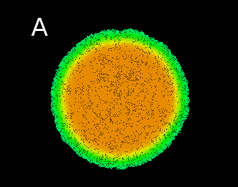
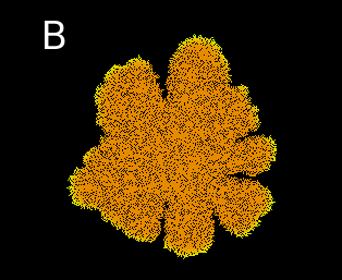
We start our simulations from either a single initial cell or a line of cells, and follow the shape of the colony after many rounds of cell replication, leading to a circular colony or a horizontal advancing front, respectively. Figure 1 shows that the morphology of a large colony of bacteria can be either smooth or branched, depending on the parameters of the model.
By performing simulations for different parameter sets we have found that the fate (smooth/branched) of the colony is determined by a dimensionless “branching parameter” , where is the initial nutrient concentration, the densely-packed cell density, and the other parameters have been defined previously.
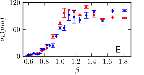
For small values of , the front of the colony remains smooth throughout the simulation (Fig. 1A,C), whereas for large values branches develop (Fig. 1B,D). Note that, as in real colonies Hallatschek and Nelson (2010), the nutrient becomes depleted within the colony so that only cells in a thin layer at the front are growing. To pinpoint the location of the transition more accurately, we compute the roughness of the front (Fig. 1E), defined as the mean square deviation of points on the front from its average position, as in Ref. Bonachela et al. (2011). The roughness measured at steady state increases by over an order of magnitude as passes through . The transition between planar and branching front is largely independent of the aspect ratio of the cells (Fig. 1E).
This transition between branched and smooth colony fronts is well known in real colonies Shapiro (1995), and has been the subject of many theoretical studies Murray (2003); Kawasaki et al. (1997), which usually attribute it to the interplay between diffusion (migration) of bacteria and diffusion of the nutrient. In our model, however, the transition is driven by the uptake of nutrient by the cells and their growth by mechanical pushing, and is unaffected by the diffusion rate of the nutrient.
To gain a better understanding of the physics of this transition, we approximate the growing colony as an incompressible cellular “fluid” 111A similar analysis has been performed in Ref. Klapper and Dockery (2002) in the context of biofilm growth.. Mass conservation in such a fluid is described by the equation , where the fluid velocity, is the growth rate, and is the dimensionless nutrient uptake function. This is coupled to an equation describing the diffusion and depletion of the nutrient. Let us begin with a one dimensional case of a colony advancing from the left and characterized by a single number which is the position of the front:
| (1) | |||||
| (2) |
Here is the nutrient diffusion constant, the rate of uptake of nutrient by cells, the cell density (constant everywhere due to incompressibility), and is the Heaviside step function. Because cells do not migrate and they are tightly packed, the density is either or zero, and hence equation (2) can be derived from the continuity equation and the incompressibility condition, assuming that . We also impose boundary conditions that and .
We first determine whether Eqs. (1,2) admit a travelling-wave solution in the limit , where the velocity of the front is constant. The resulting equations for and the front velocity are
| (3) | |||||
| (4) |
For , it is easily seen that the solution to Eq. (3) is given by (as ). For , we can rearrange the equation to yield , which, upon insertion into Eq. (4) gives
| (5) |
where we have integrated by parts, and used the fact that vanishes at , and that and must be continuous at . Therefore, a solution for exists only if (or ) exactly: we have found that in the incompressible limit the front cannot advance at a constant speed! This is in contrast to the Fisher framework, where travelling waves exist for a range of parameters. Numerical solutions of Eqs. (1, 2) fully confirm our prediction, see Supplemental Material.
The hint from this simplified 1D model is therefore that is a critical value that separates different regimes of colony growth. For , growth is limited by the nutrient diffusion rate, whereas for diffusion does not play any role. However, there are two problems here. First, the front has more freedom in 2D than in 1D - it can become branched and the profile does not have to be circularly symmetric. Since this change occurs around , it is therefore appealing to conjecture that the morphological transition in Fig. 1 is linked to the switch in growth laws described above in the theory for an incompressible colony.
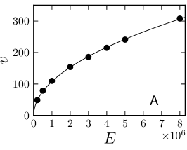
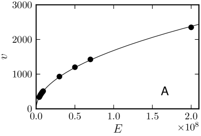
Second, incompressible theory predicts that under no conditions can growth be linear, unless . This is inconsistent with experimental results: the size of a colony of non-swimming bacteria growing on stiff agar gels does increase linearly with time Hallatschek et al. (2007). Moreover, our simulations also lead to a finite steady state speed. The speed found in simulations depends on the elasticity , as can be seen in Fig. 2A, suggesting the compressibility of the cells is important.
Generalizing the theory above to compressible cells, we now need equations for mass and momentum conservation, as well as the nutrient diffusion equation, still in the 1D geometry:
| (6) | |||||
| (7) | |||||
| (8) |
The term describes the friction between the surface and the cells. The pressure is determined by the force acting between the cells. We take to be consistent with our simulations, because the force that acts between two overlapping cells is then proportional to , where is the overlap.
Although Eqs. (6-8) cannot be solved analytically, a numerical solution (see Supplemental Material) shows that a travelling wave now exists for . The density profile close to the edge decays according to a power law towards the uncompressed cell density . This power law decay and the finite density at the very edge are in striking contrast to Fisher-Kolomogorov waves, which exhibit exponential density profiles in the wave tip Murray (2003). Many other properties of the solution to Eqs. (6-8) can be deduced without solving the equations. First, a “biomass conservation law” from Eqs. (6) and (7) states that one unit of nutrient biomass makes units of bacterial biomass, and hence the density deep in the colony must be . This explains why a travelling wave solution cannot exist in the incompressible case: unless the cell density equals exactly it will not match the density of biomass produced by the nutrient. It also explains why there is a morphological transition to branched colonies at : growth of a flat front is not possible for as it would need to have a density less than . Finally, it suggests that if bacteria are restricted to grow as a monolayer, then, when nutrient is abundant, they will grow exponentially until intermicrobial forces within the colony are so large that the bacteria in the middle are squashed to the appropriate density .
The idea that the cell population has to be compressed to a normal strain of for the front to grow at a constant speed can be turned into a simple scaling argument. At steady state the pressure profile has to rise from at the edge of the population to a maximal value in the bulk within a boundary layer of characteristic size . The characteristic length can be eliminated by estimating it to be the length by which the front moves in one generation . The bulk value of the pressure is just large enough that the density of the population is compressed down to the strain . The elastic constitutive relation of the microbial population fixes the corresponding pressure, with in our case of Hertzian contacts between cells. The pressure pushes the front population at the speed against the friction force , where acts as a friction coefficient per unit length. Force balance thus yields
| (9) |
where .
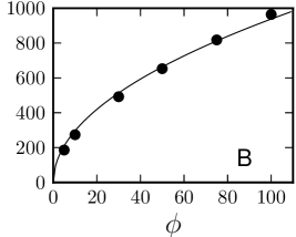
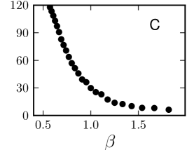
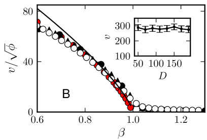
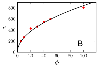
To test the above formula, we performed a fully one-dimensional version of our simulations described above, as this removed the effects of branching and was much more computationally efficient. The results are shown in Fig. 3. Figure 3A shows that the front speed grows as as predicted by Eq. (9), and Fig. 3B shows that the dependence of on is in good agreement with the numerically and theoretically predicted , although the theoretical form is only accurate for close to 1. Fig. 2 shows that the square-root dependence on and also holds in the 2D case, but the function is again different, and does not go to zero for , due to the branching. In the Supplemental Material we perform a more rigorous derivation of Eq. (9), showing that it is valid when the dimensionless parameter and is close to 1. We also show that mechanics-dominated growth is relevant for any experimentally feasible parameters.
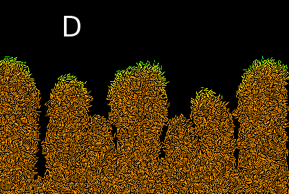
So far, our findings are relevant to bacteria growing in monolayers. On agar plates, however, cells are observed to build up vertically in the colony centre Pipe and Grimson (2008); Seminara et al. (2012). To probe how this additional degree of freedom affects our results, we simulate a colony growing in a vertical 2d plane (where the axis is perpendicular to the substrate) instead of the plane from previous simulation. We also incorporate attractive cell-cell and cell-substrate interactions, and we solve for the evolution of the nutrient field in the half-plane only, which models the agar gel on which growth occurs. As is apparent from the snapshot of the growth process in Fig. 4A, cells do now escape out of the plane they start from, due to the force exerted by neighbours. The size of the colony once again grows linearly in time. However, it is not compressibility but the possibility of escape into the vertical direction which leads to linear growth.
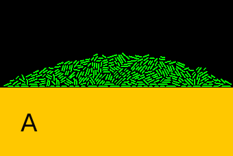
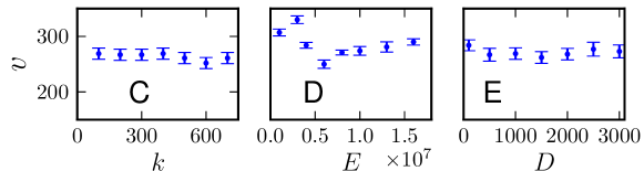
In fact, if the bulk pressure , which builds up in a strictly two-dimensional setting, is larger than some critical pressure , cells will escape into the dimension. As a consequence the pressure profile will saturate at in the bulk of the population. In our scaling argument for the speed of the front growth, we then have . Figures 4B-E show that, in contrast to the 2D case, the expansion speed and it is independent of the consumption rate , elastic modulus and the diffusion constant . Note that while the radial growth is independent of , the vertical growth will be affected by it.
In conclusion, we have reported here a joint computational and analytical study of the growth of bacterial colonies where non-motile microorganisms replicate and push each other away as they grow. We find a transition between two different growth regimes, controlled by the balance between growth and uptake of nutrients. Our model differs in that from biofilm simulations Kreft et al. (1998); Lardon et al. (2011) which do not explicitly model mechanical forces in the colony. We also find that the functional form of the density profile close to the bacterial edge qualitatively differs from those predicted by Fisher-Kolmogorov models, and predict that the speed at which the front propagates depends only weakly on the nutrient diffusion rate , for a wide range of . It would be interesting to study how the accumulation of metabolic inhibitors Hochberg and Folkman (1972), oxygen depletion Peters et al. (1987), or dependence of growth rate on the distance from the agar Wentland et al. (1996) would affect our results.
Acknowledgments. We thank R. J. Allen and M. R. Evans for helpful comments on this manuscript. O.H. thanks the Deutsche Forschungsgemeinschaft (DFG) for financial support (grant A15, SFB 937). B.W. acknowledges the support of a Leverhulme Trust Early Career Fellowship.
References
- Ramaswamy (2010) S. Ramaswamy, Annu. Rev. Cond. Matt. Phys. 1, 323–345 (2010).
- Basan et al. (2011) M. Basan, J.-F. Joanny, J. Prost, and T. Risler, Phys. Rev. Lett. 106, 158101 (2011).
- Simha and Ramaswamy (2002) R. A. Simha and S. Ramaswamy, Phys. Rev. Lett. 89, 058101 (2002).
- Sokolov et al. (2007) A. Sokolov, I. S. Aranson, J. O. Kessler, and R. E. Goldstein, Phys. Rev. Lett. 98, 158102 (2007).
- Ben-Jacob et al. (1994) E. Ben-Jacob, O. Schochet, A. Tenenbaum, I. Cohen, A. Czirók, and T. Vicsek, Nature 368, 46 (1994).
- Bonachela et al. (2011) J. Bonachela, C. Nadell, J. Xavier, and S. Levin, J. Stat. Phys. 144, 303 (2011).
- Cates et al. (2010) M. E. Cates, D. Marenduzzo, I. Pagonabarraga, and J. Tailleur, Proc. Natl. Acad. Sci. USA 107, 11715–11720 (2010).
- Kawasaki et al. (1997) K. Kawasaki, A. Mochizuki, M. Matsushita, T. Umeda, and N. Shigesada, J. Theor. Biol. 188, 177–185 (1997).
- Eden (1961) M. Eden, Proc. Fourth Berkeley Symp. on Math. Statist. and Prob 4, 223-239 (1961).
- Fujikawa and Matsushita (1989) H. Fujikawa and M. Matsushita, J. Phys. Soc. Japan 58, 3875 (1989).
- Ben-Jacob et al. (2000) E. Ben-Jacob, I. Cohen, and H. Levine, Adv. Phys. 49, 395 (2000).
- Murray (2003) J. Murray, Mathematical Biology, Vol. 2 (Springer-Verlag, Berlin, 2003), chs. 5 and 6.
- Volfson et al. (2008) F. Volfson, S. Cookson, J. Hasty, and L. Tsimring, Proc. Natl. Acad. Sci. USA 105, 15346–15351 (2008).
- Costerton et al. (1999) J. W. Costerton, P. S. Stewart, and E. P. Greenberg, Science 284, 1318 (1999).
- Hense et al. (2007) B. A. Hense, C. Kuttler, J. Müller, M. Rothballer, A. Hartmann, and J.-U. Kreft, Nat. Rev. Microbiology 5, 230 (2007).
- Xavier et al. (2009) J. B. Xavier, E. Martinez-Garcia, and K. R. Foster, Am. Nat. 174, 1 (2009).
- Hallatschek and Nelson (2010) O. Hallatschek and D. Nelson, Evolution 64, 193–206 (2010).
- Kuhr et al. (2011) J. Kuhr, M. Leisner, and E. Frey, New J. Phys. 13, 113013 (2011).
- Boyer et al. (2011) D. Boyer, W. Mather, O. Mondragón-Palomino, S. Orozco-Fuentes, T. Danino, J. Hasty, and L. S. Tsimring, Phys. Biol. 8, 026008 (2011).
- Landau and Lifschitz (2008) L. D. Landau and E. M. Lifschitz, Theory of Elasticity, 3rd ed. (Elsevier, Oxford, 2008).
- Shapiro (1995) J. A. Shapiro, BioEssays 17, 597–607 (1995).
- Hallatschek et al. (2007) O. Hallatschek, P. Hersen, S. Ramanathan, and D. Nelson, Proc. Natl. Acad. Soc. USA 104, 19926 (2007).
- Pipe and Grimson (2008) L. Z. Pipe and M. J. Grimson, Molecular BioSystems 4, 192–198 (2008).
- Seminara et al. (2012) A. Seminara, T. E. Angelini, J. N. Wilking, S. Vlamakis, H. adn Ebrahim, R. Kolter, D. A. Weitz, and M. P. Brenner, Proc. Natl. Acad. Sci. USA 109, 1116–1121 (2012).
- Kreft et al. (1998) J. U. Kreft, G. Booth, and J. W. T. Wimpenny, Microbiology 144, 3275 (1998).
- Lardon et al. (2011) L. A. Lardon, B. V. Merkey, S. Martins, A. Dötsch, C. Picioreanu, J.-U. Kreft, and B. F. Smets, Env. Microbiology 13, 2416–2434 (2011).
- Hochberg and Folkman (1972) M. S. Hochberg and J. Folkman, J. Infect. Dis. 126, 629–635 (1972).
- Peters et al. (1987) A. C. Peters, J. W. T. Wimpenny, and J. P. Coombs, J. Gen. Microbiol. 133, 1257–1263 (1987).
- Wentland et al. (1996) E. J. Wentland, P. S. Stewart, C. T. Huang, and G. A. McFeters, Biotechnol. Prog. 12, 316–321 (1996).
- Klapper and Dockery (2002) I. Klapper and J. Dockery, SIAM J. Appl. Math. 62, 853–869 (2002).