Rotating-crystal Malaria Diagnosis: Pre-clinical validation
Abstract
Improving the efficiency of malaria diagnosis is one of the main goals of current malaria research. We have recently developed a magneto-optical (MO) method which allows high-sensitivity detection of malaria pigment (hemozoin) crystals via their magnetically induced rotation in blood. Here, we validate this technique on laboratory derived blood samples infected with Plasmodium falciparum. Using two parasite cultures, the first containing mostly ring stages and the second corresponding to the end of the parasite life cycle, we demonstrate that our novel method can detect parasite densities as low as 40 and 10 parasites per microliter of blood for ring and schizont stage parasites, respectively. This detection limit exceeds the performance of rapid diagnostic tests and competes with the threshold achievable by light microscopic observation of blood smears. Our method can be performed with as little as 50 microliter of capillary blood and is sensitive to the presence of hemozoin micro-crystals down to ppm concentrations. The device, designed to a portable format for clinical and in-field tests, requires no special training of the operator or specific reagents, except for an inexpensive lysis solution to release intracellular hemozoin. Beyond diagnostics, this technique may offer an efficient tool to study hemozoin formation, trace hemozoin kinetics in the body and test susceptibility/resistance of parasites to new antimalarial drugs inhibiting hemozoin formation.
Introduction
Although there is a plethora of emerging techniques aiming at high-sensitivity diagnosis of malaria, only a few of these approaches are feasible for clinical and in-field diagnosis. Apart from purely symptom based, presumptive diagnosis, the two main diagnostic methods currently in practice are the antigen-based detection of malaria parasites using rapid diagnostic tests (RDT) and the microscopic observation of infected red blood cells in blood smears.Moody2002 ; Hanscheid1999 ; Payne1988 ; Wongsrichanalai2007 The detection limits of RDT and light microscopy have been reported to be approximately 100 parasites/L and 5-50 parasites/L, respectively.Moody2002 ; Maltha2013 ; Alonso2011 ; Prudhomme2006 Both of these methods are subject to inherent limitations: i) although RDTs are becoming more affordable, they cannot provide a quantitative measure of parasitemia and presently do not possess sufficient sensitivity to detect low-level infections which are very common in endemic settings, ii) the visual inspection of blood smears is time and labor intensive. Moreover, the detection threshold of 5 parasites/L is rather theoretical and can only be achieved under ideal conditions (good-quality blood film, highly trained microscopist, high-powered microscope, etc.). In practice, most routine diagnostic laboratories achieve approximately 50 parasites/L and detect about 50 % of malaria cases.Alonso2011 ; Okell2009 ; Perkins2008
Among molecular biology-based methods, polymerase chain reaction (PCR) assays surpass the performance of RTDs and light microscopy.Coleman2006 ; Snounou1996 However, they often require expensive equipment and reagents, highly trained laboratory personnel and are prone to contamination.Tangpukdee2009 Recent studies conclude that real-time PCR has a detection limit corresponding to a few parasites in 1 L blood,Owusu2013 ; Khairnar2009 nevertheless, it is not yet a practical method for routine diagnosis under field conditions.
The idea to take advantage of the unique magnetic properties of malaria pigment (hemozoin) and to use it as an alternative target of optical diagnosis has been proposed by several groups.Butykai2013 ; Karl2008 ; Mens2010 ; Newman2008 ; Zimmerman2006 Hemozoin is a micro-crystalline heme compound produced by malaria parasites as they detoxify free heme derived from hemoglobin digestion. Our recent study using synthetic hemozoin crystals suspended in blood demonstrated that the rotating-crystal magneto-optical (MO) diagnostic method can detect hemozoin concentrations down to 15 pg/L.Butykai2013 This threshold concentration was estimated to be equivalent to a parasite density of 30 parasites/L in infected blood provided that the whole amount of hemozoin produced by the parasites is released into the lysed cell suspension.
However, the relation between hemozoin concentration and parasite density in human infections is not straightforward. Intraerythrocytic hemozoin content is dependent on the maturity of the blood stage parasites; with the least amount of hemozoin present in erythrocytes during the ring stage and the highest amount during the schizont stage.Moore2006 ; Orjih1993 ; Becker2004 Therefore, the hemozoin concentration in blood derived from human infections depends on the parasite stage distribution at the time the blood sample was collected. In P. falciparum (the most lethal parasite species) infections often only the ring and early trophozoite stages are found in the peripheral circulation, since the later developmental stages cytoadhere to the vascular endothelium.Cooke1995
Moreover, the MO signal recorded by our technique depends not only on the amount but also on the size and morphology of the hemozoin crystals, which can be different for synthetic and naturally grown crystals.Slater1991 ; Noland2003 ; Jaramillo2009 The MO method is sensitive only to those hemozoin crystals released into suspension, which can be magnetically rotated. Correspondingly, aggregation of crystals or their binding to other components of lysed infected blood, such as cell membranes, could substantially decrease the sensitivity of our technique. Signal loss may be avoided by appropriate lysis and treatment of blood samples prior to measurement.
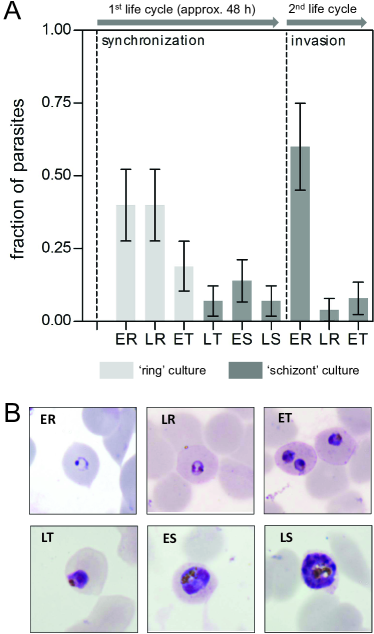
In the present study we aimed to address these key issues and to validate the rotating-crystal malaria diagnosis method using synchronized cultures of P. falciparum. (For a short description of the detection scheme see Materials and Methods section.) For this purpose, we investigated its sensitivity and detection threshold for two cultures with different maturity distributions of the parasites. The first, hereafter referred to as the ring stage culture, contained mostly ring stages and some early trophozoites with a total parasite density of P3.1105 parasites/L. It is representative to the distribution of parasite blood stages most often encountered in P. falciparum infections. The second with a lower total parasite density of P2.8104 parasites/L corresponds to the end of the parasite life cycle where some of the parasites are still in the schizont form but most of them, following an invasion, already turned to early-stage rings of the next generation. The distributions of the parasites among the different stages – early-ring, late-ring, early-trophozoite, late-trophozoite, early-schizont and late-schizont stages – in the two cultures are displayed in Fig. 1 together with light microscope images of parasites representative to these stages. Since the early-stage rings do not contain hemozoin, the hemozoin content present in the second culture is formed in the first cycle before the invasion mostly during the schizont stage.Moore2006 ; Orjih1993 ; Becker2004 For this reason, hereafter we refer to this culture as the schizont stage culture. Schizont stage parasites are not normally found in the peripheral blood during human P. falciparum infection due to their sequestration in small capillary blood vessels.Cooke1995 However, this stage restriction is not present in other non-sequestering species of human malaria parasites such as P. vivax.Carvalho2010
Results
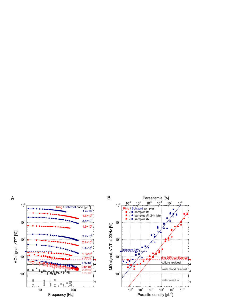
The MO signal, the measure of hemozoin content within the lysed cell suspension, is shown in Fig. 2 for dilution series of the ring and schizont stage cultures. The 20 serial 2-fold dilutions using uninfected erythrocytes allowed for MO signal to be assessed over 6 orders of magnitude of parasitemia. As a general trend in Fig. 2A, the MO signal varies proportionally to the parasitemia level and the signal for each sample shows a gradual decrease with increasing frequencies of the rotating magnetic field. This frequency dependence is in agreement with previous results obtained when using synthetic hemozoin crystals suspended in blood and originates from the viscosity of the lysed cell suspension hindering fast rotations of the crystals.
As schizont stages contain more hemzoin than ring stage-parasites, samples from the diluted schizont stage culture exhibited higher signals than ring stage samples with comparable parasitemia. Although the overall frequency dependence is similar for the two cultures, the decrease in the signal with increasing frequency is more pronounced for schizont stage samples, which is likely due to the larger crystal size in these samples. At low levels of parasite density, namely for samples with 10 parasites/L from the ring stage culture, the signal does not further drop with decreasing parasitemia. The frequency dependence of the MO signal for these ring stage samples also becomes different; the low-frequency saturation common for higher concentrations does not hold anymore. These imply a residual MO signal not related to hemozoin.
In order to determine the detection limit of our method, the results obtained for the dilution series of the ring and schizont stage cultures are summarized in Fig. 2B, where the MO signal at 20 Hz is plotted versus the parasitemia (and parasite density). Sequential measurements performed on the same sample with time delays less than one hour gave identical results. In several cases we checked the reproducibility of the protocol by repeating the measurement for both of the duplicate samples labeled as samples #1 and samples #2 in Fig. 2B. For ring stage samples with 10 parasites/L, where the MO signal shows no systematic variation with decreasing parasite density, we calculated the mean value of the residual MO signal (3.610-4 %) and its standard deviation (1.510-4 %). Assuming Gaussian distribution for the residual MO signal values, we found that the 95 % confidence level of the mean detection limit for ring stage samples is 6.610-4 %, which corresponds to a parasite density of 40 parasites/L. This is equivalent to the parasitemia level of 8 %. Since the reproducibility is poorer for schizont stage samples, in this case our rough estimate for the 95 % confidence level of the mean detection limit is considerably higher with 210-3 %, which corresponds to the parasite density of approximately 10 parasites/L and a parasitemia level of 2 %. Note that the reproducibility between duplicate samples does not significantly vary with parasite density for dilutions of either the ring or the schizont stage cultures.
The hemozoin content of the two cultures can be roughly estimated from the parasite density and the stages of parasite development specified in Fig. 1. Ring and early trophozoite stages up to 24 h were reported to convert about 3-15 % of the total hemoglobin in the infected red blood cells to hemozoin,Moore2006 ; Orjih1993 while at the schizont stage this portion is increased to approximately 50-80 %.Francis1997 ; Hackett2009 ; Weissbuch2008 According to the 3-15 % hemoglobin conversion rate reported for rings and early trophozoites, the undiluted ring stage culture with total parasite density of P=3.1105 parasites/L contained 9-48 ng/L hemozoin. The undiluted schizont stage culture, which had about 10 times lower parasite density with P=2.8104 parasites/L, contained all the hemozoin formed during the first cycle. Using the hemoglobin conversion rates quoted above, this corresponds to 14-23 ng/L hemozoin. An independent estimate, based on the MO signal yields approximately 6 ng/L and 9 ng/L for the undiluted ring and schizont stage cultures, respectively. For this estimate, we used the conversion factor cHz=1 ng/L =1.4 % between the hemozoin concentration and the low-frequency (1 Hz) MO signal previously determined for artificial hemozoin crystals suspended in blood.Butykai2013 Note that the 20-fold dilution of the samples prior to the MO measurement needs to be taken into account, since this conversion factor applies for samples with 50 % hematocrit.
We also estimate the hemozoin concentration of the two undiluted cultures based on MO signal using the conversion factor cHz=1 ng/L =1.4 % between the hemozoin concentration and the low-frequency (1 Hz) MO signal previously determined for artificial hemozoin crystals suspended in blood Butykai2013 . This yields approximately 6 ng/L and 9 ng/L for the undiluted ring and schizont stage cultures, respectively. Note that the 20-fold dilution of the samples prior to the MO measurement needs to be taken into account, since this conversion factor applies for samples with 50 % hematocrit.
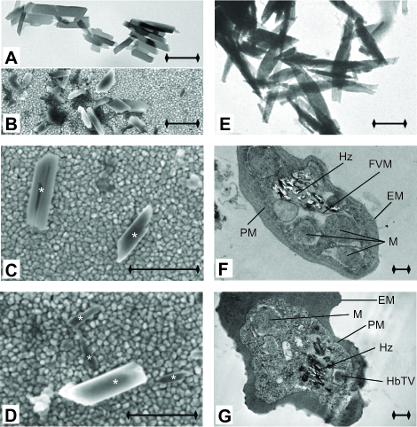
The first scenario is supported by the electron microscopy images in Fig. 3, where hemozoin crystals inside of infected erythrocytes and as extracted from the cultures are shown. These elongated crystallites are considerably smaller (with typical length of 200-500 nm) than the synthetic ones (500-900 nm) previously studied and also displayed in the figure.Butykai2013 This is also in accordance with the weaker frequency dependence of the MO signal observed in the present study, which indicates that natural crystals are able to follow the rotation of the magnetic field up to higher frequencies than the synthetic ones as a possible consequence of their reduced size. Furthermore, the comparison between the natural crystals within the parasites and those extracted from the cultures confirms no major change either in the size or in the morphology of the crystals due to our lysis-sonication protocol.
In order to minimize binding and aggregation of crystals we repeated the measurements 24 h later following another 30 min of sonication. We found modest but systematic increase of the MO signal of typically not more than 10-30 %. On this basis, we expect that most of the hemozoin is successfully released into suspension, likely in the form of individual crystals.
The presence of a frequency-dependent residual MO signal indicates that some components of lysed blood can be magnetically oriented and rotated similarly to the hemozoin crystals. We have also studied freshly drawn blood following the same lysis protocol. The residual MO signal observed in this case was considerably lower than found for either the ring or schizont stage samples (see Fig. 2), which implies better detection threshold for instant diagnosis. We suspect that due to the freeze-thaw-lysis procedure applied to the present set of samples, some portion of the hemoglobin may have been transformed to an aggregated or polymerized form – similar to the intracellular non-covalent polymerization of hemoglobin previously observed in sickle cell diseaseNoguchi1983 ; Noguchi1984 ; Ofori2001 –, which may produce the residual MO signal. These points, requiring additional systematic studies, stress the crucial role of an appropriate blood treatment prior to diagnosis.
The noise floor of our equipment, determined using pure water, is roughly frequency independent and about one order of magnitude smaller than the residual signal from fresh blood. This enables further improvement of the detection limit provided that the residual MO signal from blood can be reduced by optimizing blood treatment. Optimizing the properties of the lysis solution may also help to dissociate the crystals without the need for sonication.
Discussion
The potential of exploiting hemozoin as a magnetic biomarker for malaria diagnosis has stimulated extended research over the last few decades. Taking advantage of the paramagnetic nature of hemozoin, several approaches have been proposed to improve the sensitivity of existing methods by the magnetic separation of malaria infected erythrocytes from whole blood prior to the diagnosis.Paul1981 ; Carter2003 ; Karl2008 ; Karl2011 More recently, new techniques have been emerging, which directly use hemozoin as a target material of magnetic diagnosis. These techniques include detection of depolarized side-scatter in flow cytometry,Frita2011 electrochemical magneto immunoassays,Castilho2011 magnetically enriched surface enhanced resonance Raman spectroscopyYuen2012 ; Hobro2013 and magneto-optical detection using polarized light.Mens2010 ; Newman2008 ; Newman2010 Among them, to the best of our knowledge, our rotating crystal MO diagnostic device is the first realized in a cost-effective portable format with excellent sensitivity.
In the present study, the detection limit of our rotating-crystal MO diagnostic device was found to be 40 parasites/L and 10 parasites/L for ring and schizont stage parasites, respectively. These detection limits are below the threshold currently achievable with RDTs (100 parasites/L) and lie within the same range as the limits of conventional optical microscopy for malaria diagnosis (5-50 parasites/L).Moody2002 For the present set of blood samples kept frozen and thawed before the measurement, the performance of the method was limited by a residual MO signal due to some part of the lysed cell suspension. This residual MO signal obscures the genuine MO signal of hemozoin at parasite densities lower than the limits quoted above. Preliminary results indicate that for measurements on freshly lysed blood samples, which is the condition relevant to instant diagnosis, the detection limit of our rotating crystal MO platform could be further improved. Thus, this methodology has the potential to yield portable tools for instant diagnosis with detection limits approaching that of PCR-based platforms.
Limitations of our diagnostic technique include i) the possibility of false positive detections due to the presence of hemozoin in the blood, e.g. contained within white blood cells,Schwarzer2001 for extended periods of time after an infection has been cleared and ii) the possibility of false negative results in case an infection only contains very early ring stage parasites with little or no hemozoin. Furthermore, methods targeting only hemozoin as a marker for infection are thought to have limitations in their diagnostic capacity as they cannot distinguish between different malaria species. The specificity of our MO diagnostic scheme, owing to variations in the typical size and morphology of hemozoin crystals produced by different species, needs to be tested by a comparative study on various Plasmodium strains. We emphasize that only studies on field isolates will be able to elucidate the impact of these possible confounding factors and the present study is the basis for such field-based trials.
It is currently believed that without active case detection of asymptomatic malaria infections, malaria eradication will be impossible or very difficult to achieve.Alonso2011 However, there are no diagnostic tools for rapidly screening hundreds of people per day, on-site and with high sensitivity.Baird2010 The rotating-crystal MO diagnostic device has the potential to fulfill these requirements as it is cost-effective, rapid, highly sensitive, portable and easy to apply.
Besides on-site diagnosis, the present methodology provides an
efficient in-vitro laboratory tool to test the susceptibility of the
parasites to new antimalarial drugs by monitoring the effect of
treatment on the rate of hemozoin formation. The scope of the
technique described here may also cover the study or diagnosis of
other human diseases, such as schistosomiasis, which are also caused
by blood-feeding organisms producing hemozoin similarly to malaria
parasites.Chen2001 ; Oliveira2004 ; Oliveira2000 ; Karl2013
Materials and Methods
Parasite culture
P. falciparum parasites (laboratory adapted strain 3D7) were cultured following the method of Trager and Jensen with modifications Trager1976 . The culture medium was RPMI 1640 with L-glutamine (GIBCO cat # 31800) supplemented with 2 mg/mL NaHCO3 (Merck, cat # 106329), 25 mg/L gentamicin (Pfizer, cat # 61022027), 50mg/L hypoxanthine (Calbiochem, cat # 4010), 25 mM HEPES (SAFC, cat # 90909C) and 10 % pooled O+ human serum (mixed blood groups, Australian Red Cross Blood Service). Cultures were maintained at 4 % hematocrit with changes of culture medium every 48 h and diluted with uninfected O+ red blood cells when the parasitemia exceeded 5 %. Parasites were maintained in an atmosphere of 5 % CO2 and 1 % O2 in N2. Parasite cultures were kept in stage synchrony by applying the 5 % Sorbitol method, first described by Lambros and Vanderberg Lambros1979 . Hemozoin liberated from late stage parasites by the synchronization process was removed by washing the cells in RPMI medium after the Sorbitol induced cell lysis and before re-establishment of the culture.
Parasite densities for the the ring and schizont stage cultures described above were estimated by counting the number of parasites contained in 5000 red blood cells on Giemsa stained thin blood films. The commonly used conversion from parasitemia to parasite density based on the assumption that 1 L of blood contains 5 red blood cells at 50% hematocrit was applied Moody2002 . For both, the ring and schizont stage cultures, 2-fold dilution series were prepared in duplicate in uninfected human erythrocytes (Red Cross blood bank, Royal Melbourne Hospital, VIC, Australia). The duplicate dilution series prepared for ring and schizont cultures contained 21 and 20 dilutions, respectively, where each of the dilutions had a volume of 200 L at a hematocrit of 50 %. These dilutions were immediately frozen and thawed twice to create lysates. These lysates, hereafter referred to as ring and schizont stage samples, were subsequently frozen at -80oC and kept in a frozen state until they were thawed immediately prior to measurement. The personnel carrying out the MO measurements were blinded against the contents of each of the samples to reduce potential observer bias.
Blood treatment prior MO diagnosis
Blood samples prepared for MO measurement were thawed at room temperature and were diluted 20-fold with distilled water, resulting in a total volume of 4 mL per specimen. Additionally, 100 L of a special red cell lysis buffer, hereafter referred to as clearing solution, was added to the specimens in order to disperse the remnants of the lysed RBCs. In preliminary experiments, this clearing solution (2.5 V/V% Triton X-100 in 0.1 M NaOH) was confirmed not to cause noticeable degradation of hemozoin when added to synthetic malaria pigment suspended in lysed blood. Note that the final concentration of NaOH in the samples was only 2.5 mM. The samples were subjected to 30 min of ultrasonication to dissociate potential aggregates of hemozoin and ensure an unhindered motion of crystals in the fluid. Owing to this treatment a transmittance of 30-40 % could be achieved with no substantial light scattering observed from the blood samples. MO signals were recorded on 1 mL volumes taken from the samples subsequent to the preparation process and reproducibility was confirmed in several cases after one day of storage of the samples at 4oC and by carrying out the measurement on both of the duplicate samples.
Magneto-optical measurements
MO measurements were performed with the prototype of the rotating magnet setup described in our previous study Butykai2013 . We utilize a permanent magnetic ring, which produces a B=1 T magnetic field at the sample position and can be rotated with adjustable frequency. Polarized light from a laser diode is transmitted through the sample in the direction perpendicular to the plane of the rotating magnetic field. Owing to the magnetic alignment of the freely rotating and dichroic hemozoin crystals present in infected blood, magnetically induced linear dichroism can be observed and quantified as the difference in transmission for light polarized along and perpendicular to the magnetic field direction () divided by the average transmission (), i.e. Butykai2013 . Due to the synchronous periodic rotation of the crystals the linear dichroism gives rise to a periodic change in the transmitted intensity oscillating with twice the magnet rotation frequency. This second harmonic intensity component () can be effectively filtered by a lock-in technique. The MO signal is then obtained as the ratio of the modulated light intensity and the average intensity (); .
This detection scheme for malaria diagnosis provides the best signal-to-noise ratio in the rotation frequency regime of 10-30 Hz. In order to exclude baseline artifacts emerging from mechanical vibration or improper optical alignment of the system, the baseline is checked with pure distilled water prior to the measurement of each sample. This water baseline – due to electronic and mechanical noise – was generally found to be almost two orders of magnitudes lower than the signal from the samples with the lowest hemozoin concentration.
Electron microscopy
For transmission electron microscopy (TEM), parasite samples were fixed in resin blocks and 70-120 nm thin sections were cut using a Leica EM UC6 microtome (Leica Microsystems, North Ryde, NSW, Australia) and brought onto carbon coated copper TEM grids (ProSciTech, Thuringowa, Qld., Australia). The TEM grids were then stained with 5 % uranyl acetate for 15 min and Reynold’s lead citrate solution for 5 min. TEM was conducted on a JEOL 2100 TEM (JEOL Inc., Tokyo, Japan). For details of the method see the Supporting Information.
For scanning electron microscopy (SEM), the samples giving the highest MO signal were used and hemozoin crystals were extracted following the method of Chen and coworkers Chen2001 . The dark brown pellet obtained by this method was resuspended in 80 L water. For SEM imaging small droplets of the suspension containing the hemozoin crystals were applied to gold coated glass slides without further purification or treatment. The droplets were dried overnight at room temperature. The SEM images were acquired on a LEO 1540XB electron microscope using the in-lens detector. The accelerating voltage was set to 3 kV and the viewing angle was perpendicular to the gold surface.
Acknowledgements. The authors acknowledge fruitful
discussions with T. Hanscheid and assistance with electron
microscopy by L. Kyriliak, M. Saunders, J. Shaw and facilities of
the Centre of Microscopy, Characterization and Analysis at The
University of Western Australia. This work was supported by
Hungarian Research Funds OTKA K108918,
TÁMOP-4.2.1.B-09/1/KMR-2010-0001, by NHMRC grants GNT1021544 and
GNT1043345 awarded to IM and by NHMRC grants GNT637406 and
GNT1058665 awarded to LS. SK is supported through a NHMRC early
career research fellowship (GNT1052760).
Correspondence: Istvan Kezsmarki, Department of Physics, Budapest University of Technology and Economics, 1111-Budapest, Hungary; e-mail: kezsmark@dept.phy.bme.hu.
References
- (1) Moody A (2002) Rapid diagnostic tests for malaria parasites. Clinical Microbiology Reviews 15: 66-78.
- (2) Hanscheid T (1999) Diagnosis of malaria: a review of alternatives to conventional microscopy. Clinical and Laboratory Haematology 21: 235-245.
- (3) Payne D (1988) Use and limitations of light-microscopy for diagnosing malaria at the primary health-care level. Bulletin of the World Health Organization 66: 621-626.
- (4) Wongsrichanalai C, Barcus MJ, Muth S, Sutamihardja A, Wernsdorfer WH (2007) A review of malaria diagnostic tools: Microscopy and rapid diagnostic test (rdt). American Journal of Tropical Medicine and Hygiene 77: 119-127.
- (5) Maltha J, Gillet P, Jacobs J (2013) Malaria rapid diagnostic tests in endemic settings. Clinical Microbiology and Infection 19: 399-407.
- (6) Alonso PL, Barnwell JW, Bell D, Hanson K, Mendis K, et al. (2011) A research agenda for malaria eradication: Diagnoses and diagnostics. Plos Medicine 8: e1000396.
- (7) O’Meara WP, Remich S, Ogutu B, Lucas M, Mtalib R, et al. (2006) Systematic comparison of two methods to measure parasite density from malaria blood smears. Parasitology Research 99: 500-504.
- (8) Okell LC, Ghani AC, Lyons E, Drakeley CJ (2009) Submicroscopic infection in plasmodium falciparum-endemic populations: A systematic review and meta-analysis. Journal of Infectious Diseases 200: 1509-1517.
- (9) Perkins MD, Bell DR (2008) Working without a blindfold: the critical role of diagnostics in malaria control. Malaria Journal 7: s5.
- (10) Coleman RE, Sattabongkot J, Promstaporm S, Maneechai N, Tippayachai B, et al. (2006) Comparison of pcr and microscopy for the detection of asymptomatic malaria in a plasmodium falciparum/vivax endemic area in thailand. Malaria Journal 5: 121.
- (11) Snounou G (1996) Detection and identification of the four malaria parasite species infecting humans by pcr amplification. Methods in Molecular Biology 50: 263-291.
- (12) Tangpukdee N, Duangdee C, Wilairatana P, Krudsood S (2009) Malaria diagnosis: A brief review. Korean Journal of Parasitology 47: 93-102.
- (13) Owusu-Ofori AK, Betson M, Parry CM, Stothard JR, Bates I (2013) Transfusion-transmitted malaria in ghana. Clinical Infectious Diseases 56: 1735-1741.
- (14) Khairnar K, Martin D, Lau R, Ralevski F, Pillai DR (2009) Multiplex real-time quantitative pcr, microscopy and rapid diagnostic immuno-chromatographic tests for the detection of plasmodium spp: performance, limit of detection analysis and quality assurance. Malaria Journal 8: 284.
- (15) Butykai A, Orban A, Kocsis V, Szaller D, Bordacs S, et al. (2013) Malaria pigment crystals as magnetic micro-rotors: key for high-sensitivity diagnosis. Scientific Reports 3: 1431.
- (16) Karl S, David M, Moore L, Grimberg BT, Michon P, et al. (2008) Enhanced detection of gametocytes by magnetic deposition microscopy predicts higher potential for plasmodium falciparum transmission. Malaria Journal 7: 66.
- (17) Mens PF, Matelon RJ, Nour BYM, Newman DM, Schallig H (2010) Laboratory evaluation on the sensitivity and specificity of a novel and rapid detection method for malaria diagnosis based on magneto-optical technology (mot). Malaria Journal 9: 207.
- (18) Newman DM, Heptinstall J, Matelon RJ, Savage L, Wears ML, et al. (2008) A magneto-optic route toward the in vivo diagnosis of malaria: Preliminary results and preclinical trial data. Biophysical Journal 95: 994-1000.
- (19) Zimmerman PA, Thomson JM, Fujioka H, Collins WE, Zborowski M (2006) Diagnosis of malaria by magnetic deposition microscopy. American Journal of Tropical Medicine and Hygiene 74: 568-572.
- (20) Moore LR, Fujioka H, Williams PS, Chalmers JJ, Grimberg B, et al. (2006) Hemoglobin degradation in malaria-infected erythrocytes determined from live cell magnetophoresis. Faseb Journal 20: 747-749.
- (21) Orjih AU, Fitch CD (1993) Hemozoin production by plasmodium-falciparum - variation with strain and exposure to chloroquine. Biochimica Et Biophysica Acta 1157: 270-274.
- (22) Becker K, Tilley L, Vennerstrom JL, Roberts D, Rogerson S, et al. (2004) Oxidative stress in malaria parasite-infected erythrocytes: host-parasite interactions. International Journal for Parasitology 34: 163-189.
- (23) Cooke BM, Coppel RL (1995) Cytoadhesion and falciparum-malaria - going with the flow. Parasitology Today 11: 282-287.
- (24) Slater AFG, Swiggard WJ, Orton BR, Flitter WD, Goldberg DE, et al. (1991) An iron carboxylate bond links the heme units of malaria pigment. Proceedings of the National Academy of Sciences of the United States of America 88: 325-329.
- (25) Noland GS, Briones N, Sullivan DJ (2003) The shape and size of hemozoin crystals distinguishes diverse plasmodium species. Molecular and Biochemical Parasitology 130: 91-99.
- (26) Jaramillo M, Bellemare MJ, Martel C, Shio MT, Contreras AP, et al. (2009) Synthetic plasmodium-like hemozoin activates the immune response: A morphology - function study. Plos One 4: e6957.
- (27) Carvalho BO, Lopes SCP, Nogueira PA, Orlandi PP, Bargieri DY, et al. (2010) On the cytoadhesion of plasmodium vivax-infected erythrocytes. Journal of Infectious Diseases 202: 638-647.
- (28) Francis SE, Sullivan DJ, Goldberg DE (1997) Hemoglobin metabolism in the malaria parasite plasmodium falciparum. Annual Review of Microbiology 51: 97-123.
- (29) Hackett S, Hamzah J, Davis TME, St Pierre TG (2009) Magnetic susceptibility of iron in malaria-infected red blood cells. Biochimica Et Biophysica Acta-Molecular Basis of Disease 1792: 93-99.
- (30) Weissbuch I, Leiserowitz L (2008) Interplay between malaria, crystalline hemozoin formation, and antimalarial drug action and design. Chemical Reviews 108: 4899-4914.
- (31) Noguchi CT, Torchia DA, Schechter AN (1983) Intracellular polymerization of sickle hemoglobin - effects of cell heterogeneity. Journal of Clinical Investigation 72: 846-852.
- (32) Noguchi CT (1984) Polymerization in erythrocytes containing s-hemoglobin and non-s-hemoglobin. Biophysical Journal 45: 1153-1158.
- (33) Ofori-Acquah SF, Green BN, Davies SC, Nicolaides KH, Serjeant GR, et al. (2001) Mass spectral analysis of asymmetric hemoglobin hybrids: Demonstration of hb fs (alpha(2)gamma beta(s)) in sickle cell disease. Analytical Biochemistry 298: 76-82.
- (34) Paul F, Roath S, Melville D, Warhurst DC, Osisanya JOS (1981) Separation of malaria-infected erythrocytes from whole-blood - use of a selective high-gradient magnetic separation technique. Lancet 2: 70-71.
- (35) Carter V, Cable HC, Underhill BA, Williams J, Hurd H (2003) Isolation of plasmodium berghei ookinetes in culture using nycodenz density gradient columns and magnetic isolation. Malaria Journal 2: 35.
- (36) Karl S, Davis TME, St Pierre TG (2011) Short report quantification of plasmodium falciparum gametocytes by magnetic fractionation. American Journal of Tropical Medicine and Hygiene 84: 158-160.
- (37) Frita R, Rebelo M, Pamplona A, Vigario AM, Mota MM, et al. (2011) Simple flow cytometric detection of haemozoin containing leukocytes and erythrocytes for research on diagnosis, immunology and drug sensitivity testing. Malaria Journal 10: 74.
- (38) Castilho MD, Laube T, Yamanaka H, Alegret S, Pividori MI (2011) Magneto immunoassays for plasmodium falciparum histidine-rich protein 2 related to malaria based on magnetic nanoparticles. Analytical Chemistry 83: 5570-5577.
- (39) Yuen C, Liu Q (2012) Magnetic field enriched surface enhanced resonance raman spectroscopy for early malaria diagnosis. Journal of Biomedical Optics 17: 017005.
- (40) Hobro AJ, Konishi A, Coban C, Smith NI (2013) Raman spectroscopic analysis of malaria disease progression via blood and plasma samples. Analyst 138: 3927-3933.
- (41) Newman DM, Matelon RJ, Wears ML, Savage LB (2010) The in vivo diagnosis of malaria: Feasibility study into a magneto-optic fingertip probe. Ieee Journal of Selected Topics in Quantum Electronics 16: 573-580.
- (42) Schwarzer E, Bellomo G, Giribaldi G, Ulliers D, Arese P (2001) Phagocytosis of malarial pigment haemozoin by human monocytes: a confocal microscopy study. Parasitology 123: 125-131.
- (43) Baird JK (2010) Eliminating malaria-all of them. Lancet 376: 1883-1885.
- (44) Chen MM, Shi LR, Sullivan DJ (2001) Haemoproteus and schistosoma synthesize heme polymers similar to plasmodium hemozoin and beta-hematin. Molecular and Biochemical Parasitology 113: 1-8.
- (45) Oliveira MF, d’Avila JCP, Tempone AJ, Soares J, Rumjanek FD, et al. (2004) Inhibition of heme aggregation by chloroquine reduces schistosoma mansoni infection. Journal of Infectious Diseases 190: 843-852.
- (46) Oliveira MF, d’Avila JC, Torres CR, Oliveira PL, Tempone AJ, et al. (2000) Haemozoin in schistosoma mansoni. Molecular and Biochemical Parasitology 111: 217-221.
- (47) Karl S, Gutierrez L, Lucyk-Maurer R, Kerr R, Candido RRF, et al. (2013) The iron distribution and magnetic properties of schistosome eggshells: Implications for improved diagnostics. Plos Neglected Tropical Diseases 7: e2219.
- (48) Trager W, Jensen JB (1976) Human malaria parasites in continuous culture. Science 193: 673-675.
- (49) Lambros C, Vanderberg JP (1979) Synchronization of plasmodium-falciparum erythrocytic stages in culture. Journal of Parasitology 65: 418-420.