Noble gas excimer scintillation following neutron capture in boron thin films
Abstract
Far-ultraviolet (FUV) scintillation signals have been measured in heavy noble gases (argon, krypton, xenon) following boron-neutron capture (B(n,)Li) in B thin films. The observed scintillation yields are comparable to the yields from some liquid and solid neutron scintillators. At noble gas pressures of 107 kPa, the number of photons produced per neutron absorbed following irradiation of a 1200 nm thick B film was 14,000 for xenon, 11,000 for krypton, and 6000 for argon. The absolute scintillation yields from the experimental configuration were calculated using data from (1) experimental irradiations, (2) thin-film characterizations, (3) photomultiplier tube calibrations, and (4) photon collection modeling. Both the boron films and the photomultiplier tube were characterized at the National Institute of Standards and Technology. Monte Carlo modeling of the reaction cell provided estimates of the photon collection efficiency and the transport behavior of B(n,)Li reaction products escaping the thin films. Scintillation yields increased with gas pressure due to increased ionization and excitation densities of the gases from the B(n,)Li reaction products, increased frequency of three-body, excimer-forming collisions, and reduced photon emission volumes (i.e., larger solid angle) at higher pressures. Yields decreased for thicker B thin films due to higher average energy loss of the B(n,)Li reaction products escaping the films. The relative standard uncertainties in the measurements were determined to lie between 14 % and 16 %. The observed scintillation signal demonstrates that noble gas excimer scintillation is promising for use in practical neutron detectors.
I Introduction
Neutron detection is essential to homeland security, nuclear reactor instrumentation, neutron diffraction science, oil well logging, particle physics, radiation safety, and many other technical and commercial activities. The current shortage of He, the neutron absorber used in most gas-filled proportional counters, has created a strong incentive to develop new methods of neutron detection. Excimer-based neutron detection (END) provides an alternative with many attractive properties, including highly efficient signal, fast response time, immunity to radiation damage, unrestricted geometry, moderate gas pressure, low voltage operation, durability, and low cost of available components (e.g., no He).
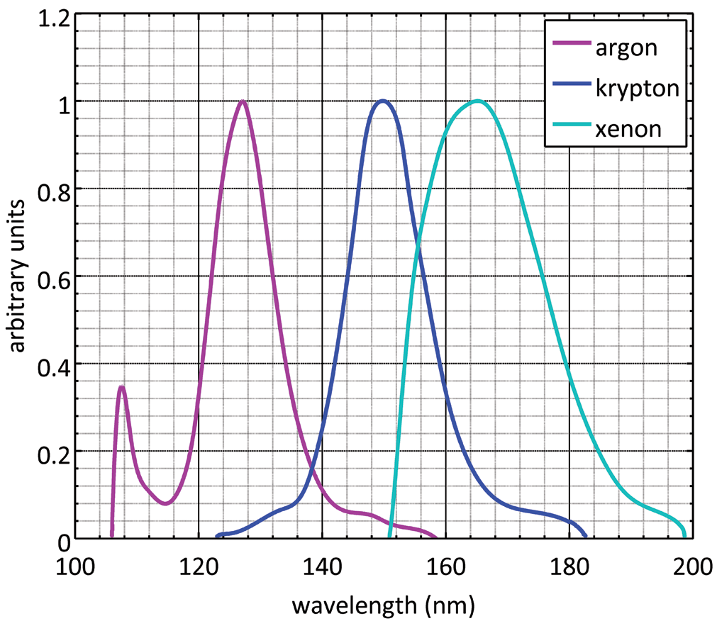
END relies on the same conversion mechanism as most traditional thermal neutron detectors. A neutron, when absorbed by specific nuclides (e.g., He, Li, B), precipitates an exothermic reaction, and the energetic charged-particle reaction products deposit their kinetic energy within a detection medium through ionization and electronic excitation. By surrounding or mixing the neutron-absorbing target with a noble gas, the charged-particle reaction products induce the formation of noble gas excimers (NGEs) as they dissipate kinetic energy. NGEs are loosely bound, diatomic molecules that exist only in an excited electronic state (e.g., Ar). NGEs are short-lived and decay by emitting far-ultraviolet (FUV) photons. This mechanism of photon emission, referred to as excimer scintillation, provides a unique method for detecting neutrons. Ar, Kr, and Xe produce excimers with wavelengths between 105 nm and 190 nm 1; 2, as shown in the emission spectra in Figure 1. A combination of features—large light output, fast decay, transparency to excimer radiation, unique decay structure, and immunity to radiation damage—make noble gases particularly suitable as radiation detection media 3.
The spectrum of the simplest excimer, He, was first analyzed by Hopfield 4, and the electronic structure was inferred from the positions and intensities of the emission lines. The molecular constants for the lowest excited singlet and triplet states are given in Herzberg 5. The excimers of Ne, Ar, Kr, and Xe were investigated by Tanaka et al. 6; 7, and later implemented in FUV radiation sources 1; 8. The use of NGE scintillation in particle detection began in the 1950s and extended into the 1980s with measurements of energy resolution and gamma sensitivity 3; 9; 10; 11; 12; 13. The mechanisms of excimer formation and decay have been investigated both experimentally 2; 14; 15 and theoretically 16; 17; 18. Recently, liquid noble gas detectors have played a significant role in efforts to detect the neutrino mass and magnetic moment, and dark matter candidates, such as weakly interacting massive particles 19. Few sources provide data on the absolute scintillation yields of gas-phase noble gas scintillators 20; 21; 22, and the data from these sources are inconsistent. For this study, we constructed an apparatus with a well-defined geometry and carefully characterized the experimental parameters. Those parameters not readily accessible to experimental measurement were modeled with standard numerical techniques.
In a previous publication, we reported on interactions of cold neutrons with mixtures of He and heavy noble gases 23. Using CaF, sapphire, and fused silica spectral filters the resulting radiation was identified as NGE emissions. In that experiment, thousands of scintillation photons per neutron absorbed were observed. Due to the growing scarcity of He, similar results were sought using other neutron absorbing targets in the presence of heavy noble gases.
Here, we present data on NGE scintillation following the boron-neutron capture reaction (B(n,)Li) in B thin films. Properties of the B films, the thermal neutron beam, and the photon detector package were characterized to derive the number of NGE photons per neutron absorbed. Additionally, Monte Carlo modeling was used to calculate charged-particle energy transfer and photon emission under the experimental conditions. Based on the experiments and modeling, we have determined the absolute number of NGE photons produced following the B(n,)Li reaction for a range of noble gas pressures and B film thicknesses.
II Experiment
II.1 Reaction cell
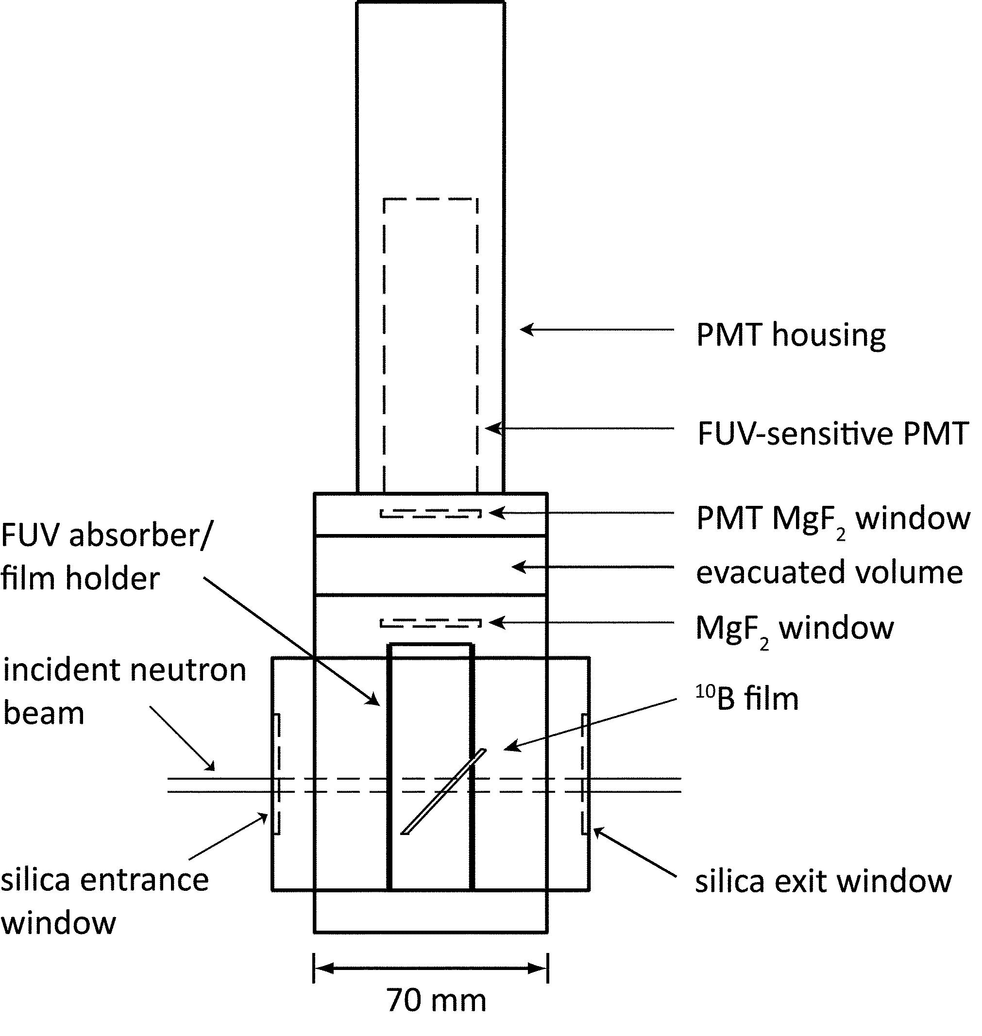
The reaction cell for the production and detection of excimer radiation consisted of a 70 mm stainless steel cube with metal-seal flange ports on all sides, attached to an FUV-sensitive photomultiplier tube (PMT). Silica entrance and exit windows allowed the neutron beam to pass through the cell without significant attenuation. Within the cell, a high-purity noble gas environment surrounded a B thin-film target held in place by a slotted aluminum cylinder beneath the PMT. The cylinder was coated with black copper oxide to reduce reflections of FUV photons. This arrangement provided a well-defined geometry for modeling the photon collection efficiency. A diagram of the apparatus appears in Figure 2.
The gas-handling system connected to the reaction cell consisted of a turbomolecular pump, noble gas cylinders, a cold cathode vacuum gauge, a digital pressure gauge, a residual gas analyzer, a gas filter, a metering valve, and a series of bellows-sealed valves. The components were connected to a stainless steel manifold with welded or metal gasket fittings. Both the Kr and Xe were research grade (99.999 % purity), while the Ar was ultra-high grade (99.9995 % purity). The gases were passed from the manifold into the reaction cell through the filter, which removed HO, O, CO, and CO to %. The filter also removed acids, organics, and refractory compounds to %, and bases to %. Before introducing inert gas into the reaction cell, the cell was baked at 100 C overnight. The base pressure in the cell was typically Pa. Upon filling, the digital pressure gauge was used to determine the pressure in the reaction cell with a relative standard uncertainty of % of full scale (206 kPa) due to non-linearity, hysteresis, and repeatability of the gauge.
II.2 B thin films
B has an isotopic abundance of 19.9 %, and a thermal neutron absorption cross section of () b 24. For nearly all thermal neutron absorptions, B undergoes an exothermic neutron-capture reaction, resulting in the emission of an -particle and a Li ion. The combined energy of these products is either 2.79 MeV or 2.31 MeV (with branching ratios of 6 % and 94 %, respectively) depending on the final state of the Li nucleus.
B thin-film targets were fabricated at the National Institute of Standards and Technology (NIST) Center for Nanoscale Science and Technology (CNST) by physical vapor deposition in an electron beam evaporator. The deposition source material was isotopically enriched in B to 92 %. The substrates were silicon wafer pieces, 25.4 mm 25.4 mm 0.525 mm. To increase adhesion and reduce film stress, thin layers of titanium and chemically deposited natural boron were added to the surfaces of the silicon wafers before depositing B. Films were fabricated at nominal thicknesses of (300, 600, 900, and 1200) nm.
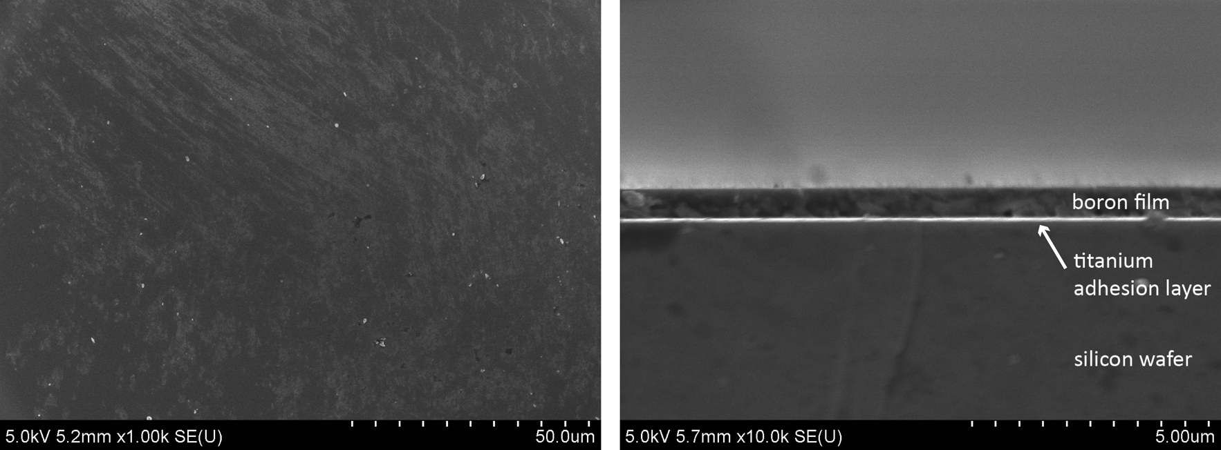
Several characterizations of the boron thin-film targets were performed to determine their neutron absorbing properties (content, thickness, density) and the stability of those properties over the course of the scintillation measurements. These characterizations included scanning electron microscopy, x-ray photoelectron spectroscopy, neutron imaging, profilometry, and x-ray diffraction. Scanning electron microscope images of a 600 nm B film appear in Figure 3. During the experiments, contamination of the B thin films from exposure to air was of particular concern. X-ray photoelectron spectroscopy scans of samples exposed to air for up to 3.5 months demonstrated no evidence of boron oxide formation within 5 nm to 10 nm of the film surface.
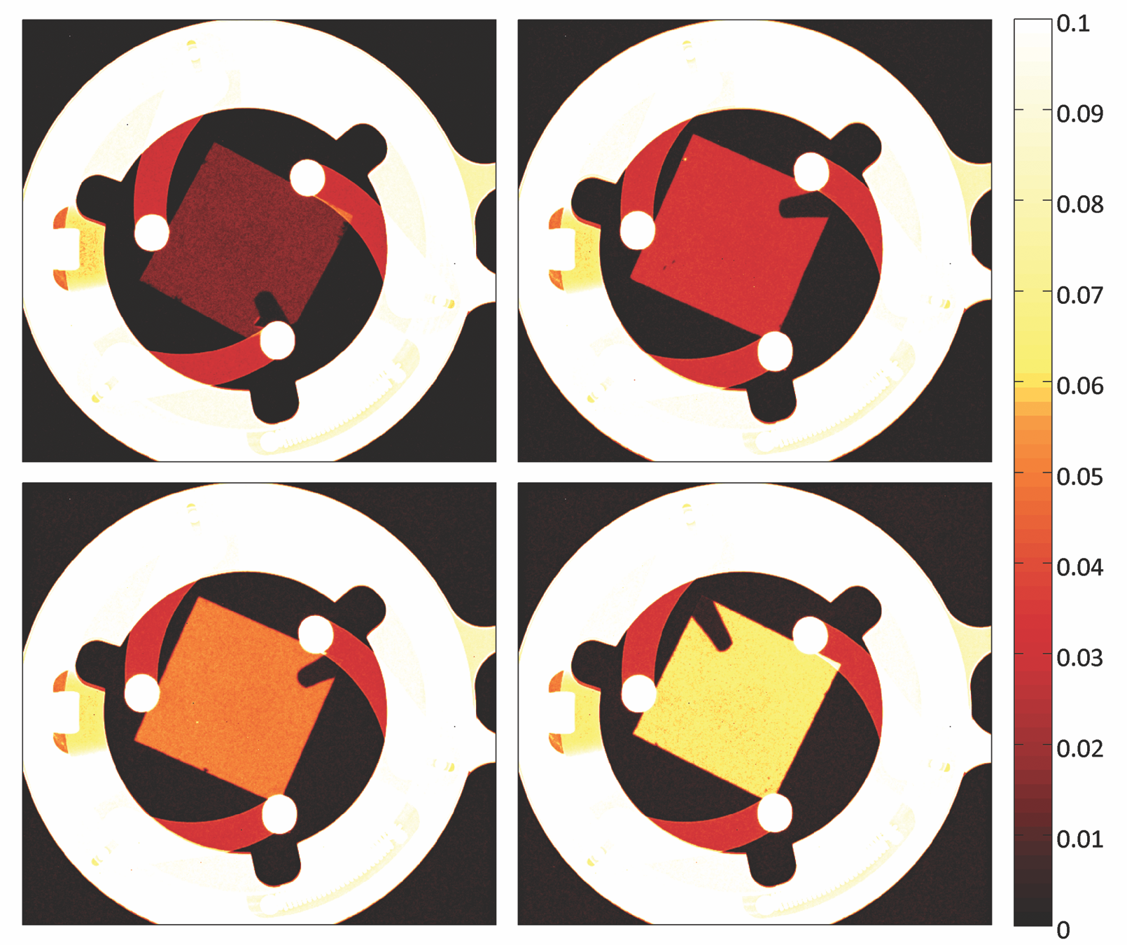
Areal densities of the B thin films were measured at the Neutron Imaging Facility (NIF) at the NIST Center for Neutron Research (NCNR). This facility uses an intense, collimated beam of thermal neutrons to obtain radiograph images of neutron absorbing samples 27. The areal densities were used to calculate the rate of neutron capture during the NGE scintillation measurements. Radiograph images of the various film targets appear in Figure 4.
II.3 Photon detector package
The photon detector package atop the reaction cell consisted of an FUV-sensitive PMT (Hamamatsu R6835 [Certain commercial equipment, instruments, or materials are identified in this paper to foster understanding. Such identification does not imply recommendation or endorsement by the National Institute of Standards and Technology, nor does it imply that the materials or equipment identified are necessarily the best available for the purpose.]) with a thin MgF entrance window, an intermediate evacuated volume, and a second MgF window separating the PMT from the reaction cell. The efficiency of the photon detector package was calibrated against an absolutely calibrated silicon photodiode at the Synchrotron Ultraviolet Radiation Facility (SURF III) at NIST. The continuous distribution of radiant power in the spectral region of interest (130 nm to 210 nm) from SURF III is well suited for radiometric calibrations 26. The efficiency of the photon detector package over this region appears in Figure 5.
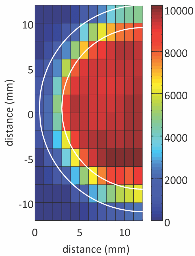
The spatial response of the photon detector package was also measured at SURF III. The response was uniform (within 3 % of the mean) over an 18 mm diameter, with a diminishing response from 18 mm to the 23 mm outer diameter of the photocathode. We attribute the diminishing response to irregularities in the MgF windows and reduced detection efficiency for photons striking the periphery of the photocathode. A color map of the spatial response appears in Figure 6. The spatial response was incorporated into the Monte Carlo model of photon collection.
During the NGE measurements, the PMT was operated at -2300 V, corresponding to a nominal gain of . Pulses from the PMT were amplified by a fast preamplifer with a gain of 200 and a rise time of ns. The amplified pulses were divided in two with a power splitter. One splitter output was sent to a counter/timer and the other to a multichannel analyzer (MCA). The MCA was used to obtain pulse-charge distributions (PCDs). These PCDs confirmed that multi-photoelectron pulses were not contributing significantly to the number of pulses counted by the counter/timer. The MCA was operated with a 20 ns integration time and a channel resolution of 0.25 pC over 1024 channels. Because the digitization time of the MCA was long with respect to the typical decay time of each scintillation event, not every pulse from the PMT was collected by the MCA. Nevertheless, the resulting PCDs were assumed to be representative of the true distributions. The counter/timer with a maximum count rate of 100 MHz and a pulse-pair resolution of ns recorded the true number of PMT pulses. A block diagram of the electronics appears in Figure 7.


II.4 Neutron beam
The Maryland University Training Reactor (MUTR) provided a source of thermal neutrons for the NGE scintillation experiments. The MUTR is a TRIGA reactor with a peak power of 250 kW. The MUTR thermal column consists of 1.5 m of graphite adjacent to the core and a custom insert to produce a collimated thermal neutron beam with a diameter of 50 mm.
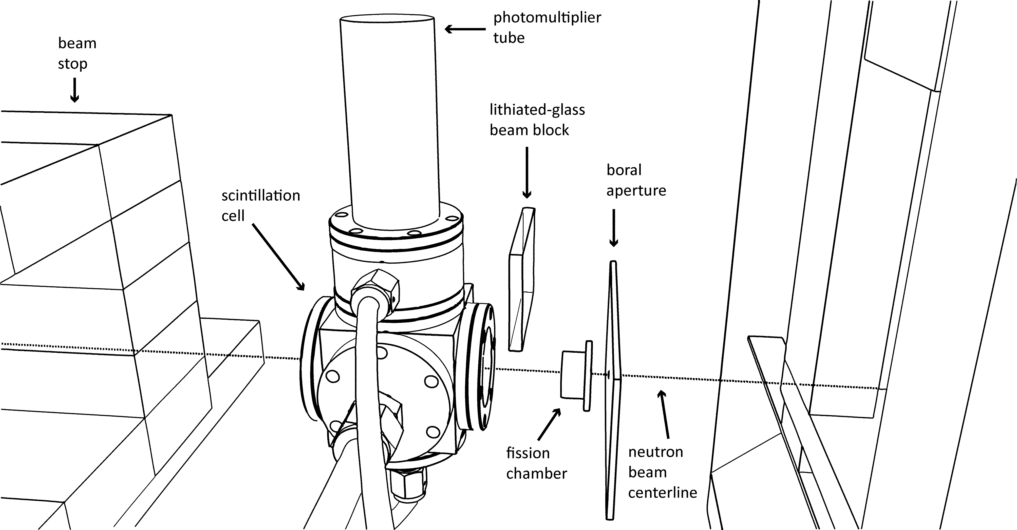
Beyond the collimator, in front of the reaction cell, a borated aluminum aperture reduced the neutron beam to a 4 mm diameter. The neutron fluence behind this aperture was monitored with a NIST-calibrated fission chamber throughout the experimental irradiations. The fission chamber was specifically designed as a beam monitor, allowing more than 99.9 % of thermal neutrons to pass through to the reaction cell unattenuated. Backgrounds were measured with a beam block of Li glass positioned between the fission chamber and the reaction cell. This beam block absorbed nearly all thermal neutrons without attenuating gamma radiation incident on the cell. In this way, the gamma-ray and dark-current contributions to the PMT signal were measured. A diagram of the neutron beam setup appears in Figure 8.
III Model Calculations
Monte Carlo techniques were employed to calculate both charged-particle transport from the B(n,)Li reaction and FUV photon emission within the reaction cell. The results of these calculations were used to determine the spatial distribution of charged particles escaping the boron films and the photon collection efficiency of the experimental apparatus.
The number of photons incident on the PMT depended upon the number of photons produced in the reaction cell, the spatial distribution of those emissions, the probability with which the photons were reflected from adjacent surfaces, and the type of reflection they underwent (i.e., specular or diffuse). Some photons intersected the PMT directly; others were reflected into the PMT from the surfaces surrounding the reaction volume; and a large majority were absorbed by the interior surfaces of the cylinder surrounding the B film without reaching the detector. The simplest model for the calculation of detector collection efficiency, assumes that all of the photons were emitted isotropically from a point source at the center of the B film surface, where
| (1) |
is the solid angle subtended by the detector, is the distance from the source to the detector photocathode (85.8 mm), and is the radius of the detector photocathode (11.5 mm). This approximation gives a collection efficiency of 0.443 %. However, the approximation does not account for the reflectivities of the surfaces surrounding the reaction volume, the extended volume over which photon emission occurs, or variations in the spatial response of the PMT.
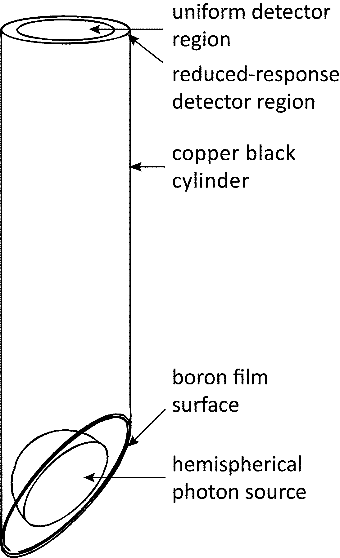
A Monte Carlo routine was developed to incorporate the extended photon emission volume, the geometry of the reaction volume, the reflectivities of the various surfaces, and the measured position-dependent sensitivity of the PMT photocathode. The model geometry appears in Figure 9. For each photon, an emission position was generated from a uniform and random distribution over the volume of a hemisphere tilted at 45 with respect to the detector plane. A direction for the photon path was then calculated from a uniform random distribution. Subsequently, the intersection of the path with one of the three surrounding surfaces (i.e., B film, black copper-oxide cylinder, and detector face) determined whether the photon was collected, reflected, or absorbed.
Photons striking the B surface were specularly reflected with a reflection coefficient of 0.35, derived from measurements in 27. Photons striking the copper-oxide cylinder were diffusely reflected with a probability of 0.01, derived from measurements in 28. The final tally was weighted according to the data in Figure 6: photons striking the uniform detector region were given a score of 1, and photons striking the outer region were given a score of 0.57. From particle histories, the collection efficiency, , was determined to be () %. The statistical uncertainty in was determined by calculating the standard deviation of 10 runs of histories each. The total uncertainty includes both the statistical uncertainty and systematic uncertainties in the model inputs (e.g., geometry, reflectivities).
The radius of the hemispherical photon emission volume in the photon collection model was approximated through modeling noble gas ionization density following the B(n,)Li reaction with the radiation transport code, TRIM 29. The TRIM input files specified the thicknesses of the boron films and the noble gas pressures, as well as the emission positions, directions, and starting energies of the B(n,)Li reaction products. The starting positions of the reaction products within the B film were sampled using an inverse transform for the depth profile and the neutron beam width.
Ionization density distributions from B(n,)Li in a 300 nm B film under various pressures of Kr appear in Figure LABEL:Figure10. The plots show the two-dimensional shape (x-y plane) and the ionization density (z axis) of the volume in which the charged particles deposited their energy. Contours are drawn at increments of eV/ per ion. Based on the results of these simulations, a radius of 10 mm was chosen for the hemispherical photon emission volume in the photon collection model. However, as shown in Figure LABEL:Figure10, the size of the emission volume was pressure dependent and the shape was non-uniform.
In summary, the photon collection model assumed the following: (1) a hemispherical excimer photon emission volume, (2) a uniform, pressure-independent distribution of NGE emissions within the volume, (3) accurate values of the reflectivities of boron and copper black, (4) negligible contributions of secondary and tertiary reflections, and (5) the absence of refraction by the MgF windows.
IV Results and Discussion
IV.1 Photon detector package efficiency
The photon detector package (i.e., MgF windows, evacuated volume, and PMT) was calibrated as a unit at SURF III, beamline 4 (BL-4). The efficiency measurements accounted for absorption by the external and PMT MgF windows, the quantum efficiency of the PMT, and the thresholds of the counting electronics. A silicon photodiode that was previously calibrated against a cryogenic radiometer 30; 31 was used to obtain the absolute response of the photon detector package. The calibration covered the FUV region common to the NGE spectra, between 130 nm and 210 nm. The PMT was operated in the pulse counting mode with electronics identical to those used in the scintillation yield measurements.
The detector calibration was performed in several stages due to the large difference in sensitivity of the photodiode and the PMT. The incident photon flux was measured with the photodiode over the wavelength range 130 nm to 210 nm in 5 nm increments, at electron beam currents of approximately (50, 40, 30, 20, and 10) mA. These measurements were used to determine a linear relationship between electron beam current in the SURF storage ring and photon flux in the detector position, at each discrete wavelength.
Following these measurements, the SURF electron beam current was reduced to (10, 5, and 1) A. At these lower beam currents, the photon flux was within the operating range of the PMT. Spectral scans over the same wavelength region were repeated at these reduced currents. The intrinsic efficiency of the photon detector package, , was determined by dividing the number of pulses observed from the PMT at each wavelength by the number of photons incident on the photon detector package at that wavelength. The results appear as discrete points in Figure 5 for three electron beam currents. The relative uncertainties in include both statistical uncertainties and uncertainties in the linear regressions.
Because NGE emissions occur over broad continua, an effective photon detection efficiency was determined for the three noble gases used in the NGE scintillation experiments. The effective efficiency, , was calculated with a continuous weighted average, in the form,
| (2) |
where is a fit of the discrete values of , and is the wavelength distribution of the NGE continua obtained by digitizing excimer emission spectra from 1. The values of for Ar, Kr, and Xe were 1.65 %, 3.14 %, and 2.61 %, respectively. The uncertainties in were derived from the average value of the relative uncertainties in .
IV.2 Areal densities of the B thin films
The thermal-neutron absorption properties of the B thin films were measured at the NIF. Thermal neutrons from the NCNR reactor passed through a cooled, single-crystal bismuth filter, a series of apertures, and an evacuated flight tube before impinging on the samples with a fluence of cms. The NIF detector consists of a Li conversion layer, a ZnS scintillation layer, and an x-ray imager made of amorphous silicon.
Each boron film used in the scintillation experiments was placed in a holder and mounted at the imaging station. The neutron beam illuminated each sample, and the detector collected a series of 1800 images with one-second exposures. This measurement was repeated to obtain a flat-field image without a sample or a sample holder in the beam. All of the images were then corrected for the point spread function (PSF) of the detector system. The PSF is a systematic additive background attributed to diffuse light in the scintillation screen of the detector.
The images of each sample were then averaged to form a single image. By dividing the averaged image of each boron film by the averaged flat-field image, the fractional absorption of each sample was determined. The fractional absorption of each pixel, , in the resulting images has the form,
| (3) |
where is the intensity of the incident neutron beam, is the intensity of the neutron beam after passing through the sample, is the macroscopic neutron absorption cross section of the sample, and is the thickness of the sample. The value of was determined by dividing each pixel value in the sample image by the corresponding pixel value in the empty flat-field image.
By rearranging Equation 3 to solve for and averaging the values of over the surface of each film, the areal density of each film, , was determined with the equation,
| (4) |
where is the average value of , is the effective neutron absorption cross section of B in the NIF neutron beam, is the molar mass of B, and is Avogadro’s number. The value of was determined with a continuous weighted average, in the form,
| (5) |
where is the wavelength-dependent, microscopic absorption cross section of B, and is the neutron wavelength distribution of the NIF beam. This neutron wavelength distribution was previously measured at the NIF with a neutron chopper and time-of-flight spectrometry. The thermal neutron absorption cross section of silicon is small (2.16 b 24); thus, the absorption of each sample was attributed completely to absorption by B. From Equation 4, the resulting areal densities of the B thin films were (73.5, 140, 209, and 260) g/cm.
A conservative 10 % uncertainty was assigned to the values of . This uncertainty arises predominantly from the uncertainty in . However, the relative root mean square (RMS) variations in ¡ 2^10