Mechanosensitive Channel Activation by Diffusio-Osmotic Force
Abstract
For ion channel gating, the appearance of two distinct conformational states and the discrete transitions between them is essential, and therefore of crucial importance to all living organisms. We show that the physical interplay between two structural elements that are commonly present in bacterial mechanosensitive channels, namely a charged vestibule and a hydrophobic constriction, creates two distinct conformational states, open and closed, as well as the gating between them. We solve the nonequilibrium Stokes-Poisson-Nernst-Planck equations, extended to include a molecular potential of mean force, and show that a first order transition between the closed and open states arises naturally from the diffusio-osmotic stress caused by the ions and water inside the channel and the elastic restoring force from the membrane. Our proposed gating mechanism is likely to be important for a broad range of ion channels, as well as for biomimetic channels and ion channel-targeting therapeutics.
Osmotic shock presents a fatal risk to unicellular organisms. A sudden increase of the environmental solute concentration, known as hypertonic shock, leads to water loss and cell volume decline, whereas a sudden decrease, referred to as hypotonic shock, causes water to enter the cell rapidly, inducing cytolysis. As a final resort in case of severe hypotonic shock, many bacteria, archaea and fungi avert cell lysis by activating non-selective membrane channels to release solutes from the cytoplasm Kung et al. (2010). In E. coli bacteria, two well-studied membrane protein channels are responsible for the release of solutes: the mechanosensitive channel of large conductance (mscl) Perozo et al. (2002) and the mechanosensitive channel of small conductance (mscs) Vásquez et al. (2008). Based on the observation that mechanosensitive channels are activated in vitro by an applied hydrostatic pressure, the prevalent model for the gating mechanism invokes a conformational change in the protein triggered by tension applied to the cell membrane Sukharev et al. (1994); Perozo et al. (2002); Vásquez et al. (2008). A free energy landscape for channel activation can be constructed by considering an elastic force proportional to the applied pressure Wiggins and Phillips (2004). However, no proposed gating hypothesis has been able to explain the appearance of two distinct conformational states and the discrete transitions between them. In E. coli mscl mutants, added charge in the pore region activates the channels also in the absence of a hydrostatic pressure difference Yoshimura et al. (2001); Bartlett et al. (2004, 2006); Batiza et al. (2002), highlighting the importance of electrostatic interactions in the activation process. Indeed, the transmembrane domains of both mscl and mscs carry a substantial net charge: Each of the ten transmembrane helices of the pentameric mscl protein carries a net charge of Gu et al. (1998), and the heptameric mscs protein carries an arginine residue with a charge of on each of its monomers Sotomayor et al. (2006). Charge-induced activation is a robust feature of mscl channels and has been used for drug delivery into mammalian cells Doerner et al. (2012). Despite its significance, however, the electrostatic contribution to the activation energy, and in particular the diffusio-osmotic force originating in the dynamic overlapping double layer at the channel’s charged surface, has not been considered up to now.
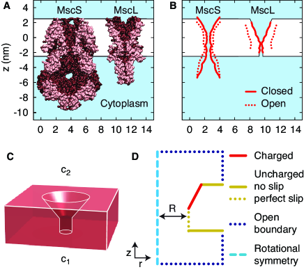
The permeation pathways of both mscl and mscs are funnel-shaped, with the conical vestibule opening to the periplasmic side Perozo et al. (2002); Vásquez et al. (2008), and the stem of the funnel lined with uncharged hydrophobic residues (Fig. 1A). Upon activation the pore walls move radially outward (Fig. 1B). In weakly polar channels, water can fill constrictions down to the size of a single water molecule Beckstein and Sansom (2004), but even strongly hydrophobic channels are intermittently filled with water Hummer et al. (2001); Beckstein and Sansom (2004); Allen et al. (2002). Ions, on the other hand, are subject to a strongly repulsive potential of mean force (pmf) up to channel radii much larger than the ionic radius, caused by their hydration shells Zhu and Hummer (2012); Richards et al. (2012); Allen et al. (2004), steric and van der Waals interactions and self energy Parsegian (1969). Using molecular dynamics simulations, the energy barrier for ion permeation through mscs has been estimated to be – Anishkin and Sukharev (2004), explaining the lack of electric conductivity of mscs in the closed state despite its relatively wide permeation pathway. A similar hydrophobic lock mechanism has been found in mscl Perozo et al. (2002) and many different membrane channels Kuo et al. (2003); Miyazawa et al. (2003); Payandeh et al. (2011). Using mutational analysis, it has been established that the hydrophobic constriction in mscl provides a threshold for channel activation Yoshimura et al. (1999).
The use of continuum hydrodynamics in nanometer-sized tubes has been shown to be justified for radii in the nanometer range Thomas and McGaughey (2009); a noteworthy result, which can be rationalized by analytic arguments Bocquet and Charlaix (2010) and has been used recently to calculate the hydrodynamic resistance of aquaporin channels Gravelle et al. (2013). Similarly, the Nernst-Planck equation for ion transport has been found to be applicable down to a radius of nm, provided that the ion concentrations are estimated accurately Song and Corry (2011). Ion concentrations at solid surfaces and lipid bilayers can be accurately calculated from mean-field theory when the ionic pmf, estimated using molecular dynamics simulations, is included as a non-electrostatic contribution to the potential Horinek et al. (2008); Bonthuis and Netz (2013). Combined, extended mean-field theory and continuum hydrodynamics reproduce the electrokinetic properties found in experiments and atomistic simulations of both hydrophilic and hydrophobic surfaces Bonthuis and Netz (2012). Capturing the dewetting transitions of water under strong hydrophobic confinement and their coupling to the ionic dynamics would require a more detailed molecular modeling Chandler (2005); Zhu and Hummer (2012). However, our primary interest here is the description of the mesoscopic electrokinetic properties of the channel, which we show to be insensitive to the hydrodynamic characteristics of the hydrophobic constriction. Although we will not be able to predict the electrolyte dynamics inside the stem area in atomic detail, this theoretical framework provides a reliable description of the electrokinetic properties at the mesoscopic scale of the protein channel. Nevertheless, solving the coupled Stokes-Poisson-Nernst-Planck equations in complex geometries has proven to be a challenging endeavor Pagonabarraga et al. (2010).
Here, we consider a model of a mechanosensitive channel consisting of the essential structural features found in mscs and mscl: a funnel-shaped pore with an uncharged hydrophobic stem and a vestibule carrying a fixed surface charge density, embedded in an impermeable membrane separating two solutions with salt concentrations and , respectively (Fig. 1C–D). This model is based directly on the experimentally determined protein crystal structure, and aims to explain experimental work showing, first, that added charge in the vestibule activates the channel Yoshimura et al. (2001); Bartlett et al. (2004, 2006); Batiza et al. (2002); Doerner et al. (2012), and second, that the hydrophobicity of the constriction provides a barrier for channel activation Yoshimura et al. (1999). As an experimental benchmark, we consider measurements showing that mscl and mscs are activated at a hypotonic shock of at least M Levina et al. (1999).
Governing equations. – We define a non-dimensional electrostatic potential , with being the potential in Volt and being the position in cylindrical coordinates. The Poisson equation relates the electrostatic potential to the ion densities ,
| (1) |
with being the Bjerrum length. At low Reynolds number, the solvent velocity is governed by the Stokes equation, which for an incompressible fluid in steady state reads
| (2) |
The components of the viscous and electrostatic stress tensors and and the force density due to the ionic pmf are given by
| (3) |
with being the hydrostatic pressure, being the viscosity and being . Inserting Eq. 3 into Eq. 2 and taking the curl results in the following equations for the vorticity ,
| (4) |
From the latter definition of it follows and , which guarantees that the incompressibility condition is satisfied. The local ion concentrations are determined by conservation of species. In steady state:
| (5) |
with being the velocity of the solvent, the concentrations of positive and negative ions, and the corresponding fluxes, given by
| (6) |
with nm2/ns being the ionic diffusion constant. We numerically solve Eqs. 1–6 in the domain shown in Fig. 1D using a finite-difference over-relaxation scheme, which allows us to analyze the diffusio-osmotic force exerted on the channel wall for the first time.
Boundary conditions. – We employ a fixed surface charge density in the conical vestibule of nm-2 and uncharged boundaries everywhere else. The hydrodynamic equations obey the no-slip boundary condition on the surface of the membrane and the vestibule, as is appropriate for hydrophilic surfaces Bocquet and Charlaix (2010); Bonthuis and Netz (2013). Inside the hydrophobic constriction, on the other hand, perfect slip is assumed, consistent with the plug-like flow found in hydrophobic nanotubes Thomas and McGaughey (2009). Note that assuming no slip inside the hydrophobic constriction instead leads to very similar results, implying that the model is robust regarding the characteristics of the hydrodynamic flow inside the constriction. The normal flux vanishes at the membrane and pore boundaries, . A fixed pressure difference between the open boundaries is achieved by adjusting the fluid flow through the box. Guided by experimental design, we set M Levina et al. (1999). Molecular dynamics simulations show that the one dimensional ionic pmf in narrow channels exhibits a peak, reaching a maximum of – in the center of the channel, which decreases with increasing channel radius Beckstein et al. (2004); Allen et al. (2004); Parsegian (1969); Richards et al. (2012); Anishkin and Sukharev (2004). At a radius of nm, is still several ’s in short nanopores Beckstein et al. (2004). Therefore, we model the ionic pmf by a repulsive potential in the stem of the funnel of height , that decreases linearly from at nm to zero at nm. This potential comprises all interactions between the ions, the water and the pore, including changes in the hydration state of the pore Zhu and Hummer (2012).
The force on the surface of the pore, consisting of the stem and the vestibule, is calculated from the normal stress, . We calculate the nonequilibrium free energy landscape as the sum of two terms: the integral over the radial force due to the electrolyte, and an elastic term due to the protein and the membrane,
| (7) |
with nm being the minimum channel radius. For the elasticity coefficient of the protein and the membrane we assume nm-2, which is well within the range of values quoted in literature Phillips et al. (2009).
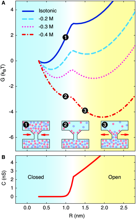
Within this theoretical framework, the tension on the channel wall results from a competition of contractile forces due to the ionic pmf and the elastic membrane, and expansile forces due to the charged vestibule. The striking result of this competition is that the nonequilibrium free energy landscape exhibits two minima, corresponding to the closed and open states (Fig. 2A). Under isotonic conditions, exclusion of ions from the hydrophobic stem at small radii (inset 1 of Fig. 2A), which is known to reduce the pressure between like-charged parallel plates Edwards and Williams (2004), gives rise to an energy barrier between the two states of . Remarkably, the energy barrier arises naturally from only electrostatic and hydrodynamic forces. The second energy minimum is caused by the expansile electrostatic force, which increases upon hypotonic shock. Whereas for nm the increased electrostatic force is partly compensated for by the reduced pressure due to the ionic pmf (inset 2 of Fig. 2A), the electrostatic force dominates when for nm, and the channel activates (inset 3 of Fig. 2A). For large the elastic term overcomes the electrostatic repulsion. The first order transition between closed and open states occurs at a hypotonic shock of approximately M, in quantitative agreement with experimental results Levina et al. (1999). The profiles show that the tension on the pore wall due to the electrolyte is sufficient to activate a mechanosensitive channel.
The channel activation is evident from the electrical conductance (Fig. 2B), which we calculate from , with being an applied potential difference across the channel and being the resulting electric current, where the integration can be carried out over any plane spanning the pore. The asymmetry in the conductance with respect to the direction of the applied potential difference, which is due to the asymmetric geometry of the channel, is negligible. The conductance is minute up to a radius of nm (Fig. 2B), owing to the repulsive pmf. Between nm, the conductance increases dramatically, before adopting linear growth with . To compare with experimental data, the salt concentration is set to M. The conductance of the open channel agrees well with the experimental values of nS measured for mscl Sukharev et al. (1997, 1994).
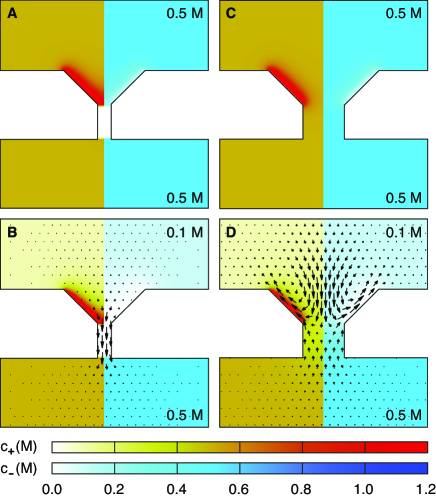
To examine the functionality of the channel, we monitor the ion concentrations and water flux throughout the activation process. In the closed state ( nm), the ionic pmf excludes both ion types from the stem of the funnel, as revealed by the concentrations (Figs. 3A–B). In the open state ( nm), on the other hand, ions flow through the channel uninhibited (Figs. 3C–D). In response to a hypotonic shock, water rushes into the cell, driven by the osmotic pressure (arrows in Fig. 3B). When the channel activates, ions flowing outward through the channel drag the fluid along, and the water flow reverses (arrows in Fig. 3D), thus reproducing the experimentally observed behavior.
In conclusion, two-state mechanosensitive channel gating emerges from the electrokinetic transport equations without phenomenological assumptions in a simplified geometry that is based directly on the experimentally determined protein crystal structure. Our proposed gating mechanism is fully supported by mutation experiments, which show a strong influence of protein surface charge and hydrophobicity on the gating kinetics. Moreover, it agrees quantitatively with experiments regarding hypotonic shock threshold and electrical conductivity. The activation mechanism can be verified further using mutation experiments, substituting charged residues for neutral ones. Although there is evidence indicating that membrane-protein interactions also play a role in the gating transition, the direct response to hypotonic shock proposed in this work provides a faster and more accurate mechanism, bypassing the inhomogeneous cell membrane. This novel modeling scheme reveals the underlying physics of the channel’s complex biological function, showing that the gating kinetics can be fully reproduced within a model consisting of only a charged vestibule and a hydrophobic constriction. Because these elements are shared features of many different ion channels, our proposed two-state gating mechanism is likely to be important for a broad range of ion channels. Moreover, this new insight into the gating mechanism constitutes an essential step toward the design of artificial mechanosensitive channels and ion channel-targeting therapeutics.
Acknowledgements.
We thank Philip Biggin and Julia Yeomans for valuable discussions of the manuscript. D.J.B. acknowledges the Glasstone Benefaction and Linacre College for funding. R.G. was supported by Human Frontier Science Program (HFSP) grant RGP0061/2013.References
- Kung et al. (2010) C. Kung, B. Martinac, and S. Sukharev, Annu. Rev. Microbiol. 64, 313 (2010).
- Perozo et al. (2002) E. Perozo, D. M. Cortes, P. Sompornpisut, A. Kloda, and B. Martinac, Nature 418, 942 (2002).
- Vásquez et al. (2008) V. Vásquez, M. Sotomayor, J. Cordero-Morales, K. Schulten, and E. Perozo, Science 321, 1210 (2008).
- Sukharev et al. (1994) S. I. Sukharev, P. Blount, B. Martinac, F. R. Blattner, and C. Kung, Nature 368, 265 (1994).
- Wiggins and Phillips (2004) P. Wiggins and R. Phillips, Proc. Nat. Acad. Sci. USA 101, 4071 (2004).
- Yoshimura et al. (2001) K. Yoshimura, A. Batiza, and C. Kung, Biophys. J. 80, 2198 (2001).
- Bartlett et al. (2004) J. L. Bartlett, G. Levin, and P. Blount, Proc. Nat. Acad. Sci. USA 101, 10161 (2004).
- Bartlett et al. (2006) J. L. Bartlett, Y. Li, and P. Blount, Biophys. J. 91, 3684 (2006).
- Batiza et al. (2002) A. F. Batiza, M. M.-C. Kuo, K. Yoshimura, and C. Kung, Proc. Nat. Acad. Sci. USA 99, 5643 (2002).
- Gu et al. (1998) L. Gu, W. Liu, and B. Martinac, Biophys. J. 74, 2889 (1998).
- Sotomayor et al. (2006) M. Sotomayor, T. A. van der Straaten, U. Ravaioli, and K. Schulten, Biophys. J. 90, 3496 (2006).
- Doerner et al. (2012) J. F. Doerner, S. Febvay, and D. E. Clapham, Nat. Commun. 3, 990 (2012).
- (13) Supplementary information.
- Beckstein and Sansom (2004) O. Beckstein and M. S. P. Sansom, Physical Biology 1, 42 (2004).
- Hummer et al. (2001) G. Hummer, J. C. Rasaiah, and J. P. Noworyta, Nature 414, 188 (2001).
- Allen et al. (2002) R. Allen, S. Melchionna, and J.-P. Hansen, Phys. Rev. Lett. 89, 175502 (2002).
- Zhu and Hummer (2012) F. Zhu and G. Hummer, Biophys. J. 103, 219 (2012).
- Richards et al. (2012) L. A. Richards, A. I. Schäfer, B. S. Richards, and B. Corry, Small 8, 1701 (2012).
- Allen et al. (2004) T. W. Allen, O. S. Andersen, and B. Roux, Proc. Nat. Acad. Sci. USA 101, 117 (2004).
- Parsegian (1969) A. Parsegian, Nature 221, 844 (1969).
- Anishkin and Sukharev (2004) A. Anishkin and S. Sukharev, Biophys. J. 86, 2883 (2004).
- Kuo et al. (2003) A. Kuo et al., Science 300, 1922 (2003).
- Miyazawa et al. (2003) A. Miyazawa, Y. Fujiyoshi, and N. Unwin, Nature 423, 949 (2003).
- Payandeh et al. (2011) J. Payandeh, T. Scheuer, N. Zheng, and W. A. Catterall, Nature 475, 353 (2011).
- Yoshimura et al. (1999) K. Yoshimura, A. Batiza, M. Schroeder, P. Blount, and C. Kung, Biophys. J. 77, 1960 (1999).
- Thomas and McGaughey (2009) J. A. Thomas and A. J. H. McGaughey, Phys. Rev. Lett. 102, 184502 (2009).
- Bocquet and Charlaix (2010) L. Bocquet and E. Charlaix, Chem. Soc. Rev. 39, 1073 (2010).
- Gravelle et al. (2013) S. Gravelle et al., Proc. Nat. Acad. Sci. USA 110, 16367 (2013).
- Song and Corry (2011) C. Song and B. Corry, Plos One 6, e21204 (2011).
- Horinek et al. (2008) D. Horinek et al., Langmuir 24, 1271 (2008).
- Bonthuis and Netz (2013) D. J. Bonthuis and R. R. Netz, J. Phys. Chem. B 117, 11397 (2013).
- Bonthuis and Netz (2012) D. J. Bonthuis and R. R. Netz, Langmuir 28, 16049 (2012).
- Chandler (2005) D. Chandler, Nature 437, 640 (2005).
- Pagonabarraga et al. (2010) I. Pagonabarraga, B. Rotenberg, and D. Frenkel, Phys. Chem. Chem. Phys. 12, 9566 (2010).
- Levina et al. (1999) N. Levina et al., EMBO J. 18, 1730 (1999).
- Beckstein et al. (2004) O. Beckstein, K. Tai, and M. S. P. Sansom, J. Am. Chem. Soc. 126, 14694 (2004).
- Phillips et al. (2009) R. Phillips, T. Ursell, P. Wiggins, and P. Sens, Nature 459, 379 (2009).
- Edwards and Williams (2004) S. A. Edwards and D. R. M. Williams, Phys. Rev. Lett. 92, 248303 (2004).
- Sukharev et al. (1997) S. I. Sukharev, P. Blount, B. Martinac, and C. Kung, Annu. Rev. Physiol. 59, 633 (1997).