Accessing defect dynamics using intense, nanosecond pulsed ion beams
Abstract
Gaining in-situ access to relaxation dynamics of radiation induced defects will lead to a better understanding of materials and is important for the verification of theoretical models and simulations. We show preliminary results from experiments at the new Neutralized Drift Compression Experiment (NDCX-II) at Lawrence Berkeley National Laboratory that will enable in-situ access to defect dynamics through pump-probe experiments. Here, the unique capabilities of the NDCX-II accelerator to generate intense, nanosecond pulsed ion beams are utilized. Preliminary data of channeling experiments using lithium and potassium ions and silicon membranes are shown. We compare these data to simulation results using Crystal Trim. Furthermore, we discuss the improvements to the accelerator to higher performance levels and the new diagnostics tools that are being incorporated.
I Introduction
Defects in materials created by ion irradiation present a multi-scale problem with defect lifetimes ranging from picoseconds to years, see Diaz de la Rubia et al., (2000) and Victoria et al., (2000). Most of the defects self-anneal with a time scale of the order of picoseconds, see Bai et al., (2010). Currently, simulation tools can only assume certain time constants since experimental verification for fast process on the time scale of picoseconds has not been possible, see Stuchbery and Bezakova, (1999) and Myers et al., (2012). However, access to these processes is important for a better understanding of materials that play an important role in the development of radiation hard electronics and the development for advanced materials for future fusion reactors and the next generation of nuclear reactors, see Zinkle, (2013) and Zinkle and Was, (2013). At the new Neutralized Drift Compression Experiment (NDCX-II) short, intense beam pulses are available that can provide a pump pulse for pump-probe experiment, see Friedman et al., (2009), Waldron et al., (2014), and Schenkel et al., (2013). We report on experimental results using beams of lithium and potassium ions to probe crystalline silicon samples. We present data from channeling experiments where the ion beam pulse is also used as a diagnostic tool. The mechanism exploited in these measurements is based on the channeling effect. Ions that channel have a longer range and lower energy loss in the material. The possibility of a channel event is strongly dependent on the crystalline integrity, or the amount of damage present in the material. A non-channeling ion hitting the material will create many interstitial-vacancy pairs in the material, temporarily blocking many channels in the material before the defects self-anneal. If the fluence of the ions is high enough, other ions will hit the same area as the remnants of the earlier ion cascade (before the self-annealing took place) and the channel transmission will be impeded. Therefore, we expect that the channeling current will change depending on the fluence, e.g., for low fluences channeling will be unobstructed (no overlapping cascades) and for high fluences the channeling current will decrease during a single beam pulse (many overlapping cascades). The transmitted beam current can be monitored with high temporal resolution and by restricting the measured ions to a small scattering angle, mostly channeled ions can be recorded. Noise from non-channeled ions can be further reduced by time-of-flight measurement, since channeled ions will have a smaller energy loss than scattered ions.
II Experiment
The source technology employed at NDCX-II currently allows the use of lithium and potassium ions. (Other alkalis, such as sodium and cesium are also feasible.) The ions are extracted from a 10 cm diameter area over a time of 600 ns. The extraction optics focus the beam to a radius of about 2-3 cm. Using an induction type accelerator the ions are then transported along a 9 m beamline where they are compressed longitudinally to a full-width-half-maximum (FWHM) beam pulse of about 20-30 ns. The last few solenoids in the accelerator are adjusted to focus the beam down to a spot size of 5 mm radius at the target. The ion energy of the beam is 135 keV for an uncompressed (600 ns long) beam and 300 keV for a compressed beam. The repetition rate of the accelerator is 2-3 shots per minute.
The peak current in the experiments is presently around 1 A, corresponding to ions ( for Li and for K) that are delivered per pulse. Ion beam transport with such high peak currents is dominated by space charge effects, and NDCX-II has been specially developed to be able to transport beams under these extreme space charge conditions, see Friedman et al., (2009) and Waldron et al., (2014).
At the target chamber the beam can be monitored using a scintillator and a fast camera, as well as a specially designed fast Faraday cup to monitor the beam current over time. We mount our silicon membranes (250 nm and 1000 nm) at the same location as the scintillator so that we can optically confirm the beam size. The transmitted beam is then recorded using a fast Faraday cup positioned 35 cm behind the membrane. This distance increases the sensitivity to channeled ions (smaller acceptance angle), since scattered ions are preferentially scattered out of the acceptance angle of the Faraday cup. We can furthermore utilize the drift distance between membrane and Faraday cup for time-of-flight measurements.
The target is mounted on a goniometer with x, y and z motion allowing for a precise alignment of the target to the beam spot recorded using the camera and scintillator. The goniometer also has the ability to rotate the sample allowing measurements of the channeling effect for different incident angles. The sample is mounted on a larger frame so that only beam passing through the sample can reach the Faraday cup even at large angles.
Improvements to the accelerator that are presently underway will allow for a beam energy of 1.2 MeV, and 1-2 ns long beam pulses with a spot size of radius. This will provide up to 400 times increase in ion fluence. It will be possible following the installation of a plasma filled drift line at the end of the accelerator. This drift-compression section will allow the beam bunch, which has an ordered head-to-tail velocity ramp, to bunch to unimpeded by its own space charge repulsion. Installation of an 8 T solenoid near the target focuses the beam to , also in the presence of a neutralizing background plasma.
III Results
Both the lithium and the potassium sources produce a very reliable beam with reproducible temporal and spatial profiles for many shots over periods of days and weeks with minor retuning.
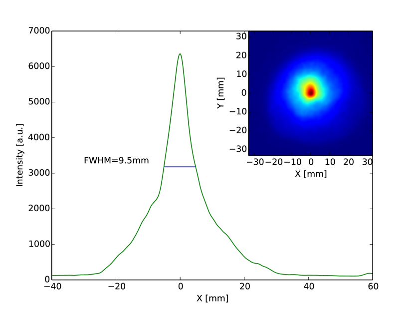
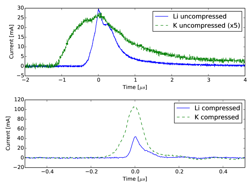
The beam has a Gaussian transverse profile. Figure 1 shows a scintillator image of a compressed potassium beam. We find a shot-to-shot variation of the peak position of 0.2 mm (rms) in the horizontal and vertical direction. Intensity variations are of the order of 5% percent and the main source for this is the variation in temperature at the filament, which currently is not regulated (the filament is voltage controlled). A feedback loop will be implement in the future which will create a more stable beam in regard to the intensity. In Figure 2 a beam profile measured on a Faraday cup at the target position is shown for a compressed beam and an uncompressed beam for both lithium and potassium ions.
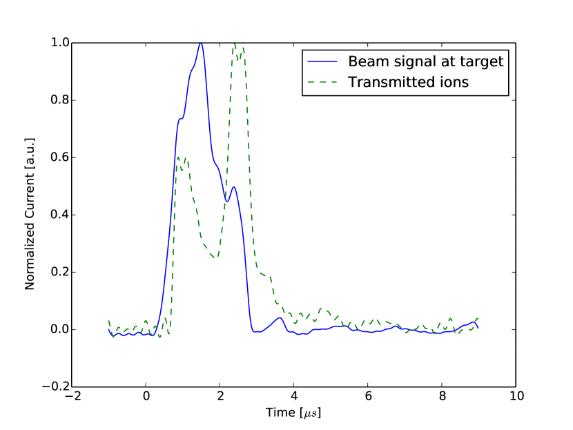
A beam current waveform for a transmitted potassium beam through a 250 nm thick silicon membrane is shown in Figure 3. We can integrate the measured current and plot the transmitted charge versus the rotation angle of the target membrane. The result is shown in Figure 4. Channeling peaks can be clearly seen around 0, 18 and 45 degree, corresponding to the 100, 310 and 110 channeling directions in the crystal.
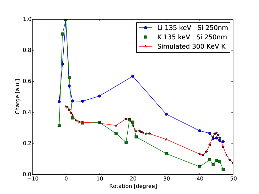
The general decrease in amplitude for the non-channeled current can be attributed to an increased thickness and a smaller effective area due to the rotation of the membrane.
Figure 4 also shows the results from a Crystal Trim simulation, see Posselt, (1994), for 300 keV K+ ions stopping in crystalline Si (with 3 nm layer of amorphous SiO2 to simulate a native oxide). The simulation uses a fluence and a beam divergence of 1 degree. The fraction of transmitted ions in Crystal Trim was calculated by taking the fraction of ions with a range larger than the membrane thickness of 250 nm compared to the total number of ions that get implanted at all depths. This will underestimate contrast from the channeling effect in the transmitted ion signal, since our experiment is set up to only accept ions with a small scattering angle. For Figure 4 the simulated data was scaled to fit the 10 degree measured data point for potassium. Furthermore a cosine dependence of the exposed area under rotation was taken into account. The simulation confirms the measured channeling peaks. Further simulations also show that we can expect changes in the time resolved transmission for potassium fluences exceeding .
As demonstrated in Guo et al., (2014) for lithium ions, the fluence is currently not high enough to observe effects from damage build-up due to overlapping cascades. Potassium ions create more damage due to their increased mass, but no effect could be observed so far which is in agreement with the simulations results for the needed fluences. We will be able to deliver the required higher fluences to samples once the additional focusing magnet is installed. With faster diagnostics, e.g. employing streak techniques, we then aim at tracking the evolution of the channeled ion fraction on a time scale.
IV Conclusion
We have shown that channeling experiments using thin membranes can be realized with intense, pulsed ion beams from NDCX-II by monitoring transmitted ion currents and that the concept of detecting defects dynamics by monitoring channeled ions is viable. However, with the current beam current, spot size, and pulse width no effect from damage build-up could be observed. We attribute this to the fact that we currently do not have high enough fluences to achieve overlapping damage cascades during the ion bombardment.
The ongoing upgrade of the accelerator aims at achieving smaller beam spot sizes (x5 in radius), shorter pulses (x20-30) and higher ion kinetic energies, up to 1.2 MeV. Thus we can expect an increase in fluence of a factor of x400. We expect to see effects of damage build-up at short timescales at these fluences as indicated by Crystal Trim simulations and further drive thin foils to temperatures approaching 1 eV.
Furthermore, we started to integrate new detection capabilities such as optical detection of defect recombination (ionoluminescence) using fast photodetectors and spectrometers coupled with a streak camera. This will allow us to resolve optical changes with a time resolution of picoseconds. Ion scattering using the channeling effect will also be implemented in backscattering geometry. This will allow us to probe a broader range of targets and remove the limitation to thin, single crystal membranes, which are currently needed for the channeling experiments and enable us to probe a much broader range of materials, such as ceramics for nuclear energy applications or materials for future fusion reactors, such as tungsten alloys.
Acknowledgements
This work was supported by the Office of Science of the U.S. DOE and by the LDRD Program at Lawrence Berkeley National Laboratory under contract no. DE-AC02-05CH11231. AM was supported by the Center for Defect Physics, an Energy Frontier Research Center funded by the U.S. DOE, Office of Science, Basic Energy Sciences.
References
- Bai et al., (2010) Bai, X. M., Voter, A. F., Hoagland, R. G., Nastasi, M., and Uberuaga, B. P. (2010). Efficient annealing of radiation damage near grain boundaries via interstitial emission. Science, 327:1631–1634.
- Diaz de la Rubia et al., (2000) Diaz de la Rubia, T., Zbib, H. M., Khraishi, T. A., Wirth, B. D., Victoria, M., and Caturla, M. J. (2000). Multiscale modeling of plastic flow localization in irradiated materials. Nature, 406:871–874.
- Friedman et al., (2009) Friedman, A., Barnard, J. J., Cohen, R. H. adn Grote, D. P., Lund, S. M., Sharp, W. M., Faltens, A., Henestroza, E., Jung, J.-Y., Kwan, J. W., Lee, E. P., Leitner, M. A., Logan, B. G., Vay, J.-L., Waldron, W. L., Davidson, R. C., Dorf, M., Gilson, E. P., and Kaganovich, I. D. (2009). Beam dynamics of the neutralized drift compression experiment-ii, a novel pulse-compressing ion accelerator. Physics of Plasmas, 17:056704.
- Guo et al., (2014) Guo, H., Persaud, A., Lidia, S., Minor, A. M., Hosemann, P., Seidl, P. A., and Schenkel, T. (2014). Dynamic investigation of defects induced by short, high current pulses of high energy lithium ions. MRS proceedings.
- Myers et al., (2012) Myers, M. T., Charnvanichborikarn, S., Shao, L., and Kucheyev, S. O. (2012). Pulsed ion beam measurement of the time constant of dynamic annealing in si. Physical Review Letters, 109:095502.
- Posselt, (1994) Posselt, M. (1994). Crystal-trim and its application to investigations on channeling effects during ion implantation. Radiation Effects and Defects in Solids, 130-131.
- Schenkel et al., (2013) Schenkel, T., Lidia, S. M., Weis, C. D., Waldron, W. L., Schwartz, J., Minor, A., Hosemann, P., and Kwan, J. W. (2013). Towards pump-probe experiments of defect dynamics with short ion beam pulses. Nucl. Inst. a Meth. In Phys. B, 315:350–355.
- Stuchbery and Bezakova, (1999) Stuchbery, A. E. and Bezakova, E. (1999). Thermal-spike lifetime from picosecond-duration preequilibrium effects in hyperfine magnetic fields following ion implantation. Physical Review Letters, 82:3637.
- Victoria et al., (2000) Victoria, M., Baluc, N., Bailat, C., Dai, Y., Luppo, M. I., Schäublin, R., and Singh, B. N. (2000). The microstructure and associated tensile properties of irradiated fcc and bcc metals. Journal of Nuclear Materials, 276:114–122.
- Waldron et al., (2014) Waldron, W. L., Abraham, W. J., Arbelaez, D., Friedman, A., Galvin, J. E., Gilson, E. P., Greenway, W. G., Grote, D. P., Jung, J.-Y., Kwan, J. W., Leitner, M., Lidia, S. M., Lipton, T. M., Reginato, L. L., Regis, M. J., Roy, P. K., Sharp, W. M., Stettler, M. W., Takakuwa, J. H., Volmering, J., and Vytla, V. K. (2014). The ndcx-ii engineering design. Nucl. Inst. a. Meth. In Phys. A, 733:226–232.
- Zinkle, (2013) Zinkle, S. J. (2013). Challenges in developing materials for fusion technology – past, present and future. Fusion Science and Technology, 64:65–75.
- Zinkle and Was, (2013) Zinkle, S. J. and Was, G. S. (2013). Materials challenges in nuclear energy. Acta Materialia, 61:735–758.