Pitfalls in the quantitative analysis of random walks and the mapping of single-molecule dynamics at the cellular scale.
Abstract
In recent years Bayesian Inference has become an efficient tool to analyse single molecule trajectories. Recently, high density single molecule tagging, Langevin Equation modelling and Bayesian Inference JBM_carte have been used to infer diffusion, force and potential fields at the full cell scale. In this short comment, we point out pitfalls holcman_arxiv ; Hoze to avoid in single molecule analysis in order to get unbiased results and reliable fields at various scales.
A response to Holcman et al holcman_arxiv
Understanding the physical and biological processes governing the trafficking of membrane proteins is an central question in cell biology and biophysics. A powerful approach to model the motion of a random walker in a complex environment is to use an overdamped Langevin equation. In the case of a coarse-grained description (corresponding to the limited spatial and temporal resolution accessible in experiments), this equation writes:
where corresponds to a local drift, is a spatially-varying friction, a spatially-varying diffusion coefficient and is a zero-averaged gaussian noise.
In past years, the advent of single molecule techniques has opened new possibilities to estimate the values of the local parameters and in the cellular context. In particular, bayesian inference tools have been developed to extract local force and diffusivity fields based on single molecule trajectories JBM1 ; JBM2 ; JBM3 ; JBM4 ; Voisinne ; Richly . Not only these tools have been extensively characterized from a methodological point of view JBM1 ; JBM2 ; Voisinne but they have also been applied to experimental systems, in order to investigate the dynamics of toxin receptors in lipid rafts JBM1 ; JBM3 ; JBM4 and to calibrate optical tweezers Richly .
More recently, the analysis of the force and diffusivity fields has been extended to the full cellular scale in Hoze and in JBM_carte thanks to high-density single molecule techniques, such as sptPALM Manley and uPAINT Gianonne . Thereby, it has been possible to quantitatively analyze the synaptic stabilization of AMPA receptors Hoze and Glycine receptors JBM_carte . Both papers similarly interpreted the role of synaptic scaffolds in terms of local confining potentials in line with a view of the neuronal membrane where neuroreceptors diffuse and transiently stabilize at synaptic sites Triller .
In their comment, Holcman et al. question the validity of the approach and description presented in JBM_carte . We reply below to the points that they raised by: (i) addressing some computational issues (statistical estimation of the model parameters, numerical simulations) and demonstrating biases in the statistical estimators used in Hoze , (ii) discussing the interpretation of the results.
Parameter estimation and numerical simulations.
A key point for the determination of the force and diffusivity fields based on single molecule data is the choice of the statistical estimators. To clearly illustrate the simplifying assumptions used in Hoze , we first remind the standard definition of the simplest likelihood used to analyze the dynamics of individual biomolecules in the absence of positional noise:
corresponding to translocations (during the sampling time ) in a subdomain characterised by a constant drift and diffusion coefficient . By differentiating with respect to and , one directly gets:
and
The maxima are obtained for:
and
The estimators used in Hoze correspond to the maxima in the limit and can thus be designated as Maximum Likelihood Estimators (MLE).
The estimators used in Hoze are biased when applied to experimentally measured trajectories for two main reasons. First, an important element in the analysis of single molecule trajectories is the positioning noise , which results from a combination of processes such as photon noise, motion blur, electronic noise and limited performance of peak fitting algorithm. However, the effect of positioning noise was entirely neglected in the analysis presented in Hoze , which, as shown below, leads to estimator biases and to improper parameter values for the synapses studied in Hoze . Second, the approximation (not discussed in Hoze ) also leads to strong bias in the force and diffusion estimation over range of values relevant for the experiments reported in Hoze . In our work, we have used Bayesian inference schemes and directly included in our computation the full positioning noise as an experimental parameter (see eq. equations 5 and 6 in JBM_carte ). We refer to Voisinne for a demonstration of the optimality of Bayesian inference methods for diffusion measurements and to JBM3 for a detailed discussion on their use in the case of confined motions.
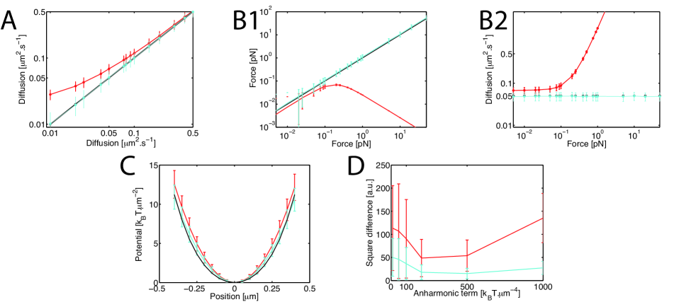
To directly compare the performances of the estimators used in Hoze and JBM_carte , we have performed numerical simulations corresponding to experimental conditions reported in Hoze ( nm, ms, about 40 points per subdomain). We first estimated the diffusion coefficient in the absence of external drift for varying between 0.01 and 0.5 m2/s (Figure 1A). For m2/s (a value comparable to the diffusion coefficient in synaptic domains), we observe a clear bias when using the MLE estimators whereas our estimator remains accurate over the entire range. The limit corresponds to the regime where is comparable or greater than .
Next, we tested the estimation of the diffusion coefficient and drift when brownian particles (with a diffusion coefficient m2/s) were submitted to a force varying between and pN. While our approach yielded accurate estimates of and , there was a systematic bias in the MLE estimation of both the force and the diffusion coefficient at low force. Furthermore, a strong deviation can be observed for forces larger than pN (Figure 1.B1,B2), a value corresponding to the the limit where the displacement due to the drift is comparable to the diffusional movement . Importantly, is comparable to the confinement force in a potential well with depth = 8 kBT and extension = 200 nm, such as the ones reported in Hoze . Overall, our simulations indicate that the estimates of the parameters (diffusion, forces and potential) for the synaptic domains are not accurate in Hoze
Note that Bayesian inference techniques have been recently applied to evaluate the experimental trapping force in optical tweezers Richly . This system is advantageous since the trap parameters can be finely tuned with the light intensity and, importantly, independently calibrated. The estimate of the trap spring constant determined with inference schemes was in good agreement with (in fact more accurate than) the results obtained by standard tools (equipartition and power-spectrum methods).
Another important point is the extraction of the potential from the force field. In Hoze , this is done by minimizing the square difference between the gradient of the potential and the force field. Such class of optimization problems, especially with a discrete set, limited number of points and harmonic approximation, are always solved with a regularization term machine_learning ; machine_learning_2 (see equation 7 in JBM_carte ). Solving these problems without any regularization, either of the force field or of the potential, leads to biases (as illustrated in Figure 1C,D).
Finally, regarding the numerical simulations (including the Gillespie scheme), we refer to FP for approximating the Fokker-Planck by the master equation and to Gillespie for simulating the master equation. We especially emphasize the importance of the Gillespie scheme for rapidly generating a large number of trajectories, which enabled us to compute fundamental descriptors of the motion (such as the propagator) as function of the time and over a large range of distance.
Interpretation and discussion of the model.
A legitimate topic of discussion is the interpretation of the diffusivity and force (or potential) fields estimated from individual trajectories. In their experiments, Hoze et al noted the existence of domains where individual translocations pointed toward a single center point Hoze . Similarly to what has been reported earlier for toxin receptors JBM1 ; JBM3 , such behavior was described in terms of a trapping potential. For AMPA receptors, the potential wells were interpreted as resulting from interactions of the receptors with scaffolding proteins PSD-95 via stargazin molecules.
Before discussing and comparing our results JBM_carte , we emphasize that our inference approach does not require the computation of a diffusion and a potential but can be performed to estimate the parameters or as it was done in JBM1 ; JBM2 ; JBM3 ; JBM4 ; Richly ; Voisinne ; silvan . The inference scheme simply provides a description of the environment in terms of the diffusivity and force fields that have the highest probability. In the specific case of our experiments on glycine receptors JBM_carte , we have favored a description in terms of a potential for several reasons. First, the force field did not exhibit rotational terms, thus justifying the computation of a potential. Second, glycine receptors are known to interact with gephyrin scaffolding proteins at inhibitory synapses. The fact that the trapping areas of the membrane proteins perfectly co-localized with the location of gephyrin clusters strongly supported the notion that the clusters acted as trapping wells. The role of the scaffolds in the observed potentials was further demonstrated experiments using membrane proteins with mutant constructs of the -loop mediating the receptor-gephyrin interactions: for a mutant with weaker affinity, we measured shallower traps, and for proteins lacking the -loop entirely, there was a complete absence of potentials. In this context, the potential depth represents the energy needed for receptors to escape gephyrin clusters. It is worth noting that it is the classical definition of the potential in physics.
In JBM_carte , we have used a spatially varying friction , reflecting the heterogeneity of the plasma membrane at the full cell scale. Furthermore, we imposed the relationship . This hypothesis was supported by the fact that the transitions between adjacent domains (determined experimentally) satisfied detailed balanced condition, thus suggesting a local equilibrium. For more advanced discussions on the FD relationship for systems with spatially varying friction, we refer to recent references FD_1 ; FD_2 .
An important difference between the results of Hoze and JBM_carte lies in the quantitative measurements of the potential depths. For the wild-type beta-loop, we found an average energy of 3.4 kBT with less than 15 of the traps above 6 kBT (measured over 69 clusters). In contrast, 25 of the traps reported in Hoze were deeper than 8 kBT, indicating a much higher level of stabilization. While it could be due to the difference between the biological systems and their molecular constituents, it seems likely that the biased estimators (see discussion above) and the limited number of data points presented in Hoze (2, 5 and 10 wells depending on the experimental conditions) also contribute to the discrepancy. Additional experiments, combined with careful statistical analysis, are needed to clarify the difference between excitatory and inhibitory scaffolds.
In our view, the main remaining challenge is to quantitatively determine how the trapping energies at scaffolding clusters relate to the biochemical properties of their individual constituents. We emphasize that there is no general procedure to go from a diffusion and force fields to biochemical characteristics of complex molecular assemblies. It will depend on many geometrical and molecular properties of the assemblies, as well as on the spatio-temporal resolution of the measurements. Fortunately, nanoscopy imaging methods now provide powerful tools to analyze the composition and structure of scaffolding complexes Specht ; Hosy and relate them to their functional roles at synapses.
Conclusion.
In summary, we stand by the methods and the approach described in JBM_carte and earlier papers, and point out to errors in the statistical and computation methods used in Hoze ; holcman_arxiv . Note also that we did not comment here on other important elements for the mapping of the diffusivity and force (or potential fields), such as the meshing procedure and the mesh size which should be adjusted to the averaged translocation length (a point not discussed in Hoze ), the effect of strong variation between the number of points between neighboring mesh domains, the effect of confinement on the accuracy of parameters extraction, the effect of heterogeneous diffusion field on the convergence of the estimators and the projection of the potential on a proper functional basis. We refer the reader to previous publications JBM1 ; JBM2 ; JBM3 ; JBM4 ; Voisinne ; Richly ; JBM_carte ; silvan (including their supplementary materials) for further discussion on all these points.
∗ Corresponding authors: jbmasson@pasteur.fr, maxime.dahan@curie.fr, triller@biologie.ens.fr.
References
- (1) D. Holcman, N. Hoze, Z. Schuss, Analysis of single particle trajectories: when things go wrong arXiv:1502.00286
- (2) N. Hoze, D. Nair, E. Hosy, C. Sieben, S. Manley, A. Herrman, J.-B. Sibarita, D. Choquet, and D. Holcman. 2012. Heterogeneity of AMPA receptor trafficking and molecular interactions revealed by superresolution analysis of live cell imaging. Proc. Natl. Acad. Sci. USA. 109:17052-17057.
- (3) Masson, J. B., D. Casanova, … A. Alexandrou. 2009. Inferring maps of forces inside cell membrane microdomains. Phys. Rev. Lett. 102:048103.
- (4) Turkcan, S., A. Alexandrou, and J.-B. Masson. 2012. A Bayesian inference scheme to extract diffusivity and potential fields from confined single-molecule trajectories. Biophys. J. 102:2288-2298.
- (5) Turkcan S, Masson J-B,… A.Alexandrou. 2012. Observing the confinement potential of bacterial pore-forming toxin receptors inside rafts with non-blinking Eu3+-doped oxide nanoparticles. Biophys J. 102: 2299-2308.
- (6) Turkcan, S., M. U. Richly, ., J. B. Masson. 2013. Probing membrane protein interactions with their lipid raft environment using single-molecule tracking and Bayesian inference analysis. PLoS ONE. 8:e53073.
- (7) Richly M. U. ,Turkcan S. , … Perronet K. , and A. Alexandrou. 2013. Optics Express. Vol 21, No 25, p31578
- (8) Voisinne, G., A. Alexandrou, and J.-B. Masson. 2010. Quantifying biomolecule diffusivity using an optimal Bayesian method. Biophys. J. 98:596-605.
- (9) Turkcan S. and J.-B Masson . 2013. Bayesian Decision Tree for the Classification of the Mode of Motion in Single-Molecule Trajectories, PloS ONE 8(12), e82799
- (10) Masson J.-B. , P. Dionne, … M. Dahan. 2014. Mapping the Energy and Diffusion Landscapes of Membrane Proteins at the Cell Surface Using High-Density Single-Molecule Imaging and Bayesian Inference: Application to the Multiscale Dynamics of Glycine Receptors in the Neuronal Membrane. Biophys. J. 106:s74?83
- (11) A. Triller and D. Choquet (2008). New Concepts in Synaptic Biology Derived from Single-Molecule Imaging. Neuron 59, 358.
- (12) Manley S, Gillette JM, Patterson GH, Shroff H, Hess HF, Betzig E, Lippincott-Schwartz J., (2008). High-density mapping of single-molecule trajectories with photoactivated localization microscopy Nat. Methods 5(2):155-7.
- (13) Giannone G., Hosy E., Levet F., Constals A., Schulze K., Sobolevsky A. I., Rosconi M. P., Gouaux E., Tampe R., Choquet D. and Laurent Cognet. 2013. Dynamic Superresolution Imaging of Endogenous Proteins on Living Cells at Ultra-High Density, Biophysical Journal Vol 99, No 4, p1303-1310
- (14) H. Risken. 1996. The Fokker-Planck Equation, Methods of Solution and Applications, Springer
- (15) Gillespie, D. 1977. Exact stochastic simulation of coupled chemical reactions. J. Phys. Chem. 81:2340-2361.
- (16) C.M. Bishop. (2007). Pattern Recognition and Machine Learning (Information Science and Statistics). Springer
- (17) Y. Wang and A. G. Yagola. (2011).Optimization and Regularization for Computational Inverse Problems and Applications. Springer
- (18) Farago O. and N. Gronbech-Jensen. (2014). Fluctuation-Dissipation Relation for Systems with Spatially Varying Friction. J. Stat. Phys. 156 (6):1093-1110
- (19) Farago O. and N. Gronbech-Jensen. (2014). Langevin dynamics in inhomogeneous media: Re-examining the Ito-Stratonovich dilemma. Phys. Rev. E 89:013301
- (20) Specht CG, Izeddin I, Rodriguez PC, El Beheiry M, Rostaing P, Darzacq X, Dahan M, Triller A. (2013). Quantitative nanoscopy of inhibitory synapses: counting gephyrin molecules and receptor binding sites. Neuron 24;79(2):308-21
- (21) Nair D, Hosy E, Petersen JD, Constals A, Giannone G, Choquet D, Sibarita JB. (2014). Super-resolution imaging reveals that AMPA receptors inside synapses are dynamically organized in nanodomains regulated by PSD95 J. of Neuroscience 7;33(32):13204-24.