Fiber networks amplify active stress
Abstract
Large-scale force generation is essential for biological functions such as cell motility, embryonic development, and muscle contraction. In these processes, forces generated at the molecular level by motor proteins are transmitted by disordered fiber networks, resulting in large-scale active stresses. While these fiber networks are well characterized macroscopically, this stress generation by microscopic active units is not well understood. Here we theoretically study force transmission in these networks, and find that local active forces are rectified towards isotropic contraction and strongly amplified as fibers collectively buckle in the vicinity of the active units. This stress amplification is reinforced by the networks’ disordered nature, but saturates for high densities of active units. Our predictions are quantitatively consistent with experiments on reconstituted tissues and actomyosin networks, and shed light on the role of the network microstructure in shaping active stresses in cells and tissue.
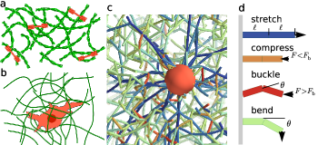
Living systems constantly convert biochemical energy into forces and motion. In cells, forces are largely generated internally by molecular motors acting on the cytoskeleton, a scaffold of protein fibers (Fig. 1a). Forces from multiple motors are propagated along this fiber network, driving numerous processes such as mitosis and cell motility Blanchoin et al. (2014); Fletcher and Mullins (2010), and allowing the cell as a whole to exert stresses on its surroundings. At the larger scale of connective tissue, many such stress-exerting cells act on another type of fiber network known as the extracellular matrix (Fig. 1b). This network propagates cellular forces to the scale of the whole tissue, powering processes such as wound healing Ehrlich (1988) and morphogenesis Heisenberg and Bellaïche (2013). Despite important differences in molecular details and length scales, a common physical principle thus governs stress generation in biological matter: internal forces from multiple localized “active units”—motors or cells—are propagated by a fiber network to generate large-scale stresses. However, a theoretical framework relating microscopic internal active forces to macroscopic stresses in these networks is lacking.
This generic stress generation problem is confounded by the interplay of network disorder and nonlinear elasticity. Active units generate forces at the scale of the network mesh size, and force transmission to larger scales thus sensitively depends on local network heterogeneities. In the special case of linear elastic networks, the macroscopic active stress is simply given by the density of active force dipoles, irrespective of network characteristics Ronceray and Lenz (2015). Importantly however, this relationship is not applicable to most biological systems, since typical active forces are amply sufficient to probe the nonlinear properties of their constitutive fibers, which stiffen under tension and buckle under compression Broedersz and MacKintosh (2014). Indeed, recent experiments on reconstituted biopolymer gels have shown that individual active units induce widespread buckling and stiffening Lam et al. (2011); Soares e Silva et al. (2011); Murrell and Gardel (2012), and theory suggests that such fiber nonlinearities can enhance the range of force propagation Shokef and Safran (2012); Notbohm et al. (2015).
Fiber networks also exhibit complex, nonlinear mechanical properties arising at larger scales, owing to collective deformations favored by the networks’ weak connectivity Broedersz and MacKintosh (2014); Onck et al. (2005); Heussinger et al. (2007); Conti and MacKintosh (2009). The role of connectivity in elasticity was famously investigated by Maxwell Maxwell (1864), who noticed that a spring network in dimension becomes mechanically unstable for connectivities . Interestingly, most biological fiber networks exhibit connectivities well below this threshold, and therefore cannot be stabilized solely by the longitudinal stretching rigidity of their fibers. Instead, their macroscopic mechanical properties are typically controlled by the fiber bending rigidity Broedersz et al. (2011). In contrast to stretching-dominated networks with connectivities above the Maxwell threshold, such weakly connected, bending-dominated networks are soft and extremely sensitive to mechanical perturbations Broedersz et al. (2011); Ulrich et al. (2013); Wyart et al. (2008); Sheinman et al. (2012). In these networks, stresses generated by active units propagate along intricate force chains Heussinger and Frey (2007); Head et al. (2005) whose effects on force transmission remain unexplored. Collections of such active units generate large stresses, with dramatic effects such as macroscopic network stiffening Koenderink et al. (2009); Jansen et al. (2013); Broedersz and MacKintosh (2011) and network remodelling Soares e Silva et al. (2011); Murrell and Gardel (2012); Bendix et al. (2008); Köhler et al. (2011); Carvalho et al. (2013).
Here we study the theoretical principles underlying stress generation by localized active units embedded in disordered fiber networks (Fig. 1c). We find that arbitrary local force distributions generically induce large isotropic, contractile stress fields at the network level, provided that the active forces are large enough to induce buckling in the network. In this case, the stress generated in a biopolymer network dramatically exceeds the stress level that would be produced in a linear elastic medium Ronceray and Lenz (2015); Ranft et al. (2010), implying a striking network-induced amplification of active stress. Our findings elucidate the origins and magnitude of stress amplification observed in experiments on reconstituted tissues Lam et al. (2011); Jen and Mclntire (1982) and actomyosin networks Koenderink et al. (2009); Carvalho et al. (2013); Lemière et al. (2015). We thus provide a new conceptual framework for stress generation in biological fiber networks.
A lattice model for elastic fiber networks
We investigate force transmission using a lattice-based fiber network model Broedersz et al. (2011); Das et al. (2007). In our model, straight fibers are connected at each lattice vertex by crosslinks that do not constrain their relative angles. Each lattice edge represents a “bond” made of two straight segments and can thus stretch, bend, or buckle (Fig. 1d). Segments have stretching rigidity and a rest length equal to one, implying a stretching energy per segment of length . The fiber bending rigidity is set to unity by penalizing angular deflections between consecutive segments through a bending energy . Consequently, individual bonds buckle under a critical force , and we consider nearly inextensible fibers by assuming (henceforth we use ).
Network disorder is introduced through bond depletion, i.e., by randomly decimating the lattice so that two neighboring vertices are connected by a bond with probability . This probability controls the network’s connectivity, giving rise to distinct elastic regimes delimited by two thresholds and . The network is stretching-dominated for , bending-dominated for , and mechanically unstable for . Here we consider 2D hexagonal lattices, for which and , and 3D FCC lattices with and . Since the network displays singular behavior in the vicinity of and , here we focus our investigations on the generic stretching- and bending-dominated regimes away from these critical points Broedersz et al. (2011).
We model active units as sets of forces exerted on network vertices with positions , and consider networks at mechanical equilibrium under the influence of these forces. We denote by the trace (i.e., the isotropic component) of the coarse-grained active stress induced in the fiber network by a density of such units.
The relationship between this active stress and local forces in homogeneous linear networks is very simple, and yields Ronceray and Lenz (2015)
| (1) |
where is the dipole moment of the forces associated with a single active unit. Equation (1) is generically violated in disordered or nonlinear networks, although it holds on average in linear networks with homogeneous disorder:
| (2) |
where denotes the average over disorder Ronceray and Lenz (2015). To quantify violations of Eq. (1), we define the far-field force dipole through the relation
| (3) |
Conceptually, this far-field dipole characterizes the apparent strength of an individual active unit renormalized by force transmission in the disordered, nonlinear network. It quantifies how contractile () or extensile () the active medium is, and the dipole amplification ratio (or equivalently the stress amplification ratio ) measures the deviation from linear homogeneous force transmission.
Contractility robustly emerges from large local forces
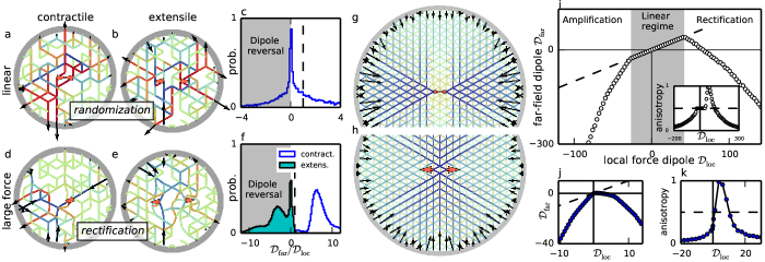
Stress generation by active units integrates mechanical contributions from a range of length scales. We first consider the immediate vicinity of the active unit. Network disorder plays a crucial role at that scale, since forces are transmitted through a random pattern of force lines determined by the specific configuration of depleted bonds (Fig. 2a-b). To understand how these patterns affect force transmission, we investigate the probability distribution of the far-field force dipole for simple active units consisting of two equal and opposite point forces of magnitude .
We first consider the linear regime , where the average dipole amplification equals unity: [see Eqs. (2-3)]. The fluctuations around this average are strikingly broad, as shown in Fig. 2c. For instance, a significant fraction () of all network geometries yield negative amplification, i.e., an effective extensility in response to a contractile dipole (Fig. 2b). Overall, the far-field response in the linear regime is only loosely correlated to the applied force dipole.
The situation is dramatically different in the large force regime (), where fibers buckle and induce nonlinear network response. This is illustrated by the distributions of dipole amplifications in two opposite cases: a large contractile and a large extensile force dipole (Fig. 2d-f). First, locally extensile dipoles predominantly undergo negative amplification, implying far-field contractility irrespective of the sign of (as in, e.g., Fig. 2e). Second, the randomization observed in the linear regime is strongly attenuated, and the sign of the amplification is very reproducible (positive for of the contractile dipoles and negative for of the extensile ones). Third, the magnitude of the average amplification is significantly larger than one (in Fig. 2f and for contractile and extensile dipoles, respectively).
To understand these three effects, we consider contractile and extensile dipoles in a simpler regular network (no bond depletion, Fig. 2g-h). Qualitatively, these uniform networks behave similarly to the randomly depleted ones described above: force dipole conservation holds for , while for dipoles are rectified towards contraction and their magnitude is amplified (Fig. 2i). The origin of these behaviors is apparent from the spatial arrangement of the forces in Figs. 2g-h. While contractile and extensile active units both induce compressive and tensile stresses in their immediate surroundings, the buckling of the individual bonds prevents the long-range propagation of the former. This results in enhanced tensile stresses in the far-field, and thus in strongly contractile far-field dipoles. In addition, this nonlinear response renders the far-field stresses uniformly tensile, and therefore more isotropic than the active unit forces. We quantify this effect in the inset of Fig 2i using an anisotropy parameter for the far-field stresses, which indeed becomes very small for both positive or negative large local dipoles.
Moving to a systematic quantification of force transmission in depleted, bending-dominated networks, we show in Fig 2j-k the same three effects of rectification, amplification and isotropization, which set in at smaller forces than in regular networks. Overall, these effects are very general and hold in both bending- and stretching-dominated depleted networks, in two and three dimensions, and for active units with complicated force distributions (see supporting Figs. S3 and S4). Thus, beyond the immediate neighborhood of the active force-generating unit, strong isotropic contractile stresses emerge in the system from a generic local force distribution due to the nonlinear force propagation properties of the fiber network.
A model for active units as isotropic pullers
While nonlinear force transmission over large length scales involves large active forces, the model for active units used above can only exert moderate dipoles in soft, weakly connected networks. Indeed, for large enough contractile dipoles the two vertices on which the forces are applied collapse to a point (Fig. 2d), preventing further contraction. In contrast, molecular motors and contractile cells continuously pull fibers in without collapsing. To reflect this, we introduce an active unit capable of exerting arbitrarily large forces without changing its size. The unit is centered on a vertex , and pulls on any vertex within a distance with a radial force
| (4) |
where is the maximum force exerted by the unit on a vertex, is the distance between and and is the associated unit vector. A strong active unit in a soft network may pull in many fibers, exerting a force on each of them. Adding the contributions of all these fibers results in a large local dipole, the magnitude of which is not well reflected by the value of . The influence of the active unit on the surrounding network is better characterized by the force , which we define as the average force per unit area exerted on the surrounding network by the active unit at its outer surface (). Finally, we assign an isotropic force distribution to the active puller defined in Eq. (4). This choice is justified by the observation that anisotropic force distributions are rectified towards isotropy by the network (Figs. 2i, k).
Contractile forces are long-ranged in bucklable media
We now study force propagation beyond the immediate vicinity of an active unit (Fig. 3) using the above-described isotropic puller. Simple theoretical arguments dictate two asymptotic regimes for this propagation. Close to the active unit, forces are large and fiber buckling affects force transmission, while beyond a crossover distance forces are weak and linear elasticity prevails.
To describe the near-field regime, we note that fiber buckling prevents the network from sustaining compressive stresses above the buckling threshold. Close to the active unit, the network is thus effectively equivalent to a network of floppy ropes. The active unit pulls on these ropes, and thus becomes the center of a radial arrangement of tensed ropes. Force balance on a small portion of a spherical shell centered on the active unit imposes that radial stresses in this rope-like medium decay as
| (5) |
where is the distance from the active unit and the dimension of space Rosakis et al. (2014). In the far field, stresses are small and buckling does not occur, implying that force transmission crosses over from rope-like to linear elasticity:
| (6) |
Stress decay is thus significantly slower in the rope-like near-field than in the linear far-field, leading to an increased range for force transmission Notbohm et al. (2015). Conceptually, the faster decay in a linear elastic medium can again be understood by balancing forces on a fraction of spherical shell centered on the active unit, where radial stresses are now partially compensated by orthoradial stresses. We expect that the crossover between these two regimes occurs when radial stresses are comparable to the buckling stress, implying that the crossover length depends on the active force:
| (7) |
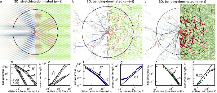
To test this two-regime scenario, we simulate force propagation away from a single active unit in both stretching- and bending-dominated networks in two and three dimensions. In all cases, rope-like radial stresses and bond buckling are predominant in the vicinity of the active unit (Fig. 3a-c). Monitoring the decay of radial stresses with , we find an apparent crossover from rope-like to linear behavior, consistent with Eqs. (5) and (6) (Fig. 3d, f, h).
Visually, the crossover length coincides with the outer boundary of the radially tensed, buckling-rich region (Fig. 3a-c, black circles). In stretching-dominated networks, our prediction of Eq. (7) captures the force dependence of this crossover length (Fig. 3e and S5). In contrast, bending-dominated networks display a more complex behavior: while the system still exhibits a transition from rope-like to linear force transmission, the crossover region is much broader (Fig. 3f, h) and forces propagate along heterogeneous patterns reminiscent of previously reported force chains (Fig. 3b-c) Heussinger and Frey (2007); Dasanayake et al. (2011). This strong concentration of the tensile stresses allows rope-like force transmission to extend much further than predicted by Eq. (7). Instead, we find behavior that is reasonably well described by a power law with anomalous exponents in 2D and in 3D (Fig. 3g, i). These exponents appear to be insensitive to the exact value of the depletion parameter within the bending-dominated regime (Supporting Fig. S5). The difference between stretching- and bending-dominated exponents suggests elastic heterogeneities qualitatively affect force transmission in such soft networks. As a result, contractile forces large enough to induce buckling benefit from an enhanced range of transmission, characterized by the mesoscopic radius of the rope-like region .
Amplification by a collection of active units
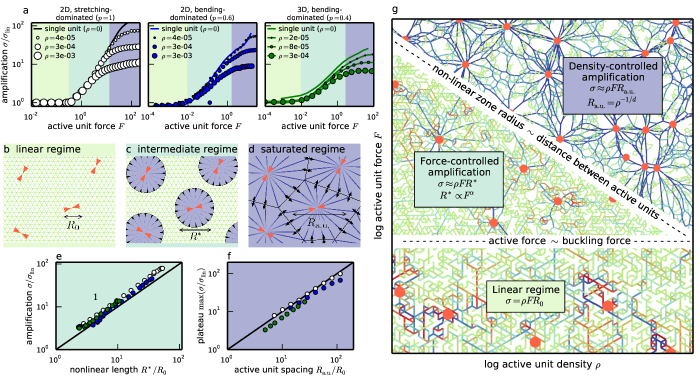
Over large length scales, active stresses in biological systems are generated by multiple active units. We thus compute the stress amplification ratio in the presence of a finite density of randomly positioned active units in 2D and 3D for various densities and depletion parameters (Fig. 4a). In all cases we observe three stress amplification regimes as a function of the unit force : a low-force plateau without amplification, an intermediate regime of increasing amplification and a saturation of the amplification at a level that depends on .
In the low-force regime, linear force transmission prevails (Fig. 4b) and the active stress is given by Eq. (1):
| (8) |
For moderate forces, the fibers in the network buckle in the vicinity of each active unit, up to a distance . Individual units are thus typically surrounded by nonoverlapping nonlinear regions of size , as illustrated in Fig. 4c. To predict the resulting active stress in the system, we model each nonlinear region as an effective active unit of size and force dipole , where we used Eq. (5) to describe force propagation within the nonlinear region. As the effective units are themselves embedded in a linear medium, linear force transmission [Eq. (1)] outside of these units implies
| (9) |
We thus predict that stress amplification in this regime scales as . We confirm this prediction in Fig. 4e. Since increases with the active unit force in this regime, the large-scale stress amplification increases with as previously observed in Fig. 4a.
For large forces, the radius of the rope-like regions becomes so large as to exceed the typical distance between adjacent active units . This causes the nonlinear regions associated to neighboring active units to overlap, and renders the whole network mechanically equivalent to a collection of tensed, inextensible ropes whose geometry does not change significantly upon further increase of the force. To estimate the resulting network stress, we approximate the system as a mosaic of effective active units of size each with a force dipole (Fig. 4d). This yields
| (10) |
The resulting prediction for the stress amplification, , is confirmed in Fig. 4f. Strikingly, the stress generated in this large-force regime has a nonlinear dependence on , again consistent with Fig. 4a. Indeed, the addition or removal of active units leads to large rearrangements of the rope network, resulting in significant local modifications of force transmission.
We summarize the physics of collective stress-generation by many active units in a phase diagram (Fig. 4g). In each regime, the magnitude of an active unit’s effective force dipole is directly proportional to one of the three length scales , and [Eqs. (8-10)]. While we have shown that depends on the dimensionality and connectivity of the network, the other two length scales are purely geometrical. An important consequence of these findings is that the active stress generated in the associated regimes is essentially independent of the detailed properties of the fiber network.
Discussion
In living organisms, microscopic units exert active forces that are transmitted by fibrous networks to generate large-scale stresses. The challenge in analyzing this force-transmission problem stems from the disordered architecture of such fibrous networks and the nonlinearities associated with the strong forces exerted by biological active units. Despite this complexity, we find surprisingly simple and robust behaviors: in response to any distribution of active forces, dramatically amplified contractile stresses emerge in the network on large scales. This remarkable property hinges only on the local asymmetry in elastic response between tensed and compressed fibers, and is enhanced by network disorder. Our simple, yet realistic description of individual fibers yields a universal scenario for force transmission: long-ranged, rope-like propagation near a strong active unit, and linear transmission in the far-field. This generic result should be contrasted with recent studies focused on fibers with special singular force-extension relation Notbohm et al. (2015); Rosakis et al. (2014) and resulting in non-universal force transmission regimes.
| System | |||||||
|---|---|---|---|---|---|---|---|
| I | 3D actomyosin Koenderink et al. (2009) | (linear regime) | |||||
| II | 2D actomyosin Carvalho et al. (2013); Lemière et al. (2015) | (force-controlled) | |||||
| III | 3D blood clot Lam et al. (2011); Jen and Mclntire (1982) | (density-controlled) |
Our generic phase diagram (Fig 4g) recapitulates our quantitative understanding of stress generation by a collection of active units based on the interplay between three length scales: active unit size , rope-like length , and typical distance between units . To validate these predictions, we compare them with existing measurements on a broad range of in vitro systems (Table 1). We first consider system I, a dense three-dimensional actin network with mesh size in the presence of myosin motors, which assemble into so-called myosin thick filaments. A thick filament—which we consider as an individual active unit—exerts a typical force , much smaller than the buckling threshold associated with a single -bond. This implies an active stress identical to the linear prediction, as confirmed by the experimental result Koenderink et al. (2009). We next consider system II, a two-dimensional actin network bound to the outer surface of a lipid vesicle. The active units are essentially the same as in System I, but are much more sparsely distributed (). The network in system II is also much looser () than in system I, resulting in a much smaller bond buckling force. The combination of a low buckling threshold and a large spacing between active units leads us to predict a significant stress amplification associated to the force-controlled regime (Fig 4c, g), in reasonable agreement with experiments Carvalho et al. (2013); Lemière et al. (2015). Finally, we consider a clot comprised of fibrin filaments and contractile platelets as active units (system III). The large forces exerted by platelets allow for long-range nonlinear effects, placing this in vitro system deep in the density-controlled regime (Figs. 4d, g). Consequently, we expect stress amplification to be controlled by the distance between active units, irrespective of the large value of the active force . We thus predict an amplification factor , in good agreement with experimental data Lam et al. (2011); Jen and Mclntire (1982). These three examples demonstrate our theory’s ability to quantitatively account for stress amplification, and recent progress in the micromechanical characterization of active fiber networks opens promising perspectives for further exploring active stress amplification Lam et al. (2011); Murrell and Gardel (2012); Carvalho et al. (2013).
Far from merely transmitting active forces, we show that fiber networks dramatically alter force propagation as contractility emerges from arbitrary spatial distributions of local active forces. This could imply that living organisms do not have to fine-tune the detailed geometry of their active units, since any local force distribution yields essentially the same effects on large length scales. This emergence of contractility sheds a new light on the longstanding debate in cytoskeletal mechanics regarding the emergence of macroscopic contraction in non-muscle actomyosin despite the absence of an intrinsic contractility of individual myosin motors Dasanayake et al. (2011); Hatano (1994); Sekimoto and Nakazawa (1998); Lenz et al. (2012a); Mizuno et al. (2007); Murrell et al. (2015). Indeed, while these motors exert equal amounts of local pushing and pulling forces Lenz et al. (2012b); Lenz (2014), our result suggests that the surrounding network rectifies pushing contributions into uniform contraction. More broadly, we suggest that this strong propensity for the emergence of contraction in fibrous materials can explain the overwhelming dominance of contractile stresses in active biological materials up to the tissue level. Clearly, this does not mean that it is impossible to generate large-scale expansion in living organisms as required for limb abduction and extension or for lung inflation. Nevertheless, in each of these examples the expansion actually results from the clever harnessing of muscle contraction through lever structures involving our skeleton.
Our results suggest a novel design principle for active fiber networks geared to maximize stress-generation. In a linear medium, the stress generated does not depend on the spatial distribution of active units. In contrast, we predict that in fiber networks, larger stresses can be obtained by clustering the active units. Such regrouping of a set number of force generators to enhance stress amplification could play a role in smooth muscle, where the number of myosins in individual thick filament is regulated dynamically Seow (2005). Similarly, at the tissue level, clustering of contractile cells occurs during wound repair da Rocha-Azevedo and Grinnell (2013).
Our findings connect widely used “active gels” phenomenological theories Prost et al. (2015) to their underlying molecular foundation, a crucial step in bringing theory and experiments together in the study of active biological matter, and calls for further progress in characterizing force transmission in more complex fiber networks. Finally, beyond biopolymer networks our work opens avenues to understand force transmission in novel metamaterials whose macroscopic properties crucially hinge on their microscopic buckling Kang et al. (2013); Florijn et al. (2014).
Acknowledgements.
We thank Cécile Sykes and Guy Atlan for fruitful discussions. This work was supported by grants from Université Paris-Sud and CNRS, the University of Chicago FACCTS program, Marie Curie Integration Grant PCIG12-GA-2012-334053 and “Investissements d’Avenir” LabEx PALM (ANR-10-LABX-0039-PALM) to ML as well as the German Excellence Initiative via the program ‘NanoSystems Initiative Munich’ (NIM) and the Deutsche Forschungsgemeinschaft (DFG) via project B12 within the SFB 1032. PR is supported by “Initiative Doctorale Interdisciplinaire 2013” from IDEX Paris-Saclay, and CPB is supported by a Lewis-Sigler fellowship. ML’s group belongs to the CNRS consortium CellTiss. Figures realized with Matplotlib Hunter (2007) and Mayavi2 Ramachandran and Varoquaux (2011).References
- Blanchoin et al. (2014) L. Blanchoin, R. Boujemaa-Paterski, C. Sykes, and J. Plastino, Physiol. Rev. 94, 235 (2014).
- Fletcher and Mullins (2010) D. A. Fletcher and R. D. Mullins, Nature 463, 485 (2010).
- Ehrlich (1988) H. P. Ehrlich, Eye 2, 149 (1988).
- Heisenberg and Bellaïche (2013) C.-P. Heisenberg and Y. Bellaïche, Cell 153, 948 (2013).
- Ronceray and Lenz (2015) P. Ronceray and M. Lenz, Soft Matter 11, 1597 (2015).
- Broedersz and MacKintosh (2014) C. P. Broedersz and F. C. MacKintosh, Rev. Mod. Phys. 86, 995 (2014).
- Lam et al. (2011) W. A. Lam, O. Chaudhuri, A. Crow, K. D. Webster, T.-D. Li, A. Kita, J. Huang, and D. A. Fletcher, Nat. Mater. 10, 61 (2011).
- Soares e Silva et al. (2011) M. Soares e Silva, M. Depken, B. Stuhrmann, M. Korsten, F. C. Mackintosh, and G. H. Koenderink, Proc. Natl. Acad. Sci. U.S.A. 108, 9408 (2011).
- Murrell and Gardel (2012) M. Murrell and M. L. Gardel, Proc. Natl. Acad. Sci. U.S.A. 109, 20820 (2012).
- Shokef and Safran (2012) Y. Shokef and S. A. Safran, Phys. Rev. Lett. 108, 178103 (2012).
- Notbohm et al. (2015) J. Notbohm, A. Lesman, P. Rosakis, D. A. Tirrell, and G. Ravichandran, Interface 12, 20150320 (2015).
- Onck et al. (2005) P. R. Onck, T. Koeman, T. van Dillen, and E. van der Giessen, Phys. Rev. Lett. 95, 178102 (2005).
- Heussinger et al. (2007) C. Heussinger, B. Schaefer, and E. Frey, Phys. Rev. E 76, 031906 (2007).
- Conti and MacKintosh (2009) E. Conti and F. C. MacKintosh, Phys. Rev. Lett. 102, 088102 (2009).
- Maxwell (1864) J. C. Maxwell, Philos. Mag. 27, 294 (1864).
- Broedersz et al. (2011) C. P. Broedersz, X. Mao, T. C. Lubensky, and F. C. MacKintosh, Nat. Phys. 7, 983 (2011).
- Ulrich et al. (2013) S. Ulrich, N. Upadhyaya, B. van Opheusden, and V. Vitelli, Proc. Natl. Acad. Sci. U.S.A. 110, 20929 (2013).
- Wyart et al. (2008) M. Wyart, H. Liang, A. Kabla, and L. Mahadevan, Phys. Rev. Lett. 101, 215501 (2008).
- Sheinman et al. (2012) M. Sheinman, C. P. Broedersz, and F. C. MacKintosh, Phys. Rev. Lett. 109, 238101 (2012).
- Heussinger and Frey (2007) C. Heussinger and E. Frey, Eur. Phys. J. E 24, 47 (2007).
- Head et al. (2005) D. A. Head, A. J. Levine, and F. C. MacKintosh, Phys. Rev. E 72, 061914 (2005).
- Koenderink et al. (2009) G. H. Koenderink, Z. Dogic, F. Nakamura, P. M. Bendix, F. C. MacKintosh, J. H. Hartwig, T. P. Stossel, and D. A. Weitz, Proc. Natl. Acad. Sci. U.S.A. 106, 15192 (2009).
- Jansen et al. (2013) K. A. Jansen, R. G. Bacabac, I. K. Piechocka, and G. H. Koenderink, Biophys. J. 105, 2240 (2013).
- Broedersz and MacKintosh (2011) C. P. Broedersz and F. C. MacKintosh, Soft Matter 7, 3186 (2011).
- Bendix et al. (2008) P. M. Bendix, G. H. Koenderink, D. Cuvelier, Z. Dogic, B. N. Koeleman, W. M. Brieher, C. M. Field, L. Mahadevan, and D. A. Weitz, Biophys. J. 94, 3126 (2008).
- Köhler et al. (2011) S. Köhler, V. Schaller, and A. R. Bausch, Nat. Mater. 10, 462 (2011).
- Carvalho et al. (2013) K. Carvalho, F.-C. Tsai, E. Lees, R. Voituriez, G. H. Koenderink, and C. Sykes, Proc. Natl. Acad. Sci. U.S.A. 110, 16456 (2013).
- Ranft et al. (2010) J. Ranft, M. Basan, J. Elgeti, J.-F. Joanny, J. Prost, and F. Jülicher, Proc. Natl. Acad. Sci. U.S.A. 107, 20863 (2010).
- Jen and Mclntire (1982) C. J. Jen and L. V. Mclntire, Cell Motil. 2, 445 (1982).
- Lemière et al. (2015) J. Lemière, M. Bussonnier, T. Betz, C. Sykes, and K. Carvalho, “Cell-sized liposome doublets reveal active cortical tension build up,” (2015), to appear.
- Das et al. (2007) M. Das, F. C. MacKintosh, and A. J. Levine, Phys. Rev. Lett. 99, 038101 (2007).
- Rosakis et al. (2014) P. Rosakis, J. Notbohm, and G. Ravichandran, arXiv , 1412.2612 (2014).
- Dasanayake et al. (2011) N. L. Dasanayake, P. J. Michalski, and A. E. Carlsson, Phys. Rev. Lett. 107, 118101 (2011).
- Hatano (1994) S. Hatano, Int. Rev. Cytology 156, 199 (1994).
- Sekimoto and Nakazawa (1998) K. Sekimoto and H. Nakazawa, “Current topics in physics,” (World Scientific, Singapore, 1998) pp. 394–405.
- Lenz et al. (2012a) M. Lenz, T. Thoresen, M. L. Gardel, and A. R. Dinner, Phys. Rev. Lett. 108, 238107 (2012a).
- Mizuno et al. (2007) D. Mizuno, C. Tardin, C. F. Schmidt, and F. C. Mackintosh, Science 315, 370 (2007).
- Murrell et al. (2015) M. Murrell, P. W. Oakes, M. Lenz, and M. L. Gardel, Nat. Rev. Mol. Cell Biol. (2015), 10.1038/nrm4012, advance online article doi:10.1038/nrm4012.
- Lenz et al. (2012b) M. Lenz, M. L. Gardel, and A. R. Dinner, New J. Phys. 14, 033037 (2012b).
- Lenz (2014) M. Lenz, Phys. Rev. X 4, 041002 (2014).
- Seow (2005) C. Y. Seow, Am. J. Physiol.-Cell Physiol. 289, C1363 (2005).
- da Rocha-Azevedo and Grinnell (2013) B. da Rocha-Azevedo and F. Grinnell, Exp. Cell Res. 319, 2440 (2013).
- Prost et al. (2015) J. Prost, F. Jülicher, and J.-F. Joanny, Nat. Phys. 11, 111 (2015).
- Kang et al. (2013) S. H. Kang, S. Shan, W. L. Noorduin, M. Khan, J. Aizenberg, and K. Bertoldi, Adv. Mater. 25, 3380 (2013).
- Florijn et al. (2014) B. Florijn, C. Coulais, and M. van Hecke, Phys. Rev. Lett. 113, 175503 (2014).
- Hunter (2007) J. D. Hunter, Comput. Sci. Eng. 9, 90 (2007).
- Ramachandran and Varoquaux (2011) P. Ramachandran and G. Varoquaux, Comput. Sci. Eng. 13, 40 (2011).
- Jülicher et al. (2007) F. Jülicher, K. Kruse, J. Prost, and J.-F. Joanny, Phys. Rep.-Rev. Sec. Phys. Lett. 449, 3 (2007).
- Joanny and Prost (2009) J.-F. Joanny and J. Prost, HFSP J. 3, 94 (2009).
- Rosenfeld et al. (2003) S. S. Rosenfeld, J. Xing, L.-Q. Chen, and H. L. Sweeney, J. Biol. Chem. 278, 27449 (2003).
- Storm et al. (2005) C. Storm, J. J. Pastore, F. C. MacKintosh, T. C. Lubensky, and P. A. Janmey, Nature 435, 191 (2005).