Biomechanical conditions of walking
Abstract
The development of rehabilitation training program for lower limb injury does not usually include gait pattern design. This paper introduced a gait pattern design by using equations (conditions of walking). Following the requirements of reducing force to the injured side to avoid further injury, we developed a lower limb gait pattern to shorten the stride length so as to reduce walking speed, to delay the stance phase of the uninjured side and to reduce step length of the uninjured side. This gait pattern was then verified by the practice of a rehabilitation training of an Achilles tendon rupture patient, whose two-year rehabilitation training (with 24 tests) has proven that this pattern worked as intended. This indicates that rehabilitation training program for lower limb injury can rest on biomechanical conditions of walking based on experimental evidence.
pacs:
87.85.Pq, 42.30.Wb.I Introduction
Gait patterns are described by gait parameters such as speed, step length and cadence 1 . Though gait parameters of each individual vary 2 ; 3 , healthy people s walking features of symmetry and economy are consistent 4 ; 5 . The inverted pendulum model typically simulates these features, and gait pattern equations were established 6 ; 7 ; 8 . Gait patterns vary when their body dysfunctions 9 . When walking is prescribed as part of rehabilitation training program 10 , it is necessary to design gait patterns to avoid further injury 11 . But the designs of gait pattern are mostly concerned with walking speed control at early rehabilitation period 12 .
Lower limb joints of ankle, knee and hip get injured easily and frequently. Training exercise is effective for such injury rehabilitation. For example, at early stage of Achilles tendon rupture (ATR) rehabilitation, controlled tensional loading to the end of tendon rupture can improve the biomechanical properties of scar tissue, reduce adhesions, stimulate fibroblast collagen fibers, and improve mechanical properties after ATR recovery. Early training can yield positive results 13 ; 14 . But not many gait patterns have been developed and included in early rehabilitation period, leading to a lack of agreed effective methods 13 ; 15 .
When injury happens at one side, that side is called the injured side and the other uninjured side. Basic early rehabilitation training requirements have been agreed: to reduce force to the injured side to avoid further injury. During walking stance phase, forces between lower limb and support surface interact. To the case of one side injury, we hypothesized that when walking, the peak values of vertical ground reaction force (VGRF) of the injured side could be reduced to be close to the weight by changing gait patterns. Gait patterns were simulated by equations. The results demonstrated that these patterns were effective. One ATR patient adopted our gait pattern. In the following two years, we took 24 tests. The results verified our hypothesis.
II Gait pattern design
II.1 Vertical ground reaction force
was set as VGRF of the injured side, as that of the uninjured side, as weight, as stride time. For walking, these quantities should meet conditions from the following equation:
| (1) |
When people walk, the VGRF forms a double-hump pattern, and the hump values exceed the weight. To meet the conditions of reducing force to the injured side and avoiding further injury, Eq. (1) presented two methods to reduce peak values of VGRF of the injured side: to increase , or to delay the stance phase of the uninjured side, because (, , where was stance phase, and swing phase). Under these conditions, inverse kinematics 16 ; 17 was used to calculate acceleration, speed and displacement of the center of mass (COM). See Fig. 1 for calculation results.
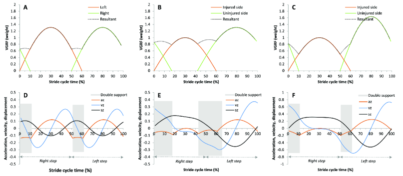 |
Fig. 1 compared these two methods to increase VGRF or to delay the stance phase of the uninjured side. We found that in a stride cycle, the speed of COM varied C the difference between the maximal and minimal value was 113.13% (, subscript 1 referring to a method to increase of the uninjured side, subscript 2 to a method to delay the stance phase of the uninjured side, and to speed of COM); the difference between the maximal and minimal value of the displacement of COM was 93.12% (, subscript 1 referring to a method to increase of the uninjured side, subscript 2 to a method to delay the stance phase of the uninjured side, and to displacement of COM). As we know, when walking, the greater the changes of speed and displacement in vertical direction, the less stable the walking, and the more risk to the injured side. A more desirable gait pattern for the injured lower limb should be: to delay the stance phase of the uninjured side.
II.2 Stride time
was set as the first double support of the uninjured side, the second double support of the uninjured side, swing phase of the injured side, i.e. single support of the uninjured side, swing phase of the uninjured side, i.e. single support of the injured side, stride time of the injured side, and stride time of the uninjured side. For walking training, these quantities should meet conditions from the following equation:
| (2) |
Eq. (2) presented two methods to increase the stance phase of the uninjured side: to only increase and ; or to increase and , and to reduce . If we only increased and , it might increase , which might bring pitfall to the injured side. Therefore, gait pattern for the injured lower limb should be: to increase and , and at the same time to reduce to increase stance phase of the uninjured side, and to reduce single support of the uninjured side.
II.3 Stride length
was set as step length of the uninjured side (the length between the heels from the injured side’s heel strike to the uninjured side’s heel strike on the same side), as step length of the injured side (the length between the heels from the uninjured side’s heel strike to the injured side’s heel strike on the same side), as stride length of the uninjured side, and as stride length of the injured side. For walking training, these quantities should meet conditions from the following equation:
| (3) |
Eq. (3) presented two methods to change the distance between the injured and uninjured side: to reduce the step length between the uninjured and injured side (i.e. to increase the step length between the injured and uninjured side); or to increase the step length between the uninjured and injured side (i.e. to shorten the step length between the injured and uninjured side). To make the uninjured side support the injured side earlier, it was more advisable to reduce .
II.4 Speed
was set as speed, as cadence. For walking, these quantities and stride length should meet conditions of the following equation:
| (4) |
Eq. (4) presented two methods to control speed: to reduce cadence or to reduce stride length. When walking, VGRF determined not only changes of speed and displacement of COM at the vertical direction, but it also determined stride length because the stride length was determined by friction, while friction was related to the VGRF. To reduce VGRF meant to reduce stride length. Therefore, it was more advisable to reduce stride length so as to slow down speed.
Based upon Eq. (1)(4), gait pattern for the rehabilitation training program of lower limb single side injury should be designed as follows: 1) To delay stance phase from the uninjured side; 2) to reduce step length from the uninjured side; and 3) to slow down speed by reducing stride length.
III Experimental verification
To examine whether the gait pattern designed for the early rehabilitation training of lower limb injury by using Equations 1-4 is scientific, we will test it.
Participant: One 50-year-old participant had an ATR when he was playing badminton without wearing badminton shoes or doing warm-up exercises in a rainy day. When he was trying to step forward on his right side to catch the ball, he felt a sudden heavy hit on his left Achilles tendon. His left foot could not support the weight, so he sat on the floor. In the hospital, the doctor diagnosed it as acute ATR, and performed a surgery the same day. After the surgery, he had to wear a cast. After he had his cast removed, he began to do rehabilitation training to stand and walk on his injured side, using assistive devices such as a walking stick. The patient and his doctor agreed to adopt our designed gait patterns. We began to do gait tests in the laboratory three days after his rehabilitation training began. In the following 98 weeks (07/2012 to 05/2014), he took 24 tests.
The test equipment was Zebris FDM System - Gait Analysis (Long platform). Gait parameters were obtained from WinFDM report (export to ASCII), including step length, stance phase, double support, single support, stride length, cadence and speed. Footprint results were shown in Fig. 2.
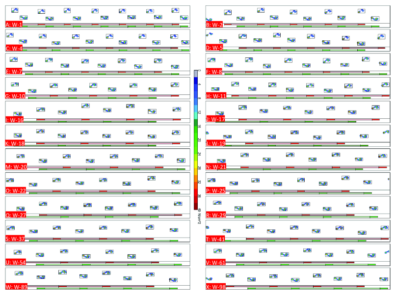 |
Fig. 2 showed that in 24 tests, the stride length of both sides was almost the same. Speed was the result of stride length dividing stride time, so the stride time was almost the same. This verified Eq. (2) and Eq. (3). The tested gait parameters were shown in Fig. 3.
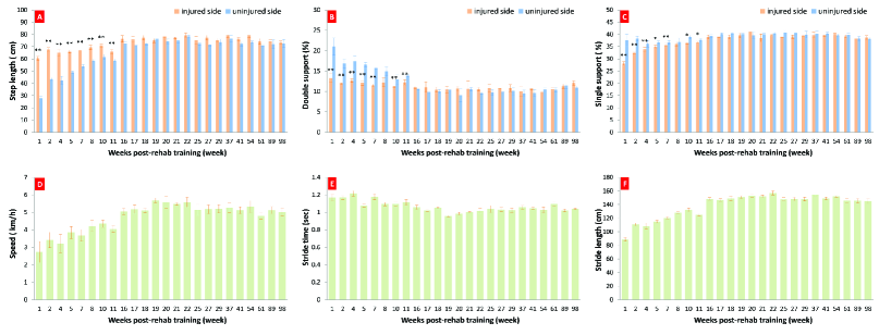 |
Fig. 3 demonstrated that at the early stage of rehabilitation, the step length of the uninjured side was shorter than that of the injured side and significant difference was found. The double support and single support of the uninjured side were greater than those of the injured side, and significant difference was found. The speed was slowed down by reducing stride length. This verified Eq. (4), which was consistent with our gait pattern design. Could such gait pattern reflect the peak values of VGRF of the injured side? VGRF was thus tested. See Fig. 4.
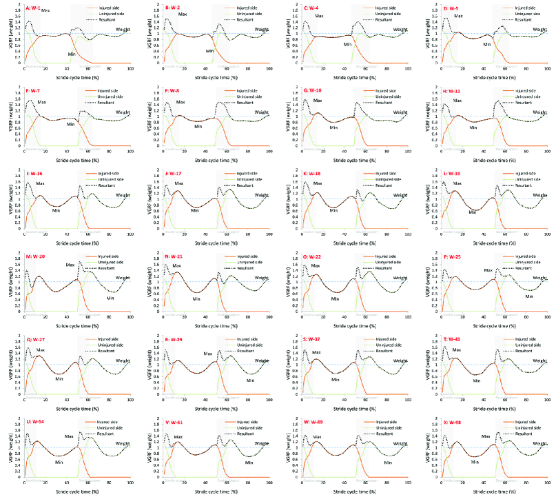 |
Fig. 4 showed that at the early stage of rehabilitation training, speed was slowed down by reducing stride length, the stance phase of the uninjured side was delayed and the step length of the uninjured side was reduced. The result was that the peak values of the injured side’s VGRF were close to the weight.
IV Conclusion
To design a reasonable walking rehabilitation training program, conditions from the above four equations should be met. The simulation of these equations verified our hypothesis to reduce VGRF of the injured side to be close to the weight by delaying the uninjured side’s stance phase, and by reducing the uninjured side’s step length. Furthermore, based upon the relation between speed, cadence and stride length, the design of gait pattern of ”reducing stride length to reduce speed, increasing the uninjured side’s stance phase and reducing the uninjured side’s step length” worked effectively. This also verified our hypothesis that when walking, by changing gait pattern, the injured side’s VGRF peak values could be reduced to be close to the weight. It is advisable to design a gait pattern of lower limb injury rehabilitation training program according to these biomechanical conditions.
Acknowledgments
This project was funded by the National Natural Science Foundation of China under the Grant Number . The authors would like to acknowledge the support from the participant.
References
- (1) D. C. Shapiro, R. F. Zernicke, and R. J. Gregor, Journal of motor behavior 13 (1), 33 (1981).
- (2) M. R. Bennett, J. W. K. Harris, B. G. Richmond, D. R. Braun, E. Mbua, P. Kiura, D. Olago, M. Kibunjia, C. Omuombo, A. K. Behrensmeyer, D. Huddart, and S. Gonzalez, Science 323 (5918), 1197 (2009).
- (3) N. L. W. Keijsers, N. M. Stolwijk, and T. C. Pataky, Gait and Posture 31 (1), 140 (2010).
- (4) H. Sadeghi, P. Allard, F. Prince, and H. Labelle, Gait and Posture 12 (1), 34 (2000).
- (5) B. Brouwer, K. Parvataneni, and S. J. Olney, Clinical Biomechanics 24 (9), 729 (2009).
- (6) N. F. Troje, Journal of Vision 2 (5), 371 (2002)
- (7) A. D. Kuo, J. M. Donelan, and A. Ruina, Exercise and Sport Sciences Reviews 33 (2), 88 (2005).
- (8) M. Srinivasan, and A. Ruina, Nature 439 (7072), 72 (2006).
- (9) N. Olsson, K. Nilsson-Helander, J. Karlsson, B. I. Eriksson, R. Thomee, E. Faxen, and K. G. Silbernagel, Knee Surgery Sports Traumatology Arthroscopy 19 (8), 1385 (2011).
- (10) Nirmal C. Tejwani, James Lee, Justin Weatherall, and Orrin Sherman, American journal of orthopedics (Belle Mead, N.J.) 43 (10), E221 (2014).
- (11) C. Rosso, P. Vavken, C. Polzer, D. M. Buckland, U. Studler, L. Weisskopf, M. Lottenbach, A. M. Muller, and V. Valderrabano, Knee Surgery Sports Traumatology Arthroscopy 21 (6), 1369 (2013).
- (12) F. Hahn, C. Maiwald, Th Horstmann, and P. Vienne, Clinical Biomechanics 23 (1), 109 (2008).
- (13) D. M. van der Eng, T. Schepers, C. J. Goslings, and N. W. L. Schep, Journal of Foot and Ankle Surgery 52 (5), 622 (2013).
- (14) T. Schepull, and P. Aspenberg, American Journal of Sports Medicine 41 (11), 2550 (2013).
- (15) A. Soroceanu, F. Sidhwa, S. Aarabi, A. Kaufman, and M. Glazebrook, Journal of Bone and Joint Surgery-American Volume 94A (23), 2136 (2012).
- (16) L. Ren, R. K. Jones, and D. Howard, Journal of Biomechanics 41 (12), 2750 (2008).
- (17) S. Winiarski, and A. Rutkowska-Kucharska, Acta of Bioengineering and Biomechanics 11 (1), 53 (2009).