Corresponding author:] djhuang@nsrrc.org.tw
Jahn-Teller distortion driven magnetic polarons in magnetite
Abstract
The first known magnetic mineral, magnetite (Fe3O4), has unusual properties which have fascinated mankind for centuries; it undergoes the Verwey transition at 120 K with an abrupt change in structure and electrical conductivity. The mechanism of the Verwey transition however remains contentious. Here we use resonant inelastic X-ray scattering (RIXS) over a wide temperature range across the Verwey transition to identify and separate out the magnetic excitations derived from nominal Fe2+ and Fe3+ states. Comparison of the RIXS results with crystal-field multiplet calculations shows that the spin-orbital excitons of the Fe2+ sites arise from a tetragonal Jahn-Teller active polaronic distortion of the Fe2+O6 octahedra. These low-energy excitations, which get weakened for temperatures above 350 K but persist at least up to 550 K, are distinct from optical excitations and best explained as magnetic polarons.
Since its first X-ray structural elucidation by W. H. Bragg a hundred years agoBragg15 and the discovery of the Verwey transition Verwey39 ; Walz02 , Fe3O4 has received much attention for decades. Even today, it attracts significant scientific and technological interest for its applications in ultrafast magnetic sensorsJong13 , palaeomagnetismAlmeida14 ; Jacob16 , nanomedicine carriersVeintmillas14 , etc. Fe3O4 becomes ferrimagnetic below Tc 850 K, followed by an abrupt decrease in its electrical conductivity by two orders of magnitude as the temperature is cooled below . Verwey first suggested a Fe2+-Fe3+ charge-ordering (CO) model as the driving force of this transition. There are two major schools of interpretation: the first one interprets the Verwey transition as a transition driven by charge/orbital ordering Wright01 ; Wright02 ; Jeng04 ; Leonov04 ; Huang06a ; Jeng06 ; Nazarenko06 ; Schlappa08 ; Senn12a ; Senn12b ; Senn15 . The second one exploits the mechanism of a lattice distortion driven electron-phonon interaction which opens a gap at the Fermi energy when the temperature is lowered below the transition temperature Garcia00 ; Piekarz06 ; Subias04 ; Chainani95 ; Park97 ; Taguchi15 ; Schrupp05 .
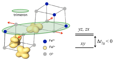
Although numerous investigations have been carried out to verify the charge localization on the octahedrally () coordinated B sites, the charge-ordering pattern of magnetite is subtle and still elusive Garcia00 ; Subias04 . While it is agreed that the charge disproportionation involves changes in the nominal Fe2+ and Fe3+ states associated with the B-sites, recent X-ray diffraction studies of the low-temperature phase of magnetite microcrystals Senn12b ; Senn15 revealed that the electrons of the B-site are not fully localized in the form of Fe2+ states. Instead, the electrons are distributed over linear three-Fe-site units termed “trimerons,” (consisting of one Fe2+δ and two Fe3-δ sites) which are coupled to the tetragonal () Jahn-Teller distortion of B-site Fe2+O6 octahedra, as illustrated in Fig. 1. To the first approximation, the B-site Fe3+O6 octahedra are Jahn-Teller inactive. The tetragonal distortion of B-site Fe2+O6 octahedra removes the degeneracy of orbitals, in going from symmetry to symmetry. In the absence of spin-orbit coupling, an effective energy separation between and is created if the four Fe-O bonds in the plane are elongated. The trimeron scenario then indicates that the Verwey transition is essentially an ordering of trimerons. The authors further conclude that trimeron correlations might persist in the cubic phase at temperatures above , in line with the existence of the short-range order above from results of neutron/X-ray diffuse scattering Bosak14 , X-ray absorption Subias05 , optical conductivity Park98 , photoemission Chainani95 ; Taguchi15 and anomalous phonon broadening Hoesch13 .
To the best of our knowledge, the relation of the local tetragonal distortion field of Fe2+ ions with the magnetic excitations of magnetite has not been reported to date. The issue of the quenched/unquenched orbital moment at the Fe2+ sites also remains controversialHuang04 ; Goering06 . Here we present measurements of resonant inelastic X-ray scattering (RIXS)Ament11 at the Fe -edge on magnetite to reveal the low-energy spin-orbital excitations of Fe2+ ions in both the monoclinic and cubic phases. In combination with crystal-field multiplet calculations, we show the the existence of magnetic polarons in magnetite which is driven by Jahn-Teller distortion.
Results
RIXS at the Fe -edge. Figure 2(a) shows the Fe -edge absorption spectrum (XAS) of magnetite. By comparing with crystal-field multiplet calculations (See Supplementary Fig. 6.), it is understood that the absorption-energy centroid of Fe2+ ions is lower than that of Fe3+ ions by eV, consistent with earlier workKuiper97 ; Chen04 ; Arenholz06 . Accordingly, the features labelled “” and “” at X-ray energies of 706.0 eV and 707.5 eV originate from the absorption of octahedrally coordinated B-site Fe2+ states, while the maximum intensity feature “” is dominated by intensity from the the Fe3+ ions of both the B-site octahedral and A-site tetrahedral symmetries.
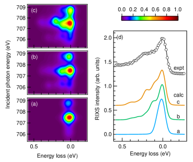
The color map of RIXS intensity in the plane of incident photon energy vs. energy loss shown in Fig. 2(b) presents the evolution of the RIXS spectral profile associated with Fe2+ and Fe3+ ions as detailed in the following. When the incident X-ray energy set to below , we observed excitations of Fe2+ with energy loss at 2.8 eV, 1.65 eV and 1.16 eV shown in Fig. 2(c), and also a broad excitation centered at 200 meV shown in Fig. 2(d). If the incident X-ray energy goes beyond , the 1.16-eV excitation of Fe2+ begins to evolve into a fluorescence that has a constant X-ray emission energy independent of incident energy, as indicated by the dashed line in Fig. 2(e). With the incident X-ray energy set to , RIXS excitations arise mostly from Fe3+ ions of octahedral or tetrahedral symmetry.
Figure 2(d) shows two RIXS features centered at 90 meV and 200 meV in a magnified plot of energy loss below 0.7 eV. Measurements carried out by varying the scattering angle suggested that these two low-energy excitations do not disperse in momentum space. (See Supplementary Fig. 3.) The 200-meV excitation has a full width at half maximum (FWHM) larger than the instrumental energy resolution. This broad RIXS feature resonates near the -edge of Fe2+ and almost disappears for incident energy above 708 eV, at which the other excitation centered at 90 meV emerges. The 90-meV excitation has a FWHM nearly equal to the instrumental energy resolution and resonates at 708.4 eV. The distinct incident X-ray energies for these resonant excitations indicates that the 200-meV and 90-meV features arise from Fe2+ and Fe3+ states, respectively.
Crystal-field multiplet calculations. To characterize the origin of the observed excitations centered at 200 meV, we undertook crystal-field multiplet calculations by using simulation programs CTM4RIXS CTM4RIXS and MISSING MISSING . Figures 3(a), 3(b) and 3(c) show the low-energy RIXS excitations of Fe2+ in the form of incident photon energy vs. energy loss maps, calculated using the same geometry as that of the experiments.
The calculated spectra are obtained as an average of the spectra calculated for magnetic domains with the easy axis along the [100], [010] and [001] directions. The crystal field parameter was set to 1.13 eV and the Slater integral was reduced to 79% for accurately reproducing the excitation energies. With only the spin-orbit coupling strength = 52 meV included, there exists low-energy excitation at 64 meV, but the 200 meV is not reproduced (Fig. 3(a)). If an effective exchange field meV is included without the tetragonal distortion, these excitations are split further with the excitation energy centroid at 132 meV, but still the 200 meV feature is not obtained (Fig. 3(b)). We, therefore, need to either increase the effective exchange field to nearly 200 meV, or include the effect of the tetragonal distortion of FeO6 octahedra. It is, however, unreasonable to use an exchange field much larger than the spin wave energy or the exchange field of Fe3+, 90 meV. Hence, we included a tetragonal distortion for calculating the RIXS spectrum of Fe2+ in magnetite. Figure 3(c) reveals that the calculated RIXS obtained on including the tetragonal distortion, exchange interaction and spin-orbit coupling matches fairly well with the experimental data. The results further indicate that within an excitation energy of the first 400-meV, there are effectively 15 separate states from Fe2+, as the ground state is split by the combination of these interactions. Further improvements in energy resolution is expected to reveal these low-lying excitations in ever increasing detail.
Discussion
Because the observed RIXS excitations exist in the cross-polarization geometry of a 90∘ scattering (See Supplementary Fig. 3(a).), it rules out the orbital excitations of the same symmetryVeenendaal06 . The origin of the 90-meV and 200-meV excitations is thus different from those of optical gapPark98 , photoemission gapChainani95 ; Park97 ; Taguchi15 ; Schrupp05 and the low-temperature activation energy obtained from electrical resistivityKuipers76 . Like magnetic excitations observed in the RIXS of cupratesAment11 ; Jia14 ; Huang16 and nickelates deGroot98 ; Haverkort10 , the observed 90-meV excitation results from spin-flip excitations of Fe3+ ions because its energy scale nearly corresponds to the energy of spin waves observed in inelastic neutron scattering McQueeney06 ; McQueeney07 and the RIXS cross section for phonons is much smaller than that of magnetic excitation.
For the broad 200-meV RIXS feature associated with the octahedral Fe2+ states, the excitation energy is too large to be explained in terms of only spin-flip excitations. In order to understand the nature of this feature, we carefully checked the effect of the local Jahn-Teller distortion to explain the energy of the observed excitations. According to Hund’s rule, out of the six electrons of the Fe2+ ion, five electrons occupy spin-up states (, ); the remaining one electron occupies one of the three spin-down orbitals (). The ground state of the octahedral Fe2+ ion is a high-spin state with . When the spin-orbit effect of electrons couples a pseudo-orbital angular momentum to , the state splits into three manifolds of pseudo-angular momenta and 3 with degeneracies 3, 5 and 7, respectively.
From an extensive set of RIXS calculations of Fe2+ with varied tetragonal distortions (See Supplementary Fig. 5.), we found that calculations using meV and about meV explain the measured RIXS spectra most satisfactorily. Figure 3(d) presents calculated RIXS of Fe2+ in comparison with measurements of the incident X-ray energy set to 707 eV, at which the 200-meV RIXS feature is most pronounced. The negative value of signifies that the energy of is lower than that of , i.e., tetragonally distorted Fe2+O6 octahedra with elongated Fe-O bonds in the plane. This shows that the tetragonal distortion is directly related to a polaronic distortion of the Fe2+O6 octahedra, which in turn couple to the neighbouring Fe3+O6 octahedra constituting the trimerons, although, as mentioned earlier, they are Jahn-Teller inactive in the first approximation. In short, the observation of spin-orbital excitations driven by a tetragonal Jahn-Teller polaronic distortion provides evidence for magnetic polarons in magnetite.
The magnitude of obtained is comparable with the spin-orbit coupling strength and thus confirms the observation of the unquenched orbital moment Huang04 , which is known from work on Fe2+ impurities in MgO thin films Haupricht10 . These results are also consistent with conclusions of band-structure calculations Jeng04 ; Leonov04 ; Jeng06 and the recent X-ray diffraction study on magnetiteSenn12a ; Senn15 . Band-structure calculations using the monoclinic crystal structure of magnetite Jeng04 also give the energy splitting 50 meV between minority-spin and bands at the point, conforming to the deduced .
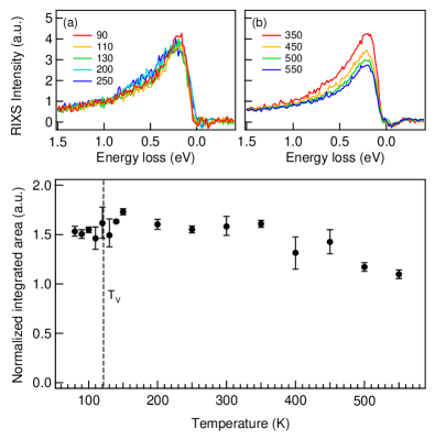
Figure 4 plots the temperature-dependent RIXS spectra with the incident X-ray energy set to the pre-edge absorption at 706 eV which is denoted , an energy below by 1.5 eV and at which the RIXS arises predominantly from octahedral Fe2+ ions with a negligible contribution from Fe3+ and the elastic component is weak. The results show that, when the temperature is varied across , the spin-orbital excitation of 200 meV does not abruptly change its intensity and persists at least up to 550 K, albeit with a gradual decrease above 350 K. We interpret this as a gradual weakening of the polarons. RIXS results shown here serve as a fast probe to snapshot the dynamic lattice-spin-orbital excitations of Fe3O4. These temperature-dependent RIXS results indicate that the FeO6 octahedra are already locally distorted in the cubic phase of magnetite, with the existence of the short-range order above Bosak14 .
To summarize, our results demonstrate the usefulness of RIXS to unravel the local electronic structure of a mixed-valence compound by selecting the energy and polarization of incident X-rays. We revealed excitons in magnetite that have an energy centroid 200 meV and arise from polaronic distortion driven spin-orbital excitations, which are best explained as magnetic polarons. We also applied crystal-field multiplet calculations to obtain the crystal field meV induced by the tetragonal Jahn-Teller distortion. These results are consistent with the trimeron mechanism for the Verwey transition. It would be interesting to carry out RIXS experiments with an improved energy resolution to study the change of spin-orbital excitations across the Verwey transition.
Methods
Crystal growth. Single-crystal growth of magnetite was carried out in an infrared image furnace in high-purity argon gas (99.999% purity) atmosphere. Measurements of the temperature-dependent specific heat and resistivity of the synthesized magnetite crystal showed that it exhibits a sharp first order Verwey transition at K . The synthesized single crystal has a chemical composition of Fe3(1-δ)O4 with , indicative of a nearly ideal chemical stoichiometry. See Supplementary Figs. 1 and 2 for the sample characterization.
RIXS measurements. Using the AGM-AGS spectrometer at beamline 05A1 of the National Synchrotron Radiation Research Center (NSRRC) in Taiwan, we measured RIXS on a single-crystal Fe3O at incident photon energies set to specific energies about the () X-ray absorption edge of Fe. Both the scattering angle defined as the angle between the incident and the scattered X-rays, and the incident angle from the crystal plane were variable. The polarization of the incident X-ray was switchable between and polarizations, i.e. the polarization within and perpendicular to the scattering plane, respectively, and the polarization of scattered X-rays was not analyzed. The energy bandwidth of the incident X-rays was 500 meV and the total RIXS energy resolution was 80 meV because the energy compensation method was used to ensure a high-resolution measurement in the energy loss scheme Lai14 . The beam diameter of incident X-ray at the sample is about 0.5 mm. See Supplementary Fig. 3(a) for the scattering geometry.
Crystal-field multiplet calculationsGroot05 . The starting point of the crystal field model is to approximate the transition metal Fe as an isolated atom surrounded by a distribution of charges that mimic the solid around the transition metal. The crystal field Hamiltonian is regarded as a perturbation to the atomic Hamiltonian in terms of an electrostatic potential that describes the surroundings. The atomic Hamiltonian include the spin-orbit coupling of electrons and the electron-electron interaction is parameterized by the Slater-Condon parameters and through a reduction factor. The Fe -edge RIXS spectral intensity is calculated using the Kramers-Heisenberg formula
| (1) |
in which is the total Hamiltonian and , and respectively represents the ground state of electrons in the orbitals, intermediate states in which one core electron is promoted to the orbital, and the final state of the excitation, i.e. an excited state of . The corresponding energies of these three states are , , and . and are the incident photon energy and the energy loss, respectively. The transition operator (and h.c.) dictates the dipole transition process from Fe to the level (or from Fe to ), with the X-ray polarization ( for outgoing photon) either or ; and is the inverse core-hole lifetime.
References
- (1) Bragg, W. H. The Structure of Magnetite and the Spinels. Nature 95, 561 (1915).
- (2) Verwey, E. J. W. Electronic Conduction of Magnetite and its Transition Point at Low Temperatures. Nature , 327 (1939).
- (3) Walz, F. The Verwey transition–a topical review. J. Phys. Condens. Matter , R285 (2002).
- (4) De Jong, S. et al. Speed limit of the insulator-metal transition in magnetite. Nature Mat. 12, 882 (2013).
- (5) Almeida, T. P. et al. Visualized effect of oxidation on magnetic recording fidelity in pseudo-single-domain magnetite particles. Nat. Commun. 5, 5154 (2014).
- (6) Jacob, D. E. et al. Redox-freezing and nucleation of diamond via magnetite formation in the Earth’s mantle. Nat. Commun. 7, 11891 (2016).
- (7) Veintemillas-Verdaguer, S. et al. Magnetic nanocrsyatls for biomedical applications. Progress in Crystal growth and Characterization of Materials 60, 80 (2014).
- (8) Wright, J. P., Attfield, J. P. & Radaelli, P. G. Long range charge ordering in magnetite below the Verwey transition. Phys. Rev. Lett. 87, 266401 (2001).
- (9) Wright, J. P., Attfield, J. P. & Radaelli, P. G. Charge ordered structure of magnetite below the Verwey transition. Phys. Rev. B 66, 214422 (2002).
- (10) Jeng, H.-T., Guo, G. Y. & Huang, D. J. Charge-orbital ordering and Verwey transition in magnetite. Phys. Rev. Lett. , 156403 (2004).
- (11) Leonov, I., Yaresko, A. N., Antonov, V. N., Korotin, M. A., & Anisimov, V. I. (2004). Charge and Orbital Order in Fe3O4. Phys. Rev. Lett. , 146404 (2004).
- (12) Jeng, H. T., Guo, G. Y., & Huang, D. J. Charge-orbital ordering in low-temperature structures of magnetite: GGA+ U investigations. Phys. Rev. B , 195115 (2006).
- (13) Huang, D. J. et al. Charge-orbital ordering and Verwey transition in magnetite measured by resonant soft X-ray scattering. Phys. Rev. Lett. 96, 096401 (2006).
- (14) Nazarenko, E., Lorenzo, J. E., Joly, Y., Hodeau, J. L., Mannix, D., & Marin, C. (2006). Resonant X-ray diffraction studies on the charge ordering in magnetite. Phys. Rev. Lett. , 056403 (2006).
- (15) Schlappa, J. et al. Direct observation of orbital ordering in magnetite. Phys. Rev. Lett. 100, 026406 (2008).
- (16) Senn, M. S., Wright, J. P., & Attfield, J. P. (2012). Charge order and three-site distortions in the Verwey structure of magnetite. Nature , 173 (2012).
- (17) Senn, M. S., Loa, I., Wright, J. P., & Attfield, J. P. Electronic orders in the Verwey structure of magnetite. Phys. Rev. B , 125119 (2012).
- (18) Senn, M. S., Wright, J. P., Cumby, J., & Attfield, J. P. (2015). Charge localization in the Verwey structure of magnetite. Phys. Rev. B , 024104 (2015).
- (19) J. García, J. et al. Resonant ”forbidden” reflections in magnetite. Phys. Rev. Lett. 85, 578 (2000).
- (20) Subías, G. et al. Magnetite, a model system for mixed-valence oxides, does not show charge ordering. Phys. Rev. Lett. , 156408 (2004).
- (21) Piekarz, P., Parlinski, K., & Oleś, A. M. Mechanism of the Verwey transition in magnetite. Phys. Rev. Lett. 97, 156402 (2006).
- (22) Chainani, A., Yokoya, T., Morimoto, T., Takahashi, T., & Todo, S. High-resolution photoemission spectroscopy of the Verwey transition in Fe3O4. Phys. Rev. B , 17976 (1995).
- (23) Park, J. H., Tjeng, L. H., Allen, J. W., Metcalf, P., & Chen, C. T. Single-particle gap above the Verwey transition in Fe3O4. Phys. Rev. B , 12813 (1997).
- (24) Schrupp, D. et al. High-energy photoemission on Fe3O4: Small polaron physics and the Verwey transition. Europhys. Lett. 70, 789.(2005)
- (25) Taguchi, M. et al. Temperature Dependence of Magnetically Active Charge Excitations in Magnetite across the Verwey Transition. Phys. Rev. Lett. 115, 256405 (2015).
- (26) Bosak, A. et al. Short-range correlations in magnetite above the Verwey temperature. Phys. Rev. X , 011040 (2014).
- (27) Subías, G. J. García, J. & J. Blasco, J. EXAFS spectroscopic analysis of the Verwey transition in Fe3O4. Phys. Rev. B , 155103 (2005).
- (28) Park, S. K., Ishikawa, T., & Tokura, Y. Charge-gap formation upon the Verwey transition in Fe3O4. Phys. Rev. B , 3717 (1998).
- (29) M. Hoesch, P. Piekarz, A. Bosak, M. Le Tacon, M. Krisch, A. Kozlowski, A. M. Oleś, and K. Parlinski, Phys. Rev. Lett. , 207204 (2013).
- (30) Huang, D. J. et al. Spin and Orbital Magnetic Moments of Fe3O4. Phys. Rev. Lett. , 077204 (2004).
- (31) Goering, E., Gold, S., Lafkioti, M., & Schütz, G. Vanishing Fe 3d orbital moments in single-crystalline magnetite. Europhys. Lett. , 97 (2006).
- (32) Ament, L. J. P., van Veenendaal, M., Devereaux, T. P., Hill, J. P. & van den Brink, J. Resonant inelastic x-ray scattering studies of elementary excitations. Rev. Mod. Phys. 83, 705 (2011).
- (33) Lai, C. H. et al. Highly efficient beamline and spectrometer for inelastic soft X-ray scattering at high resolution. J. Synchrotron Radiat. , 325 (2014).
- (34) Kuiper, P., Searle, B. G., Duda, L. C., Wolf, R. M., & Van der Zaag, P. J. (1997). Fe linear and circular magnetic dichroism of Fe3O4. J. Electron Spectrosc. Relat. Phenom. , 107 (1997).
- (35) Chen, J., Huang, D. J., Tanaka, A., Chang, C. F., Chung, S. C., Wu, W. B., & Chen, C. T. (2004). Magnetic circular dichroism in Fe resonant photoemission of magnetite. Phys. Rev. B , 085107 (2004).
- (36) Arenholz, E., van der Laan, G., Chopdekar, R. V., & Suzuki, Y. Anisotropic X-ray magnetic linear dichroism at the Fe edges in Fe3O4. Phys. Rev. B , 094407 (2006).
- (37) Verble, J. L. Temperature-dependent light-scattering studies of the Verwey transition and electronic disorder in magnetite. Phys. Rev. B , 5236 (1974).
- (38) Gasparov, L. V., Rush, A., Guntherodt, G., & Berger, H. Electronic Raman scattering in magnetite: Spin versus charge gap. Phys. Rev. B , 144303 (2009).
- (39) Kumar, A., Chaudhary, S., Pandya, D. K., & Sharma, S. K. Evidence of electron-phonon and spin-phonon couplings at the Verwey transition in Fe3O4. Phys. Rev. B , 024302 (2014).
- (40) Jia, C. J. et al. Persistent spin excitations in doped antiferromagnets revealed by resonant inelastic light scattering. Nat. Commun. 5, 3314 (2014).
- (41) Huang, H. Y. et al. Raman and fluorescence characteristics of resonant inelastic X-ray scattering from doped superconducting cuprates. Sci. Rep. , 19657 (2016).
- (42) van Veenendaal, M. Polarization dependence of L-and M-edge resonant inelastic X-ray scattering in transition-metal compounds. Phys. Rev. Lett. 96, 117404 (2006).
- (43) Kuipers, A. J. M., & Brabers, V. A. M. Thermoelectric properties of magnetite at the Verwey transition. Phys. Rev. B 14, 1401 (1976).
- (44) de Groot, F. M. F., Kuiper, P., & Sawatzky, G. A. Local spin-flip spectral distribution obtained by resonant x-ray Raman scattering. Phys. Rev. B 57, 14584 (1998).
- (45) Haverkort, M. W. Theory of resonant inelastic X-ray scattering by collective magnetic excitations. Phys. Rev. Lett. 105, 167404 (2010).
- (46) McQueeney, R. J., Yethiraj, M., Montfrooij, W., Gardner, J. S., Metcalf, P., & Honig, J. M. Investigation of the presence of charge order in magnetite by measurement of the spin wave spectrum. Phys. Rev. B , 174409 (2006).
- (47) McQueeney, R. J., Yethiraj, M., Chang, S., Montfrooij, W., Perring, T. G., Honig, J. M., & Metcalf, P. Zener double exchange from local valence fluctuations in magnetite. Phys. Rev. Lett. , 246401 (2007).
- (48) Stavitski, E., & de Groot, F. M. F. The CTM4XAS program for EELS and XAS spectral shape analysis of transition metal L edges. Micron , 687 (2010).
- (49) Claudia Dallera and Riccardo Gusmeroli, http://www.esrf.eu/computing/scientic/MISSING/.
- (50) Haupricht, T. et al. Local electronic structure of Fe2+ impurities in MgO thin films: Temperature-dependent soft X-ray absorption spectroscopy study Phys. Rev. B , 035120 (2010).
- (51) de Groot, F. M. F. Multiplet effects in X-ray spectroscopy. Coord. Chem. Rev. , 31 (2005).