Photodissociation of trapped Rb: Implications for simultaneous trapping of atoms and molecular ions
Abstract
The direct photodissociation of trapped 85Rb (rubidium) molecular ions by the cooling light for the 85Rb magneto-optical trap (MOT) is studied, both experimentally and theoretically. Vibrationally excited Rb ions are created by photoionization of Rb2 molecules formed photoassociatively in the Rb MOT and are trapped in a modified spherical Paul trap. The decay rate of the trapped Rb ion signal in the presence of the MOT cooling light is measured and agreement with our calculated rates for molecular ion photodissociation is observed. The photodissociation mechanism due to the MOT light is expected to be active and therefore universal for all homonuclear diatomic alkali metal molecular ions.
The spatially overlapped trapping of cold atoms and ions Smith et al. (2005); Zipkes et al. (2010); Hall et al. (2011); Ravi et al. (2012a); Ray et al. (2014); Grier et al. (2009); Rellergert et al. (2011) has significantly expanded our ability to study interactions in cold, dilute gas ensembles. In particular, atomic ion-atom collisions, charge exchange collisions Grier et al. (2009); Lee et al. (2013); Rellergert et al. (2011); Haze et al. (2015); Sivarajah et al. (2012), sympathetic cooling of ions by ultracold trapped atoms Ravi et al. (2012b); Hall and Willitsch (2012); Sivarajah et al. (2012); Ray et al. (2014); Dutta et al. (2015), three body reactions Härter et al. (2012, 2013); Krükow et al. (2016) and molecular ion formation processes Hall et al. (2011); da Silva Jr et al. (2015) have been investigated. Two complementary directions motivate key goals for future work, (a) the low partial wave ion-atom collisions which explores quantum scattering and many particle physics and (b), the controlled collisions between the cold molecular ions produced in the ion-atom traps with co-trapped neutral atoms Rellergert et al. (2013) and with light.
A critical question which arises is whether the molecular ions can be trapped for a substantial extent of time simultaneously with an ensemble of cold atoms in order to study the interaction between them. In this letter, we address the possibility of simultaneous trapping of 85Rb molecular ions with ultracold 85Rb atoms in a magneto-optical trap (MOT). Our experimental observation shows that the cooling light for the Rb MOT leads to rapid destruction of the measured Rb ion signal. We measure the lifetime of trapped Rb in the presence of 780.2413 nm ( 12816.54 cm-1) light to be 49580 ms. We discuss possible dissociation mechanisms and show that the experimental observation is in agreement with our theoretical calculations for the photodissociation of Rb molecular ions.The observed photodissociation mechanism is expected to be universal to all diatomic homonuclear alkali molecular ions as they exhibit similar potential energy characteristics.
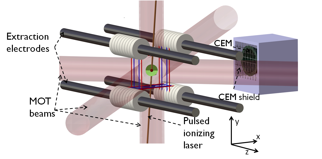
The experimental system consists of an ion-atom hybrid trap assembly as shown in Fig. 1. The detailed description of the experimental system can be found in earlier work Ray et al. (2013, 2014); Jyothi et al. (2015). Briefly, the hybrid trap consists of a MOT for atoms and a modified spherical Paul trap made of four tungsten wire loops in a square shape geometry for trapping ions. The ion trap radio frequency (rf) of 500 kHz with 150 V amplitude is applied to the inner pair of wires and a small (-5 V) constant potential is applied on the outer wires. The ion trap is operated at the optimal trapping voltage for Rb ions.
For the experiment, 85Rb atoms are cooled and trapped in the MOT using three pairs of mutually orthogonal counter-propagating laser beams of mm diameter intersecting at the center of a gradient magnetic field. The cooling laser beam is red detuned from the (F=3) 5 =4) transition by 12 MHz. The repumper light is on resonance with the (F=2) 5 =3) transition. Approximately 40 mW of cooling and 3 mW of repumper power is used and is equally distributed over the six laser beams.
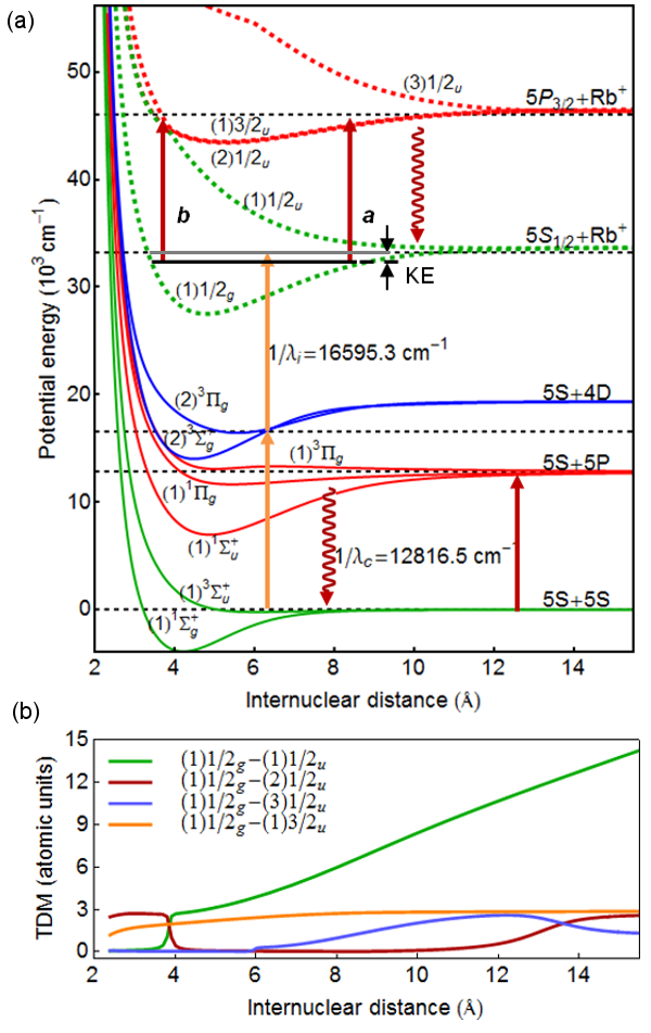
The Rb molecular ions are created by ionizing neutral Rb2 molecules produced by photoassociation in the MOT Gabbanini et al. (2000); Caires et al. (2005). The potential energy curves (PEC) of Rb2 Allouche and Aubert-Frécon (2012) and Rb All molecules relevant for this experiment are shown in Fig. 2(a). Two Rb atoms in the ground state photoassociate in the presence of a cooling laser photon cm to form a loosely bound molecule in the excited electronic state. The excited molecule spontaneously decays either to a highly vibrationally excited bound molecule in the electronic ground states ( or ) or to two free ground state atoms by emitting a photon. These loosely bound neutral Rb2 molecules are ionized by two photons of 602.5 nm cm-1) to produce Rb ions in their electronic ground state (or in Hund’s case ”a” notation) as previously shown by Gabbanini et. al. Gabbanini et al. (2000). The ionization laser is a pulsed dye laser, pumped by the second harmonic of a Nd:YAG laser (10 Hz repetition rate).
Energy consideration allows only those vibrational levels of Rb which have binding energies 500.2 cm-1 (= Ionization potential(Rb) 33690.8 cm-1 - 2) to be populated. Based on the calculation of vibrational energies Roy (2007) using the ab initio potentials All , the vibrational level with binding energy close to 500.2 cm-1 is 174 of electronic state, which essentially implies that the ionization process can create Rb in levels 174. The kinetic energy (KE) of the electron determines the initial vibrational levels in which the Rb ions are created.
Since the molecular ions are created from the MOT, they are created at the centre of the ion trap. The ion trap voltages are on during the ionization process so that the ions are trapped as they are produced. The trapped ions are detected by extracting them onto a channel electron multiplier (CEM), by switching the voltages appropriately on a set of electrodes Ray et al. (2014). Prior to the extraction, the trapping rf field is switched off to mass separate any Rb+ ions from Rb ions Jyothi et al. (2015).
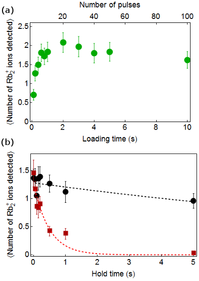
The process of loading of Rb into the ion trap is monitored by extracting the ions at different times during the loading process as shown in Fig. 3(a). In our experiment, the molecular ions created per shot fluctuate due to the energy fluctuation of the ionizing dye laser pulse. For this reason, we repeated the experiment at each hold time more than 40 times. A steady state is reached when the ion production rate equals the ion loss rate and with a loading time of 10s, the ion trap has 1.620.22 Rb ions. In principle, the ion loss rate depends on factors such as rf heating, collisions with background gases, collisions with ultracold atoms and on the presence of light. In order to determine the dominant loss channel, we load the trap to steady state and hold the ions in the ion trap for variable hold time in different scenarios followed by ion extraction onto the CEM to count the number of ions survived.
The effect of Rb D2 light on the population of Rb molecular ions is studied by measuring the lifetime of Rb when held in the presence of MOT cooling light. Each experimental cycle consists of the following sequence: loading the MOT to steady state, turning on the ion trap, turning on the pulsed dye laser to create Rb, turning off the pulsed dye laser (thus stopping further creation of Rb), removing the MOT atoms by blocking the repumper light and then holding the Rb ions in the ion trap for a predetermined hold time(t) either in presence or absence of the MOT cooling light. At the end of the hold time, the ions are extracted out of the ion trap and detected by the CEM. The hold time is varied and the sequence is repeated. Fig. 3(b) shows the mean number of Rb ions detected, for different ion trap hold times either in presence (red squares) or absence (black circles) of the MOT cooling light. It should be mentioned that, during the Rb ion creation process a large number of Rb+ ions are also created. However the ion trap parameters favor molecular ions and atomic ions escape from the ion trap within a few tens of milliseconds.To avoid systematic effects due the presence of atomic ions, we restrict the our analysis to t50 ms. We fit the average number of Rb ions to A exp(-t/) to obtain the lifetime of Rb in the ion trap. In the absence of MOT lights the lifetime is 168 s and is dramatically reduced to 49580 ms in the presence of the cooling light. The single exponential fit allows a comparison between the with and without light decay. While different vibrational levels decay at different rates in the presence of MOT light (see below), the resolution of the present experiment is not enough to distinguish between them, and so the single exponential is representative of a rate for the disintegration process.
We consider two possible channels for the disintegration of the Rb ground state molecules induced by the MOT cooling light. The dipole allowed transitions from the initial state lead to the states , , and (written in Hund’s case c notation including spin-orbit interaction), with the relevant electric transition dipole moment (TDM) All shown in Fig. 2(b). Of these states, the is energetically not accessible. The state is of character below 4 , and changes into before 4 due to an avoided crossing induced by the spin-orbit interaction. This explains the crossing of TDM curves in Fig. 2(b) around 4 . The possible dissociation channels are shown in Fig. 2(a) and are:
(a) bound-to-bound excitation (indicated by a in Fig. 2(a)) followed by spontaneous decay to ion-atom pair.
Rb(RbRb + Rb
(b) direct photodissociation (PD) from to (indicated by b in Fig. 2(a)).
Rb Rb + Rb+
The possibility for the dissociation of Rb ions through these excitation channels are investigated by calculating the transition rates.
In the case a, the energy of the cooling light , where is the frequency, is insufficient to excite the molecule to the continuum of the first excited electronic state (dissociation to Rb+ asymptote). However, one can consider a resonant bound to bound transition and subsequent de-excitation to the ground state. If the de-excitation to the ground state continuum dominates over that to the bound levels, this process can be considered as a dissociation channel. We first calculate the bound-to-bound excitation by the MOT cooling light from various possible initial vibrational levels of the state to the and states. The accuracy of the ab initio PECs is not high enough to predict the vibrational levels exactly and it is thus not possible to say if the excitation is resonant or off-resonant, although the latter is more likely. Assuming resonant excitation to the nearest vibrational level determined from photon energy consideration, the maximum excitation rate is calculated to be of the order of MHz or smaller for the and transitions. The spontaneous emission rate to the bound and free states of is then calculated for both the and states. For both electronic states we find that the bound-to-free spontaneous emission rate is negligibly small compared to the bound-to-bound spontaneous emission rate, eliminating the possibility of this dissociation channel.
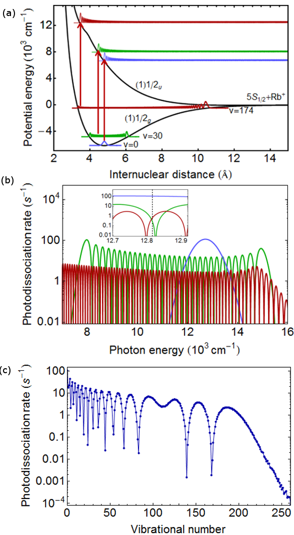
In the direct photodissociation process (case b), a bound Rb molecule in absorbs a photon (the slower rate determining step) to reach the state and dissociates immediately to the continuum of to form Rb and Rb+ as shown in Fig. 4(a). The photodissociation cross section is proportional to the photon energy and the Franck-Condon overlap between the levels involved Lefebvre-Brion and Field (2004); Dunn (1969). The rate of photodissociation is given by
| (1) | ||||
| (2) |
where is the dissociation cross section, is the dissociating light flux, I is the intensity of the light, is the Bohr radius, 2 is the Planck’s constant, c is the velocity of light, is the photon energy and d(r) is the transition dipole moment between and states All . The continuum wavefunction is calculated using Cooley-Numerov method Levine (2009) and the bound wavefunction is calculated using LEVEL code Roy (2007).
The variations of the calculated PD rates vs the photon energy reproduce the oscillations of the initial bound wave functions, as shown for few vibrational levels in Fig. 4(b) Blange et al. (1996); Dunn (1969). Fig. 4(c) shows the PD rate (at 12816.5 cm-1 with 0.16 W/cm2 of intensity) for all the vibrational levels of Rb ground state. The results show that molecular ions in different vibrational levels dissociate at different rates in presence of the MOT cooling light with the trend that the lower vibrational levels dissociate at faster rates.
In the experiment, we cannot determine precisely the vibrational level of the initially created Rb because the details of the Rb2 photoionization process are unknown and difficult to compute. The two limits concerning the vibrational level of the initially created Rb are determined by the KE of the ejected electron. (i) In case the electron is ejected with zero KE, the Rb ions are created in/near 174 which dissociates at the calculated rate of 0.7 s-1. However, we note that the dissociation rate for v = 174 could be in the range 0.7 to 2.1 s-1, considering the uncertainty in the dissociation energy of the ab initio PEC compared to the experimental dissociation energy Bellos et al. (2013), which is higher by 120 cm-1 compared to theory, and causes shift of the calculated vibrational levels and hence the PD rates. (ii) In case of electron ejection with all possible KE, all vibrational levels of Rb from 0 to 174 may be populated. An average of calculated PD rates (Fig. 4(c)) over all vibrational levels below 174 yields 5.9 s-1 but the actual rate could be anywhere between 0.001 to 100 s-1 as shown in Fig. 4(c).The experimentally measured rate 2.00.3 s-1is consistent with either of the above cases. Further detailed experimental and theoretical work is thus needed to establish the initial vibrational distribution of Rb. However, even in the absence of such information, the experimental and theoretical results presented above firmly establish that light near D2 transition induces dissociation of Rb. It should also be noted that the commonly used 1064 nm light for optical dipole trapping would dissociate loosely bound Rb (10) by a similar process but have no significant effect on deeply bound Rb (e.g. 0).
The above experiments and calculations show that the direct photodissociation of Rb ion by the cooling light of the MOT is the dominant mechanism for the loss of trapped Rb molecular ions in all vibrational states. Since all homonuclear alkali (X) molecular ion dimers, X, have a similar arrangement of their molecular PECs T.E.Sharp (1971), we conclude through preliminary inspection that this photodissociation process will be active and influential for all such molecular ion systems. This would imply that in the presence of the MOT cooling light, long lived trapping of X will be challenging, and creating sizable steady state ensembles of X ions and parent alkali atoms would be experimentally difficult. A possible solution to the problem is the use of a far-detuned optical dipole trap for ultracold atoms instead of a MOT.
S. J. and S. A. R. acknowledge Krishna Rai Dastidar for assistance in the setting up of the molecular level calculations. S. D. acknowledges support in the form a Pancharatnam Fellowship from RRI. A. R. A. acknowledges the access to the HPC resources of the FLMSN,”Féderation Lyonnaise de Modélisation et Sciences Numeriques”, partner of EQUIPEX EQUIP@MESO and HPC of the ”Centre de calcul CC-IN2P3” at Villeurbanne. O. D. and S. A. R. acknowledge support from the Indo-French Centre for the promotion of Advanced Research-CEFIPRA project 5404-1.
References
- Smith et al. (2005) W. W. Smith, O. P. Makarov, and J. Lin, J. Mod. Opt. 52, 2253 (2005).
- Zipkes et al. (2010) C. Zipkes, S. Palzer, C. Sias, and M. Köhl, Nature 464, 388 (2010).
- Hall et al. (2011) F. H. J. Hall, M. Aymar, N. Bouloufa-Maafa, O. Dulieu, and S. Willitsch, Phys. Rev. Lett. 107, 243202 (2011).
- Ravi et al. (2012a) K. Ravi, S. Lee, A. Sharma, G. Werth, and S. Rangwala, Appl. Phys. B 107, 971 (2012a).
- Ray et al. (2014) T. Ray, S. Jyothi, N. Ram, and S. Rangwala, Appl. Phys. B 114, 267 (2014).
- Grier et al. (2009) A. T. Grier, M. Cetina, F. Oručević, and V. Vuletić, Phys. Rev. Lett. 102, 223201 (2009).
- Rellergert et al. (2011) W. G. Rellergert, S. T. Sullivan, S. Kotochigova, A. Petrov, K. Chen, S. J. Schowalter, and E. R. Hudson, Phys. Rev. Lett. 107, 243201 (2011).
- Lee et al. (2013) S. Lee, K. Ravi, and S. A. Rangwala, Phys. Rev. A 87, 052701 (2013).
- Haze et al. (2015) S. Haze, R. Saito, M. Fujinaga, and T. Mukaiyama, Phys. Rev. A 91, 032709 (2015).
- Sivarajah et al. (2012) I. Sivarajah, D. S. Goodman, J. E. Wells, F. A. Narducci, and W. W. Smith, Phys. Rev. A 86, 063419 (2012).
- Ravi et al. (2012b) K. Ravi, S. Lee, A. Sharma, G. Werth, and S. Rangwala, Nat. Commun. 3, 1126 (2012b).
- Hall and Willitsch (2012) F. H. J. Hall and S. Willitsch, Phys. Rev. Lett. 109, 233202 (2012).
- Dutta et al. (2015) S. Dutta, R. Sawant, and S. A. Rangwala, arXiv:1512.04197 (2015).
- Härter et al. (2012) A. Härter, A. Krükow, A. Brunner, W. Schnitzler, S. Schmid, and J. Hecker Denschlag, Phys. Rev. Lett. 109, 123201 (2012).
- Härter et al. (2013) A. Härter, A. Krükow, M. Deiß, B. Drews, E. Tiemann, and J. Hecker Denschlag, Nat. Phys. 9, 512 (2013).
- Krükow et al. (2016) A. Krükow, A. Mohammadi, A. Härter, J. H. Denschlag, J. Pérez-Ríos, and C. H. Greene, Phys. Rev. Lett. 116, 193201 (2016).
- da Silva Jr et al. (2015) H. da Silva Jr, M. Raoult, M. Aymar, and O. Dulieu, New J. Phys. 17, 045015 (2015).
- Rellergert et al. (2013) S. S. T. Rellergert, Wade G. and, S. J. Schowalter, S. Kotochigova, K. Chen, and E. R. Hudson, Nature 495, 490 (2013).
- Ray et al. (2013) T. Ray, A. Sharma, S. Jyothi, and S. A. Rangwala, Phys. Rev. A 87, 033832 (2013).
- Jyothi et al. (2015) S. Jyothi, T. Ray, and S. Rangwala, Appl. Phys. B 118, 131 (2015).
- Gabbanini et al. (2000) C. Gabbanini, A. Fioretti, A. Lucchesini, S. Gozzini, and M. Mazzoni, Phys. Rev. Lett. 84, 2814 (2000).
- Caires et al. (2005) A. R. L. Caires, V. A. Nascimento, D. C. J. Rezende, V. S. Bagnato, and L. G. Marcassa, Phys. Rev. A 71, 043403 (2005).
- Allouche and Aubert-Frécon (2012) A.-R. Allouche and M. Aubert-Frécon, J. Chem. Phys. 136, 114302 (2012).
- (24) Supplementary material.
- Roy (2007) R. J. L. Roy, LEVEL 8.0: A Computer Program for Solving the Radial Schrodinger Equation for Bound and Quasibound Levels, University of Waterloo Chemical Physics Research Report CP-663 (2007), see http://leroy.uwaterloo.ca/programs/.
- Lefebvre-Brion and Field (2004) H. Lefebvre-Brion and R. W. Field, The Spectra and Dynamics of Diatomic Molecules (Elsevier Academic Press, 2004).
- Dunn (1969) G. H. Dunn, Phys. Rev. 172, 1 (1969).
- Levine (2009) I. N. Levine, Quantum Chemistry (Pearson Education Inc., New Jersey,U.S.A., 2009).
- Blange et al. (1996) J. J. Blange, X. Urbain, H. Rudolph, H. A. Dijkerman, H. C. W. Beijerinck, and H. G. M. Heideman, J. Phys. B: At. Mol. Opt. Phys. 29, 2763 (1996).
- Bellos et al. (2013) M. A. Bellos, R. Carollo, J. Banerjee, M. Ascoli, A.-R. Allouche, E. E. Eyler, P. L. Gould, and W. C. Stwalley, Phys.Rev.A 87, 012508 (2013).
- T.E.Sharp (1971) T.E.Sharp, At.Data 2, 119 (1971).