The first two authors contribute equally to this work
\altaffiliationThe first two authors contribute equally to this work
\alsoaffiliationDepartment of Electrical and Computer Engineering
Michigan State University, MI 48824, USA
\alsoaffiliationDepartment of Biochemistry and Molecular Biology
Michigan State University, MI 48824, USA
Accurate, robust and reliable calculations of Poisson-Boltzmann binding energies
Abstract
Poisson-Boltzmann (PB) model is one of the most popular implicit solvent models in biophysical modeling and computation. The ability of providing accurate and reliable PB estimation of electrostatic solvation free energy, , and binding free energy, , is important to computational biophysics and biochemistry. Recently, it has been warned in the literature (Journal of Chemical Theory and Computation 2013, 9, 3677-3685) that the widely used grid spacing of produces unacceptable errors in estimation with the solvent exclude surface (SES). In this work, we investigate the grid dependence of our PB solver (MIBPB) with SESs for estimating both electrostatic solvation free energies and electrostatic binding free energies. It is found that the relative absolute error of obtained at the grid spacing of compared to at averaged over 153 molecules is less than 0.2%. Our results indicate that the use of grid spacing ensures accuracy and reliability in calculation. In fact, the grid spacing of appears to deliver adequate accuracy for high throughput screening.
keywords:
Accurate coarse grid Poisson Boltzmann solver, reaction field energy, electrostatic binding free energy1 Introduction
Electrostatics is ubiquitous in biomolecular and cellular systems and of paramount importance to biological processes. Accurate and reliable prediction of electrostatic binding free energy, , is crucial to biophysical modeling and computation. The prediction of plays a vital role in the study of many cellular processes, such as signal transduction, gene expression, and protein synthesis. Additionally, many pharmaceutical applications, specially in the final stage of the drug design, rely on the accurate and reliable calculation of binding free energy. Technically, the accuracy and reliability of electrostatic binding energy prediction depend essentially on the quality of electrostatic solvation () estimation, which can be achieved by solving the Poisson-Boltzmann (PB) equation in the implicit solvent model 1, 2, 3, 4, 5. In past decades, the development of a robust PB solver catches much attention in computational biophysics and biochemistry. Mathematically, most PB solvers reported in the literature are based on three major approaches, namely, the finite difference method (FDM) 6, the finite element method (FEM) 7, and the boundary element method (BEM) 8, 9. Among them, the FDM is prevalently used in the field due to its simplicity in implementation. The emblematic solvers in this category are Amber PBSA 10, 11, Delphi 12, 13, APBS 14 and CHARMM PBEQ 6.
Recently, it has been warned that “the widely used grid spacing of 0.5 Å produces unacceptable errors in ” 15. Although all results were obtained with the adaptive Cartesian grid (ACG) finite difference PB equation solver 16 in this work, similar results were reported in a later study 17 by using APBS, DelPhi and PBSA. Therefore, these studies have arisen serious concerns about the validity of using PB model for biomolecular electrostatic binding analysis at an affordable grid spacing of 0.5 Å.
In the past few years, there have been many attempts to develop highly accurate PB solvers using advance techniques for interface treatments 16, 18, 19, 20. The later verison of the ACG solver 16, 18 has somewhat remedied the grid-dependence issue for estimates of binding energy. However, no confirmation for the reliable use of grid spacing of 0.5 Å in has been given. In this work, we investigate the grid dependence of our PB solver (MIBPB) 21, 22 in estimating both electrostatic solvation free energies and electrostatic binding free energies. The MIBPB solver is by far the only existing method that is second-order accurate in norm for solving the Poisson-Boltzmann equation with discontinuous dielectric constants, singular charge sources, and geometric singularities from the solvent excluded surfaces (SESs) of biomolecules 21. Here the norm means the maxmum absolute error measure and “second order accurate” means that the error reduces four times when the grid spacing is halved. Contrary to the findings in the literature 15, our results indicate that the use of grid spacing 0.6 Å ensures accuracy and reliability in calculation. In fact, a grid spacing of 1.1 Å appears to deliver adequate accuracy for high throughput screening. We therefore believe that when it is used properly, the PB methodology is able to deliver accurate and reliable electrostatic binding analysis.
2 Methods
2.1 MIBPB package
In the current work, we employ the our MIBPB package 21, 22 to predict the electrostatic solvation free energy. The MIBPB package is a second-order convergence PB solver for dealing with the SESs of biomolecules. Numerically, there are three major obstacles in constructing accurate and reliable PB solvers. First, commonly used solvent-solute interfaces, i.e., the van der Walls (vdW) surface, solvent accessible surface (SAS), and the solvent excluded surface (SES) 23, 24 admit geometric singularities, such as sharp tips, cusps and self-intersecting surfaces 25, which make the rigorous enforcement of interface jump conditions a formidable task in PB solvers. An advanced mathematical interface techniques, the matched interface and boundary (MIB) method 26, 27, 28, 29, 30, 31, is employed in the MIBPB package to achieve the second order accuracy in handling biomolecular SESs. Additionally, the atomic singular charges described by the delta functions give rise to another difficulty in constructing highly accurate PB solver. A Dirichlet-to-Neumann map technique has been developed in the MIBPB package to avoid the numerical approximation of singular charges by using the analytical Green’s functions 32. Finally, the nonlinear Boltzmann term can affect solver efficiency when handled inappropriately, particularly for BEMs. A quasi-Newton algorithm is implemented in the MIBPB package 21, 22 to take care the nonlinear term 21, 22.
2.2 Interface generation
Many studies suggest that SES is able to deliver the state of the art accurate modeling of the solvated molecule 7, 13, 9. As a result, much effort has been paid to developing an accurate and robust SES software 25, 33. However, the MSMS software 25 generates a Lagrangian representation of the SES and is inconvenient for the Cartesian domain implementation of PB solvers. A Lagrangian to Eulerian transformation is required to convert MSMS surfaces for our Cartesian based MIBPB solver 4. Most recently, we have developed a new SES software, Eulerian solvent excluded surface (ESES), to directly generate the SESs in the Eulerian representation 34. Our ESES software enables the MIBPB solver to produce a reliable . Both MSMS and ESES are supported by our MIBPB software. By increasing the MSMS surface density, the electrostatic solvation free energies calculated by using MSMS converge to those obtained by using ESES 34. Therefore, only results employing ESES are shown in this work.
2.3 Data sets
In the present work, we adopt three sets of biomolecular complexes employed in the literature 15 for solvation free energy and binding free energy estimations. Specifically, the first set, Data Set 1, is a collection of DNA-minor groove drug complexes having a narrow range of . The Protein Data Bank (PDB) IDs (PDBIDs) for this set are as follows: 102d, 109d, 121d, 127d, 129d, 166d, 195d, 1d30, 1d63, 1d64, 1d86, 1dne, 1eel, 1fmq, 1fms, 1jtl, 1lex, 1prp, 227d, 261d, 164d, 289d, 298d, 2dbe, 302d, 311d, 328d, and 360d. The second set, Data Set 2, includes various wild-type and mutant barnase-barstar complexes. Its PDBIDs are as follows: 1b27, 1b2s, 1b2u, 1b3s, 2aza4, 1x1w, 1x1y, 1x1u, and 1x1x. In the last set, Data Set 3, we investigate RNA-peptide complexes with following PDBIDs: 1a1t, 1a4t, 1biv, 1exy, 1g70, 1hji, 1i9f, 1mnb, 1nyb, 1qfq, 1ull, 1zbn, 2a9x, and 484d. The detail of the structural prepossessing can be found in Ref. Harris et al.. The data sets can be downloaded from website http://www.sb.fsu.edu/~mfenley/convergence/downloads/convergence_pqr_sets.tar.gz. They are also available from the present authors upon request.
2.4 Poisson-Boltzmann calculation details
The electrostatics binding free energy is a measure of binding affinity of two compounds due to the electrostatics interaction. Based on the free energy cycle, the electrostatics binding free energy can be calculated by the following formula 35
| (1) |
where is the electrostatic solvation free energy of the bounded complex AB, and are the electrostatic solvation free energies of the unbounded components A and B, and is the electrostatic binding free energy of the two components in vacuum.
The electrostatic solvation free energies are obtained by using MIBPB software 21, 22 while the binding energy is easily evaluated analytically via the following formula
| (2) |
where and are the corresponding charges of the given pair of atoms, and is the distance between this pair. Here, is the dielectric constant of the solute region. Table S3 (in the supporting information) lists values of 51 studied complexes.
In all our calculations, the absolute temperature of the ionic solvent is chosen to be , the dielectric constants for solute and solvent are 1 and 80, and the ionic strength is M NaCl. The PBE is solved by the linearized solver, but the nonlinear one does not produce any notably differences. The incomplete LU biconjugate gradient squared (ILUBGS) solver is employed to solve all linear systems risen by the MIBPB approach. To maintain consistent computations of the PB solver at different grid sizes, the criteria convergence of ILUBGS solver measured by -norm is set to be , and the maximum iteration number is set to 100,000. The predictions of MIBPB solver on and are confirmed by other solvers such as PBSA 10, 11, Delphi 12, 13, and APBS 14 at the grid size of 0.2 Å, see Table S2 of Supporting Information.
3 Results and discussion
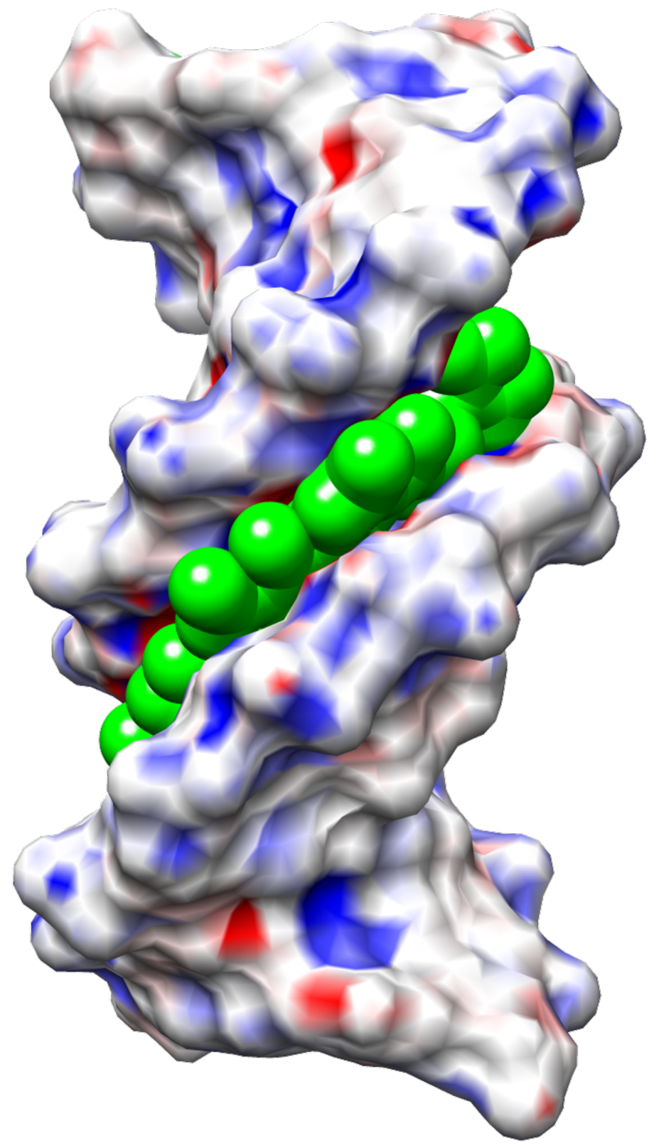
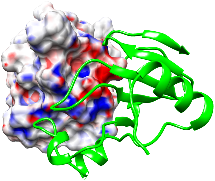
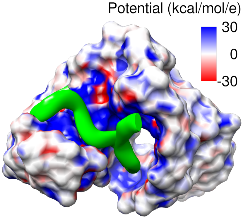
As described above, we consider three sets of binding complexes, namely, drug-DNA, barnase-barstar and RNA-peptide systems. For the sake of illustration, three sample surface electrostatic potentials, each from one distinct set, are depicted in Fig. 1. PDBIDs for these three complexes are respectively 121d (in Drug-DNA complexes), 1b3s (in barnase-barstar complexes), and 1biv (in RNA-peptide complexes). In the rest of this section, we explore the influence of grid spacing in Poisson-Boltzmann equation solvation and binding free energy estimations using our MIBPB solver.
3.1 The influence of grid spacing in estimation


| Grid sizes (pair) | Best fitting line | ||
|---|---|---|---|
| DNA-drug | (0.2,0.3) | 1.0000 | |
| (0.2,0.4) | 1.0000 | ||
| (0.2,0.5) | 1.0000 | ||
| (0.2,0.6) | 1.0000 | ||
| (0.2,0.7) | 1.0000 | ||
| (0.2,0.8) | 1.0000 | ||
| (0.2,0.9) | 1.0000 | ||
| (0.2,1.0) | 1.0000 | ||
| (0.2,1.1) | 1.0000 | ||
| Barnase-barstar | (0.2,0.3) | 1.0000 | |
| (0.2,0.4) | 1.0000 | ||
| (0.2,0.5) | 1.0000 | ||
| (0.2,0.6) | 1.0000 | ||
| (0.2,0.7) | 1.0000 | ||
| (0.2,0.8) | 1.0000 | ||
| (0.2,0.9) | 0.9999 | ||
| (0.2,1.0) | 0.9999 | ||
| (0.2,1.1) | 0.9997 | ||
| RNA-peptide | (0.2,0.3) | 1.0000 | |
| (0.2,0.4) | 1.0000 | ||
| (0.2,0.5) | 1.0000 | ||
| (0.2,0.6) | 1.0000 | ||
| (0.2,0.7) | 1.0000 | ||
| (0.2,0.8) | 1.0000 | ||
| (0.2,0.9) | 1.0000 | ||
| (0.2,1.0) | 1.0000 | ||
| (0.2,1.1) | 1.0000 |
We first examine the accuracy and robustness of our MIBPB solver in predicting the electrostatic solvation free energies of the aforementioned three data sets. Some previous literature 37, 38 has recognized that a grid size of is small enough to produce a reliable . Such an observation certainly remains for the MIBPB solver. In fact, our PB solver is able to deliver a very well-convergent calculations of electrostatic solvation free energies at as coarse grid sizes as and .
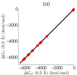
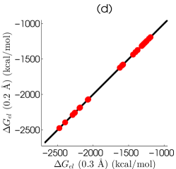
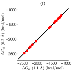
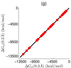
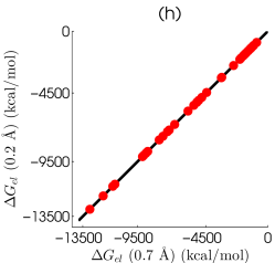
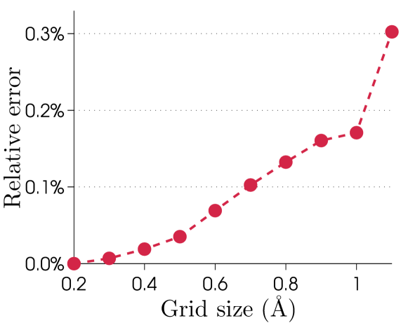
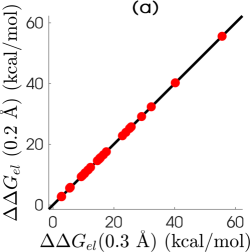
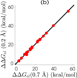
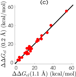
In the current calculations, the finest grid size is chosen to be , and the coarser grid sizes are between and . Figure 2 depicts the correlations of at various meshes for all complexes and unbounded components of three data sets. The electrostatic solvation free energies obtained at the finest grid spacing of are plotted against those computed from coarser grid spacings of , and . Obviously, the best fitting lines for these data at various coarse grid spacings produce near perfect alignments between the finest mesh results and those from coarse meshes. As shown in Table 1, and slope values at the pair of grid sizes for DNA-drug, barnase-barstar and RNA-peptide are, respectively, , , and . These results indicate the accuracy and robustness in the MIBPB prediction of electrostatic solvation free energies (). Table S1, in the Supporting Information, reports the values for all the 51 complexes and associated 102 components studied in this work. Finally, we examine the performance of our solver by considering the relative absolute error, the difference between results obtained with coarser and the finest grid spacings, defined as follows
| (3) |
Figure 3 illustrates the averaged relative absolute errors, i.e., the average of relative absolute errors designated in Eq. (3) over all the 153 discussed molecules, at different mesh sizes. It can be seen from Fig. 3 that the averaged relative absolute errors at all studied cases are less than , and for any grid spacing smaller than , these errors are always below . This behavior further indicates the grid size independence of our PB solver over the normal grid-size range in molecular biophysical applications.
3.2 The influence of grid spacing in estimation
Motivated by well-converged estimations of electrostatic solvation free energies at very coarse grid spacings as previously discussed, we are interested in predicting the binding free energies for all RNA-drug, barnase-barstar, and RNA-peptide complexes using our MIBPB package.


| Grid sizes (pair) | Best fitting line | ||
|---|---|---|---|
| DNA-drug | (0.2,0.3) | 1.0000 | |
| (0.2,0.4) | 0.9999 | ||
| (0.2,0.5) | 0.9998 | ||
| (0.2,0.6) | 0.9991 | ||
| (0.2,0.7) | 0.9982 | ||
| (0.2,0.8) | 0.9966 | ||
| (0.2,0.9) | 0.9906 | ||
| (0.2,1.0) | 0.9875 | ||
| (0.2,1.1) | 0.9747 | ||
| Barnase-barstar | (0.2,0.3) | 0.9999 | |
| (0.2,0.4) | 0.9995 | ||
| (0.2,0.5) | 0.9923 | ||
| (0.2,0.6) | 0.9946 | ||
| (0.2,0.7) | 0.9932 | ||
| (0.2,0.8) | 0.9883 | ||
| (0.2,0.9) | 0.9493 | ||
| (0.2,1.0) | 0.9384 | ||
| (0.2,1.1) | 0.8002 | ||
| RNA-peptide | (0.2,0.3) | 1.0000 | |
| (0.2,0.4) | 1.0000 | ||
| (0.2,0.5) | 1.0000 | ||
| (0.2,0.6) | 1.0000 | ||
| (0.2,0.7) | 0.9999 | ||
| (0.2,0.8) | 1.0000 | ||
| (0.2,0.9) | 0.9999 | ||
| (0.2,1.0) | 0.9997 | ||
| (0.2,1.1) | 0.9998 |
[b] complexes DNA-drug 102d 9.45 10.50 8.73 10.01 10.21 10.76 10.53 10.45 10.34 10.31 109d 3.61 2.18 2.30 3.72 2.63 2.07 2.66 2.69 2.82 2.72 121d 23.95 23.99 23.80 24.05 22.84 23.65 24.10 23.94 23.96 23.93 127d 27.60 28.80 28.45 28.89 28.88 28.93 29.27 29.16 29.12 29.12 129d 37.58 40.23 39.92 39.03 39.52 39.90 40.04 40.15 40.20 40.24 166d 15.04 16.60 13.97 14.93 14.91 15.49 15.47 15.62 15.67 15.67 195d 2.74 3.73 2.80 2.63 2.77 2.77 2.63 2.69 2.72 2.73 1d30 11.27 10.01 10.71 9.31 10.26 10.12 10.40 10.75 10.53 10.59 1d63 15.51 12.83 7.08 12.56 12.07 11.24 12.10 12.29 12.34 12.39 1d64 14.98 14.11 14.26 14.03 14.86 14.24 14.51 14.57 14.59 14.58 1d86 27.37 25.53 24.50 25.88 25.04 25.57 25.39 25.44 25.50 25.54 1dne 22.26 22.73 22.32 22.92 22.48 22.62 22.58 22.74 22.83 22.81 1eel 16.71 17.06 14.94 14.82 14.77 14.60 15.08 14.85 15.15 15.07 1fmq 12.35 13.40 14.28 14.72 15.27 15.09 15.30 15.36 15.36 15.37 1fms 27.08 26.14 25.17 24.52 25.41 25.93 25.82 25.75 25.71 25.74 1jtl 11.62 10.99 11.80 11.47 11.30 11.28 11.37 11.28 11.41 11.45 1lex 13.47 10.37 9.79 9.44 8.74 9.74 9.81 9.70 9.70 9.70 1prp 11.30 11.93 10.78 10.88 11.01 11.55 11.55 11.49 11.61 11.61 227d 6.28 4.79 5.96 3.79 5.47 5.16 5.80 5.75 5.46 5.58 261d 1.91 3.00 1.75 1.55 2.60 2.79 2.97 2.76 2.80 2.85 264d 33.64 32.35 31.57 30.83 32.09 31.97 32.07 32.20 32.31 32.34 289d 15.32 15.71 17.94 16.70 16.57 16.21 16.22 16.56 16.59 16.56 298d 11.65 15.94 14.81 15.89 14.87 14.88 15.38 15.45 15.50 15.41 2dbe 3.48 5.06 3.49 6.14 5.51 5.77 5.76 5.88 5.68 5.81 302d 23.28 25.22 24.91 25.49 24.95 25.00 24.87 25.29 25.17 25.19 311d 11.76 3.36 7.15 9.72 11.12 8.17 8.98 9.34 9.30 9.32 328d 14.25 16.21 17.38 17.85 16.79 17.14 17.84 17.68 17.54 17.54 360d 54.41 54.72 56.63 54.59 55.38 54.56 55.44 55.80 55.61 55.57 Barnase-barstar 1b27 86.80 83.08 95.40 82.48 87.05 89.40 87.35 87.96 86.96 87.05 1b2s 67.80 66.33 68.81 71.75 70.03 71.47 72.32 71.93 72.25 72.12 1b2u 85.97 76.39 74.78 78.29 75.29 77.35 77.15 78.38 78.87 78.57 1b3s 48.41 56.58 49.61 46.02 49.87 48.17 53.44 49.38 49.07 49.25 1x1u 61.75 66.86 74.53 75.07 77.56 76.56 75.06 76.41 76.06 75.95 1x1w 90.62 87.91 99.13 93.67 94.65 93.55 95.53 95.32 95.47 95.30 1x1x 115.79 110.40 110.45 112.19 113.51 114.45 115.30 114.65 114.62 114.65 1x1y 67.27 88.13 89.80 90.39 87.87 87.24 88.54 88.91 89.20 89.21 2za4 74.13 70.86 70.64 69.80 72.45 73.22 73.26 74.18 74.00 74.35 RNA-peptide 1a1t* 62.24 58.24 61.71 61.89 62.94 63.18 62.88 62.75 62.95 63.00 1a4t* 72.37 72.44 69.76 69.94 71.19 72.46 72.41 72.39 72.24 72.27 1biv* 41.80 40.73 42.07 44.90 45.66 44.70 44.69 44.75 44.86 44.76 1exy* 178.36 178.17 178.16 176.29 178.70 176.91 177.52 177.50 177.70 177.36 1g70 133.22 131.38 132.83 132.75 132.83 133.85 133.94 134.37 134.34 133.53 1hji 46.78 46.06 49.80 50.70 51.51 51.26 51.21 51.42 51.17 51.23 1i9f -19.78 -22.55 -22.49 -19.44 -19.31 -19.20 -19.18 -19.18 -19.20 -19.22 1mnb 126.65 129.00 127.74 126.95 127.80 127.82 128.30 128.15 128.15 128.20 1nyb -14.63 -13.07 -13.62 -12.58 -11.16 -13.04 -12.79 -12.45 -12.41 -12.61 1qfq 18.09 19.84 16.64 20.52 21.19 20.03 22.60 20.66 20.19 20.32 1ull* -53.38 -58.11 -54.20 -53.61 -51.66 -53.28 -52.52 -52.76 -52.66 -52.75 1zbn* 216.31 214.29 215.60 214.96 214.84 216.05 215.70 215.95 215.94 215.74 2a9x 385.99 384.41 385.18 385.36 385.05 385.20 385.36 385.36 385.43 385.44 484d* 129.72 129.65 133.42 132.09 131.34 132.03 132.79 133.20 133.23 133.38
-
*
Results are significantly different (>50 kcal/mol) from those in Ref. 15.
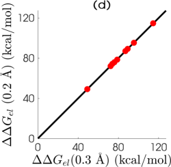
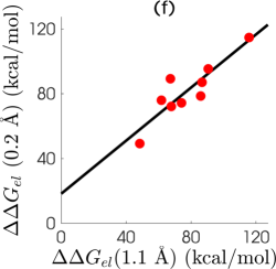
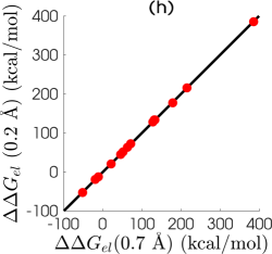
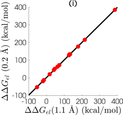
Similar to the study of the convergence of , we correlate the binding free energy calculated at the finest grid spacing, , and ones estimated at coarser mesh sizes, . Figure 4 illustrates these relationships with the regression lines whose parameters are revealed in Table 2. Since the previous discussion confirms MIBPB solver can produce very good R-squared values even at very coarse grid spacings, it is interesting to explore whether a similar behavior can be found for binding energy estimation. Indeed, the PB binding energy estimation behaves the same as the PB solvation calculation in our MIBPB technique. Specifically, is always at the fine mesh, . Moreover, these values are still satisfactory at relatively coarser mesh sizes. For example, at the grid spacing of , the and slope of the regression line for DNA-drug, barnase-barstar, and RNA-peptide complexes are, respectively, (0.9747,1.0081), (0.8002,0.8187), and (0.9998, 0.9937). In contrast, the R-squared values reported in Ref. 15, computed between and , are unacceptable for SESs, and usually less than . Our statistical measures strongly support the reliable binding energy prediction of our solver at coarse grid sizes. Table 3 displays the binding free energy for all complexes with different grid spacings. As can be seen from Table 3, the difference between binding energies at coarse meshes and the finest mesh, , is mostly less than 10 kcal/mol for all complexes.
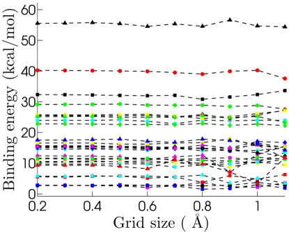
The trend of binding free energy at different grid spacings can be seen clearly in Figs. 5 which plots against grid sizes varying between and for DNA-drug complexes. Similar figures for barnase-barstar and RNA-peptide complexes can be referred to Figs. S1 and S2 in the Supporting Information. Based on these figures, our solver can rank the binding free energy for DNA-drug complexes at grid spacing of , barnase-barstar complexes at grid spacing of , and RNA-peptide complexes at significantly coarse grid spacing of . To further assess the reliable estimates of binding energy of our MIBPB solver, we consider the absolute difference between results computed at a coarser grid spacing and the finest grid spacing defined by
| (4) |
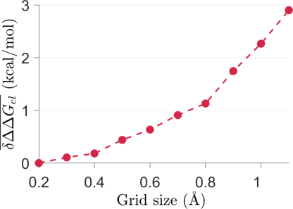
Figure 6 plots the averaged absolute errors, , i.e., the average of absolute errors defined in Eq. (4) over all 51 complexes, at different mesh sizes. It is seen that even the use of grid spacing of still delivers an averaged binding calculation error under 1 kcal/mol for this set of complexes. Therefore, we can draw a conclusion that the common use of grid size being is still adequate for predicting the binding energy free without producing a misleading result.
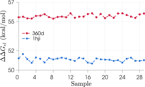
Grid positioning error is another feature to validate the robustness and accuracy of a PB solver. To examine such numerical error for our MIBPB solver, we consider two protein complexes with PDBIDs: 360d and 1hji. To estimate the standard deviation, in , we randomly generate 29 grid positions around the initial origin with the amplitude of the random seed being , where is the grid spacing. Then is evaluated at all of the 30 grid positions. Figure 7 plots electrostatic binding energies at 30 distinct samples of grid positions, including the original one marked by Sample 0 on the graph. The values of complexes 360d and 1hji are found to be 0.18 and 0.21, respectively. These results indicate that the MIBPB solver is not sensitive to grid position.
Note that in Table 3, some binding energies obtained in our calculations, all from RNA-peptide complexes, differ significantly from those reported in Ref. 15, i.e., more than 50 kcal/mol at the finest grid spacing, under the same parametrization and data inputs. However, overall, our results have a good agreement with those in Ref. 15 with for RNA-peptide complexes. To further support our calculations, we have employed PBSA, Delphi, and APBS for electrostatic energy calculations at the grid size of 0.2 Å. We note that results obtained from these solvers are in excellent agreements, i.e., , with ours. The electrostatic energies calculated by PBSA, Delphi, and APBS solvers are listed in Table S2 of Supporting Information.
4 Concluding remarks
Poisson-Boltzmann (PB) theory is an established model for biomolecular electrostatic analysis and has been widely used in electrostatic solvation and binding energy estimations. However, doubt has been cast on the validity of the commonly used grid spacing of 0.5 Å for producing converged estimates of due to the unacceptable errors observed in the calculation using the solvent excluded surface (SES) and the adaptive Cartesian grid (ACG) finite difference PB equation solver 15. Three sets of biomolecular complexes, namely, DNA-drug complexes, barnase-barstar complexes, and RNA-peptide complexes, are employed in the study. The discrepancies between results obtained from different surface definitions were also utilized to support the general pessimism for the PB methodology.
In this work, we employ the MIBPB software 21, 32 to estimate electrostatic solvation free energy, , and binding free electrostatic energy, , for the three sets of biomolecular complexes used in Ref. 15. The popular SES is adopted in the present work. In our estimation, the averaged relative absolute error computed at a relatively coarse grid size of against the finest grid size of 0.2 Å over 153 studied biomolecules is less than 0.31%. The same error obtained at the grid size of is less than 0.2%. These results indicate the reliability of using the MIBPB solver at the grid spacing of or even for electrostatic solvation analysis. The robustness and accuracy of MIBPB solver for estimates of have been reported for 24 proteins in the literature 21, 32. This characteristics has been confirmed again in the present work for DNA-drug complexes, barnase-barstar complexes, and RNA-peptide complexes.
The well-converged produced by our solver enables a promising performance in predicting at a coarse grid spacing. Indeed, numerical estimates of in the current work reveals that obtained at a grid spacing mostly differ by less than 10 kcal/mol from that achieved by using a grid spacing. Moreover, MIBPB solver conducted at grid size of perfectly produces a well-converged , and qualitatively ranks the complexes in term of their binding free energies. Therefore, the current results support an opinion that the widely used grid size of can give reliable and accurate enough predictions of both electrostatic free energy 39, 40 and binding free energy.
To develop highly accurate, robust and reliable PB solvers for biomolecular electrostatics, it is crucial to validate one’s numerical methods by appropriate norms and against realistic problems. We emphasize that as an elliptic interface problem, it is important to measure the convergence of PB solvers in the norm, or maximum absolute error, because integral norms, such as and , are insensitive to the performance of numerical methods near the interface. Additionally, the convergence should be tested by solving the PB equation, rather than by calculating the solvation free energy. Finally, validation should be carried out by using the SESs of proteins, rather than smooth surfaces, such as a sphere.
Electrostatic free energies calculated by different solvers, namely MIBPB, DELPHI, PBSA and APBS; Coulombic binding energies; binding energy plots for barnase-barstar and RNA-peptide complexes (filename: \urljctc_si_bdenergy.pdf).
This work was supported in part by NSF Grant Nos. IIS- 1302285 and DMS-1160352, NIH Grant No. R01GM-090208 and MSU Center for Mathematical Molecular Biosciences Initiative. DDN and GWW thank the Mathematical Biosciences Institute for its hospitality and support during their visit in Ohio State University, where this manuscript was finalized. This manuscript was reviewed by Professors Emil Alexov and Ray Luo prior its submission.
References
- Gilson et al. 1987 Gilson, M. K.; Sharp, K.; Honig, B. Calculating the Electrostatic Potential of Molecules in Solution: Method and error assessment. J. Comp. Chem. 1987, 9, 327–335
- Gilson et al. 1993 Gilson, M. K.; Davis, M. E.; Luty, B. A.; McCammon, J. A. Computation of electrostatic forces on solvated molecules using the Poisson-Boltzmann equation. Journal of Physical Chemistry 1993, 97, 3591–3600
- Talley et al. 2008 Talley, K.; Ng, C.; Shoppell, M.; Kundrotas, P.; Alexov, E. On the electrostatic component of protein-protein binding free energy. PMC Biophysics 2008, 1:2, 1–23
- Zhou et al. 2008 Zhou, Y. C.; Feig, M.; Wei, G. W. Highly accurate biomolecular electrostatics in continuum dielectric environments. Journal of Computational Chemistry 2008, 29, 87–97
- Ren et al. 2012 Ren, P.; Chun, J.; Thomas, D. G.; Schnieders, M. J.; Marucho, M.; Zhang, J.; Baker, N. A. Biomolecular electrostatics and solvation: a computational perspective. Quarterly reviews of biophysics 2012, 45(04), 427–491
- Jo et al. 2008 Jo, S.; Vargyas, M.; Vasko-Szedlar, J.; Roux, B.; Im, W. PBEQ-Solver for online visualization of electrostatic potential of biomolecules. Nucleic Acids Research 2008, 36, W270 –W275
- Baker et al. 2001 Baker, N. A.; Sept, D.; Holst, M. J.; Mccammon, J. A. The adaptive multilevel finite element solution of the Poisson-Boltzmann equation on massively parallel computers. IBM Journal of Research and Development 2001, 45, 427–438
- Geng and Krasny 2013 Geng, W. H.; Krasny, R. A treecode-accelerated boundary integral Poisson-Boltzmann solver for continuum electrostatics of solvated biomolecules. J. Comput. Phys. 2013, 247, 62–87
- Lu et al. 2013 Lu, B.; Cheng, X.; Huang, J.; McCammon, J. A. AFMPB: An Adaptive Fast Multipole Poisson-Boltzmann Solver for Calculating Electrostatics in Biomolecular Systems. Comput. Phys. Commun. 2013, 184, 2618–2619
- Wang et al. 2008 Wang, J.; Tan, C. H.; Tan, Y. H.; Lu, Q.; Luo, R. Poisson-Boltzmann solvents in molecular dynamics Simulations. Communications in Computational Physics 2008, 3(5), 1010–1031
- Cai et al. 2009 Cai, Q.; Hsieh, M. J.; Wang, J.; Luo, R. Performance of Nonlinear Finite-Difference Poisson-Boltzmann Solvers. Journal of Chemical Theory and Computation 2009, 6(1), 203–211
- Li et al. 2012 Li, L.; Li, C.; Sarkar, S.; Zhang, J.; Witham, S.; Zhang, Z.; Wang, L.; Smith, N.; Petukh, M.; Alexov, E. Delphi: a comprehensive suite for Delphi software and associated resources. BMC Biophysics 2012, 5:9, 2046–1682
- Rocchia et al. 2002 Rocchia, W.; Sridharan, S.; Nicholls, A.; Alexov, E.; Chiabrera, A.; Honig, B. Rapid grid-based construction of the molecular surface and the use of induced surface charge to calculate reaction field energies: Applications to the molecular systems and geometric objects. Journal of Computational Chemistry 2002, 23, 128 – 137
- Baker et al. 2001 Baker, N. A.; Sept, D.; Joseph, S.; Holst, M. J.; McCammon, J. A. Electrostatics of nanosystems: Application to microtubules and the ribosome. Proceedings of the National Academy of Sciences of the United States of America 2001, 98, 10037–10041
- Harris et al. 2013 Harris, R. C.; Boschitsch, A. H.; Fenley, M. O. Influence of Grid Spacing in Poisson-Boltzmann Equation Binding Energy Estimation. Journal of Chemical Theory and Computation 2013, 9, 3677–3685
- Boschitsch and Fenley 2015 Boschitsch, A. H.; Fenley, M. O. Chapter 4, The Adaptive Cartesian Grid-Based Poisson–Boltzmann Solver: Energy and Surface Electrostatic Properties, in Computational Electrostatics for Biological Applications: Geometric and Numerical Approaches to the Description of Electrostatic Interaction Between Macromolecules, Rocchia, W., Spagnuolo, M., Eds. Cham, 2015 2015, 73–110
- Sorensen et al. 2015 Sorensen, J.; Fenley, M. O.; Amaro, R. E. Chapter 3, A Comprehensive Exploration of Physical and Numerical Parameters in the Poisson-Boltzmann Equation for Applications to Receptor-Ligand Binding, in Computational Electrostatics for Biological Applications: Geometric and Numerical Approaches to the Description of Electrostatic Interaction Between Macromolecules, Rocchia, W., Spagnuolo, M., Eds. Cham, 2015 2015, 39–71
- Fenley et al. 2015 Fenley, M. O.; Harris, R. C.; Mackoy, T.; Boschitsch, A. H. Features of CPB: A Poisson–Boltzmann solver that uses an adaptive cartesian grid. J. Comput. Chem. 2015, 36, 235–243
- Wang et al. 2014 Wang, C.; Wang, J.; Cai, Q.; Li, Z.; Zhao, H. K.; Luo, R. Exploring accurate Poisson–Boltzmann methods for biomolecular simulations. Comput. Theor. Chem. 2014, 1:2, 34–44
- Mirzadeh et al. 2011 Mirzadeh, M.; Theillard, M.; Gibou, F. A second-order discretization of the nonlinear Poisson–Boltzmann equation over irregular geometries using non-graded adaptive Cartesian grids. J. Comput. Phys. 2011, 230, 2125–2140
- Chen et al. 2011 Chen, D.; Chen, Z.; Chen, C.; Geng, W. H.; Wei, G. W. MIBPB: A software package for electrostatic analysis. J. Comput. Chem. 2011, 32, 657 – 670
- Wang et al. 2015 Wang, B.; Nguyen, D. D.; Zhao, Z.; Cang, Z.; Wei, G.-W. MIBPB: An accurate and reliable software package for electrostatic analysis. Preprint 2015,
- Richards 1977 Richards, F. M. Areas, Volumes, Packing, and Protein Structure. Annual Review of Biophysics and Bioengineering 1977, 6, 151–176
- Connolly 1983 Connolly, M. L. Analytical molecular surface calculation. Journal of Applied Crystallography 1983, 16, 548–558
- Sanner et al. 1996 Sanner, M. F.; Olson, A. J.; Spehner, J. C. Reduced surface: An efficient way to compute molecular surfaces. Biopolymers 1996, 38, 305–320
- Zhou and Wei 2006 Zhou, Y. C.; Wei, G. W. On the fictitious-domain and interpolation formulations of the matched interface and boundary (MIB) method. J. Comput. Phys. 2006, 219, 228–246
- Zhou et al. 2006 Zhou, Y. C.; Zhao, S.; Feig, M.; Wei, G. W. High order matched interface and boundary method for elliptic equations with discontinuous coefficients and singular sources. J. Comput. Phys. 2006, 213, 1–30
- Yu et al. 2007 Yu, S. N.; Geng, W. H.; Wei, G. W. Treatment of geometric singularities in implicit solvent models. Journal of Chemical Physics 2007, 126, 244108
- Yu et al. 2007 Yu, S. N.; Zhou, Y. C.; Wei, G. W. Matched interface and boundary (MIB) method for elliptic problems with sharp-edged interfaces. J. Comput. Phys. 2007, 224, 729–756
- Yu and Wei 2007 Yu, S. N.; Wei, G. W. Three-dimensional matched interface and boundary (MIB) method for treating geometric singularities. J. Comput. Phys. 2007, 227, 602–632
- Xia et al. 2014 Xia, K. L.; Zhan, M.; Wei, G. W. MIB Galerkin method for elliptic interface problems. Journal of Computational and Applied Mathematics 2014, 272, 195– 220
- Geng et al. 2007 Geng, W.; Yu, S.; Wei, G. W. Treatment of charge singularities in implicit solvent models. Journal of Chemical Physics 2007, 127, 114106
- Decherchi and Rocchia 2013 Decherchi, S.; Rocchia, W. A general and robust ray-casting-based algorithm for triangulating surfaces at the nanoscale. PLoS ONE 2013, 8, e59744
- Liu et al. 2015 Liu, B.; Wang, B.; Zhao, R.; Tong, Y.; Wei, G. W. ESES: software for Eulerian solvent excluded surface. Preprint 2015,
- Harris et al. 2011 Harris, R. C.; Bredenberg, J. H.; Silalahi, A. R. J.; Boschitsch, A. H.; Fenley, M. O. Understanding the physical basis of the salt dependence of the electrostatic binding free energy of mutated charged ligand-nucleic acid complexes. Biophysical Chemistry 2011, 156, 79–87
- Pettersen et al. 2004 Pettersen, E. F.; Goddard, T. D.; Huang, C. C.; Couch, G. S.; Greenblatt, D. M.; Meng, E. C.; Ferrin, T. E. UCSF Chimera–a visualization system for exploratory research and analysis. J. Comput. Chem. 2004, 25, 1605–1612
- Feig et al. 2004 Feig, M.; Onufriev, A.; Lee, M. S.; Im, W.; Case, D. A.; Brooks, I., C. L. Performance comparison of generalized Born and Poisson methods in the calculation of electrostatic solvation energies for protein structures. Journal of Computational Chemistry 2004, 25, 265–284
- Reyes and Kollman 2000 Reyes, C. M.; Kollman, P. A. Structure and thermodynamics of RNA-protein binding: using molecular dynamics and free energy analyses to calculate the free energies of binding and conformational change. Journal of molecular biology 2000, 297, 1145–1158
- Nicholls et al. 2008 Nicholls, A.; Mobley, D. L.; Guthrie, J. P.; Chodera, J. D.; Bayly, C. I.; Cooper, M. D.; Pande, V. S. Predicting small-molecule solvation free energies: an informal blind test for computational chemistry. Journal of Medicinal Chemistry 2008, 51, 769–779
- Shivakumar et al. 2010 Shivakumar, D.; Williams, J.; Wu, Y.; Damm, W.; Shelley, J.; Sherman, W. Prediction of absolute solvation free energies using molecular dynamics free energy perturbation and the OPLS force field. Journal of Chemical Theory and Computation 2010, 6, 1509–1519
![[Uncaptioned image]](/html/1603.04054/assets/x26.png)