A quantum spectrum analyzer enhanced by a nuclear spin memory
Abstract
We realize a two-qubit sensor designed for achieving high spectral resolution in quantum sensing experiments. Our sensor consists of an active “sensing qubit” and a long-lived “memory qubit”, implemented by the electronic and the nitrogen-15 nuclear spins of a nitrogen-vacancy center in diamond, respectively. Using state storage times of up to 45 ms, we demonstrate spectroscopy of external ac signals with a line width of 19 Hz () and of carbon-13 nuclear magnetic resonance (NMR) signals with a line width of 190 Hz (). This represents an up to 100-fold improvement in spectral resolution compared to measurements without nuclear memory.
Quantum sensors based on nitrogen-vacancy centers in diamond show promise for a number of fascinating applications in condensed matter physics, materials science and biology Schirhagl et al. (2014); Rondin et al. (2014). By embedding them in a variety of nanostructures, such as tips Degen (2008); Balasubramanian et al. (2008); Rondin et al. (2012); Maletinsky et al. (2012), nanocrystals Dussaux et al. (2016) or surface layers Kolkowitz et al. (2015); Dussaux et al. (2016), local properties of samples can be investigated with high sensitivity and spatial resolution. In particular, diamond chips with near-surface NV centers have enabled pioneering experiments in nanoscale detection of nuclear magnetic resonance (NMR), potentially enabling structural analysis of individual molecules with atomic resolution Ajoy et al. (2015); Lazariev and Balasubramanian (2015); Kost et al. (2015).
A key feature of many quantum sensing experiments is the ability to record time-dependent signals and to reconstruct their frequency spectra. The canonical approach uses dynamical decoupling sequences, which are sensitive to frequencies commensurate with the pulse spacing while efficiently rejecting all other frequencies Cywinski et al. (2008); Lange et al. (2011); Kotler et al. (2011). The spectral resolution of dynamical decoupling spectroscopy, however, is inherently limited by the inverse of the decoherence time , which is about for shallow NV centers Loretz et al. (2014). It has recently been recognized that by correlating two consecutive decoupling sequences, separated by a variable waiting time , the spectral resolution can be extended to the inverse state life time , which can be longer than (Refs. Laraoui et al., 2011, 2013). Correlation spectroscopy has been applied to both generic ac magnetic fields and to nuclear spin detection, and spectral resolutions of a few 100 Hz have been demonstrated Staudacher et al. (2015); Kong et al. (2015); Boss et al. (2016).
Despite these impressive advances there is a strong motivation to further extend the spectral resolution. For example, many proposed nanoscale NMR experiments Ajoy et al. (2015); Lazariev and Balasubramanian (2015); Kost et al. (2015) require discrimination of fine spectral features, often in the few-Hz range. In addition, atomic-scale mapping of nuclear spin positions strongly relies on precise measurements of NMR frequencies and hyperfine coupling constants Boss et al. (2016). Therefore, methods to acquire frequency spectra with even higher spectral resolution are highly desirable.
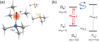
In this study, we implement a two-qubit sensor designed to further refine the spectral resolution by a factor of . Our two-qubit sensor consists of an active sensing qubit and an auxiliary memory qubit, formed by the electronic spin and the 15N nuclear spin of the NV center in diamond. By intermittently storing the state information in the nuclear – rather than electronic – spin qubit, we extend the maximum waiting time from to the nuclear , with a corresponding gain in spectral resolution. In addition, we use the nuclear memory to enhance sensor readout efficiency through repeated readout Jiang et al. (2009); Lovchinsky et al. (2016), which would otherwise result in untenably long acquisition times. The presented two-qubit system is particularly useful because it is intrinsic to the NV center, with no need for additional sensor engineering.
The advantage of one or more “auxiliary” qubits has been recognized in several recent works. In particular, auxiliary nuclear spins have been used to increase the effective coherence time of an electronic sensor spin by quantum error correction Unden et al. (2016), quantum feedback Hirose and Cappellaro (2016) or by exploiting double-quantum coherence Zaiser et al. (2016). Moreover, ancillary nuclei have been used to enhance the readout efficiency Jiang et al. (2009); Lovchinsky et al. (2016). In our study we utilize the auxiliary nuclear spin as a long-lived memory for the electron qubit’s state.
Our two-qubit sensor exploits the four-level system formed by the subspace of the electronic spin and the two states of the nuclear spin. This pair has four allowed spin-flip transitions (see Fig. 1). Due to the hyperfine interaction (), all four transitions are spectrally resolved and can be addressed individually using frequency-selective microwave or rf pulses. Driving a selective rotation on either of theses transitions leads to a conditional inversion, depending on the state of the other spin. This realizes controlled-NOT gates on the electronic and nuclear spins, respectively, which we denote by c-NOTe and c-NOTn .
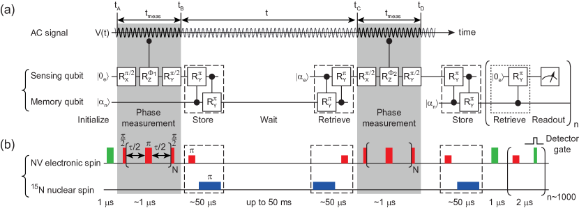
To implement the “store” and “retrieve” operations, we combine a c-NOTe gate and c-NOTn gate (dashed boxes in Fig. 2). Assuming the electronic spin is initially in the state and the nuclear spin in an idle (unspecified) state , the effect of the two gates is , where etc. denote product states. Likewise, if the electronic spin is initially in the state, . As a result, the state of the electronic spin is stored in the state of the nuclear spin. To retrieve the state, the order of the c-NOT gates simply needs to be reversed (see Fig. 2). Alternatively, the state can also be retrieved by initializing the electronic spin followed by a single c-NOTe gate (dotted box in Fig. 2). Opposite to the double c-NOT implementation, this protocol leaves the nuclear memory relatively unperturbed such that the memory state can be retrieved and read out many times Jiang et al. (2009); Neumann et al. (2010).
We assess the performance of the nuclear spin memory under a set of store, retrieve and hold operations. To characterize the efficiency of the store and retrieve operations, we perform selective Rabi rotations on all four spin-flip transitions, and find efficiencies for c-NOTe and for c-NOTn , respectively sup . The memory access time is between for the double c-NOT implementation, limited by the duration of the rf pulse, and for the single c-NOT implementation. We further test the complete memory by performing an electronic Rabi oscillation, storing the result in the nuclear memory, clearing the electronic qubit by an initialization step, and retrieving the Rabi signal sup . Lastly, we assess the memory hold time – given by the nuclear – in the absence and presence of laser illumination, with typical values of (no laser) and (under periodic readout) at a bias field of . This bias field supports non-destructive read outs of the memory (dotted box in Fig. 2) before the nuclear spin becomes repolarized sup ; Neumann et al. (2010).
We compose the full spectroscopy protocol from a correlation sequence Laraoui et al. (2013); Boss et al. (2016) and several storage and retrieval operations (Fig. 2). In a first step, we initialize the electronic sensor spin into the state. An initial phase measurement is then performed using a multipulse sensing sequence approximately tuned to the frequency of the ac field (see Fig. 2(b)). During the multipulse sequence, the ac signal imprints a phase on the electronic qubit, leaving it in a superposition of states and with a probability amplitude . Next, we store in the nuclear memory, wait for a variable delay time (which can be very long), and read it back. A second phase measurement is then used to acquire a further phase . In a last step, we read out the final state of the electronic qubit via storing it in the nuclear memory and performing periodic readouts. By averaging the protocol over many repetitions, the probability of finding the sensor in the initial state can be precisely estimated.
Because and depend on the relative phase of the ac signal , the total phase acquired by the qubit oscillates with . As detailed in the Supplemental Material sup , the resulting state probability then also oscillates with ,
| (1) | ||||
| (2) |
where we assume that the ac signal is not synchronized with the acquisition. Eq. (2) is for small signals where and , resulting in an oscillation amplitude where (see Fig. 2). In order to obtain a frequency spectrum of , we can therefore simply measure for a series of values followed by a Fourier transform.
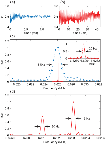
We demonstrate the performance of the memory-enhanced spectrometer for two experimental scenarios. In a first experiment, we expose the sensor to an external ac test signal with a nominal frequency of and an amplitude of . The test signal is produced on an auxiliary function generator not synchronized with the acquisition, and coupled into the same waveguide structure used for spin control. Two measurements are carried out: in a first acquisition (Fig. 3(a)) we perform a regular spectroscopy measurement without the nuclear memory. We can clearly observe an oscillation in the time trace due to the ac signal. The signal decays on a time scale of , limited by the electronic of this NV center. In Fig. 3(b) we repeat the measurement, now making use of the nuclear memory. The oscillation persists beyond , overcoming the limitation due to the electronic by almost two orders of magnitude.
Fourier spectra of the two time signals (Fig. 3(c)) show that the peak width reduces from () without memory to () with memory. This corresponds to an improvement in spectral resolution by . Fig. 3(d) shows a second example of nuclear-memory-assisted spectroscopy, where two ac test signals separated by about are applied. Both peaks can be clearly distinguished, demonstrating that the method is effective in precisely resolving spectral features. A narrow line width of only () is observed, and peak positions are defined with 8 digits of precision. The absolute accuracy of the frequency measurement is governed by the internal clock of the microwave pulse generator.
The spectral resolution in Fig. 3(c,d) is limited by the memory hold time given by the nuclear , here . Since the nuclear relaxation is dominated by a flip-flop process with the NV center’s electron spin and slows down for higher bias fields Neumann et al. (2010), there is scope for an additional improvement in spectral resolution at Tesla bias fields Pfender et al. .
We further apply the two-qubit sensor to detect NMR spectra from nearby 13C nuclear spins that are naturally present at in the diamond chip. This experiment represents an important test case towards the detection of more complex NMR spectra, such as those from molecules deposited on the chip Mamin et al. (2013); Staudacher et al. (2013); Loretz et al. (2014). The detection of nuclear spin signals is considerably more involved compared to external ac signals because the electronic sensor spin remains coupled to the nuclear spins during and causes back-action on the nuclear evolution. In addition, NMR spectroscopy is known to be very sensitive to drifts in the external bias field.
Fig. 4 shows a set of four NMR spectra recorded from the same 13C nuclear spin using the memory-assisted spectroscopy protocol. The four panels represent increasing refinements in spectrum acquisition. Fig. 4(a) shows an initial 13C spectrum that displays features over a wide frequency range of several kHz (). We find these features to be linked to small changes in the bias field, probably caused by temperature-induced drifts in the magnetization of the permanent magnet in our setup. By carefully tracking the electron spin resonance during the experiment and using post-correction, these drifts can be eliminated (Fig. 4(b)) sup .
The remaining line width of the 13C resonance is on the order of , which corresponds to of this NV center. This reflects the fact that due to the hyperfine interaction between the electronic and 13C spins. Thus, even if our spectrometer is technically capable of achieving a better spectral resolution, this improvement does not carry over to the 13C spectrum.
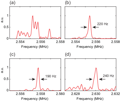
To further reduce the line width of the 13C resonance, we have explored several decoupling protocols, neither of which turned out to be effective. A first protocol (Fig. 4(b)) includes a series of pulses on the transition. However, this protocol does not decouple the and transitions, and only a marginal improvement can be expected. Indeed, no significant change is observed in the 13C line width without and with dynamical decoupling even when using many hundred decoupling pulses (Fig. 4(c)). A more effective approach would be to simultaneously decouple both the and transitions using double-frequency irradiation Mamin et al. (2014), but this control is not currently supported by our hardware. Instead, we use a series of laser pulses to periodically repolarize the NV center into the state (Fig. 4(d)) or the state (data not shown). No narrowing of the 13C resonance is observed with either protocol. Hence, we speculate that the residual line width is either because decoupling is ineffective due to the relatively strong hyperfine coupling sup , or due to interactions with other 13C nuclei in the diamond chip. The latter could be addressed by homonuclear decoupling sequences in future experiments Slichter (1990); Maurer et al. (2012).
In summary, we implemented a two-qubit quantum sensor based on the electronic and 15N nuclear spins in diamond. By operating the nuclear spin as a long-lived quantum memory, we achieve exceptionally high spectral resolution, with a best effort of or . The 15N spin forms a particularly suitable memory qubit because the nucleus is a natural part of the NV center, and because long storage times are possible combined with rapid memory access. The ability to sense signals with high spectral resolution supports recent strides at detecting nuclear spin signals of nanoscale sample volumes, with possible applications in single-molecule NMR spectroscopy.
The authors thank Kristian Cujia for fruitful discussions and Nicole Raatz, Sebastien Pezzagna and Jan Meijer for help with sample preparation. This work was supported by Swiss NSF Project Grant , the NCCR QSIT, and the DIADEMS programme 611143 of the European Commission.
References
- Schirhagl et al. (2014) R. Schirhagl, K. Chang, M. Loretz, and C. L. Degen, Annu. Rev. Phys. Chem. 65, 83 (2014).
- Rondin et al. (2014) L. Rondin, J. P. Tetienne, T. Hingant, J. F. Roch, P. Maletinsky, and V. Jacques, Rep. Prog. Phys. 77, 056503 (2014).
- Degen (2008) C. L. Degen, Appl. Phys. Lett. 92, 243111 (2008).
- Balasubramanian et al. (2008) G. Balasubramanian, I. Y. Chan, R. Kolesov, M. Al-Hmoud, J. Tisler, C. Shin, C. Kim, A. Wojcik, P. R. Hemmer, A. Krueger, T. Hanke, A. Leitenstorfer, R. Bratschitsch, F. Jelezko, and J. Wrachtrup, Nature 455, 648 (2008).
- Rondin et al. (2012) L. Rondin, J. P. Tetienne, P. Spinicelli, C. dal Savio, K. Karrai, G. Dantelle, A. Thiaville, S. Rohart, J. F. Roch, and V. Jacques, Appl. Phys. Lett. 100, 153118 (2012).
- Maletinsky et al. (2012) P. Maletinsky, S. Hong, M. S. Grinolds, B. Hausmann, M. D. Lukin, R. L. Walsworth, M. Loncar, and A. Yacoby, Nat. Nanotechnol. 7, 320 (2012).
- Dussaux et al. (2016) A. Dussaux, P. Schoenherr, K. Koumpouras, J. Chico, K. Chang, L. Lorenzelli, N. Kanazawa, Y. Tokura, M. Garst, A. Bergman, C. L. Degen, and D. Meier, Nature Communications 7, 12430 (2016).
- Kolkowitz et al. (2015) S. Kolkowitz, A. Safira, A. A. High, R. C. Devlin, S. Choi, Q. P. Unterreithmeier, D. Patterson, A. S. Zibrov, V. E. Manucharyan, H. Park, and M. D. Lukin, Science 347, 1129 (2015).
- Ajoy et al. (2015) A. Ajoy, U. Bissbort, M. D. Lukin, R. L. Walsworth, and P. Cappellaro, Physical Review X 5, 011001 (2015).
- Lazariev and Balasubramanian (2015) A. Lazariev and G. Balasubramanian, Scientific Reports 5, 14130 (2015).
- Kost et al. (2015) M. Kost, J. Cai, and M. B. Plenio, Scientific Reports 5, 11007 (2015).
- Cywinski et al. (2008) L. Cywinski, R. M. Lutchyn, C. P. Nave, and S. D. Sarma, Phys. Rev. B 77, 174509 (2008).
- Lange et al. (2011) G. D. Lange, D. Riste, V. V. Dobrovitski, and R. Hanson, Phys. Rev. Lett. 106, 080802 (2011).
- Kotler et al. (2011) S. Kotler, N. Akerman, Y. Glickman, A. Keselman, and R. Ozeri, Nature 473, 61 (2011).
- Loretz et al. (2014) M. Loretz, S. Pezzagna, J. Meijer, and C. L. Degen, Appl. Phys. Lett. 104, 33102 (2014).
- Laraoui et al. (2011) A. Laraoui, J. S. Hodges, C. A. Ryan, and C. A. Meriles, Phys. Rev. B 84, 104301 (2011).
- Laraoui et al. (2013) A. Laraoui, F. Dolde, C. Burk, F. Reinhard, J. Wrachtrup, and C. A. Meriles, Nature Commun. 4, 1651 (2013).
- Staudacher et al. (2015) T. Staudacher, N. Raatz, S. Pezzagna, J. Meijer, F. Reinhard, C. A. Meriles, and J. Wrachtrup, Nature Commun. 6 (2015), 10.1038/ncomms9527.
- Kong et al. (2015) X. Kong, A. Stark, J. Du, L. P. McGuinness, and F. Jelezko, Phys. Rev. Applied 4, 024004 (2015).
- Boss et al. (2016) J. M. Boss, K. Chang, J. Armijo, K. Cujia, T. Rosskopf, J. R. Maze, and C. L. Degen, Phys. Rev. Lett. 116, 197601 (2016).
- Jiang et al. (2009) L. Jiang, J. S. Hodges, J. R. Maze, P. Maurer, J. M. Taylor, D. G. Cory, P. R. Hemmer, R. L. Walsworth, A. Yacoby, A. S. Zibrov, and M. D. Lukin, Science 326, 267 (2009).
- Lovchinsky et al. (2016) I. Lovchinsky, A. O. Sushkov, E. Urbach, N. P. de Leon, S. Choi, K. de Greve, R. Evans, R. Gertner, E. Bersin, C. Muller, L. McGuinness, F. Jelezko, R. L. Walsworth, H. Park, and M. D. Lukin, Science 351, 836 (2016).
- (23) See Supplemental Material accompanying this manuscript .
- Gullion et al. (1990) T. Gullion, D. B. Baker, and M. S. Conradi, J. Magn. Res. 89, 479 (1990).
- Unden et al. (2016) T. Unden, P. Balasubramanian, D. Louzon, Y. Vinkler, M. B. Plenio, M. Markham, D. Twitchen, A. Stacey, I. Lovchinsky, A. O. Sushkov, M. D. Lukin, A. Retzker, B. Naydenov, L. P. McGuinness, and F. Jelezko, Physical Review Letters 116, 230502 (2016).
- Hirose and Cappellaro (2016) M. Hirose and P. Cappellaro, Nature 532, 77 (2016).
- Zaiser et al. (2016) S. Zaiser, T. Rendler, I. Jakobi, T. Wolf, S. Lee, S. Wagner, V. Bergholm, T. Schulte-herbruggen, P. Neumann, and J. Wrachtrup, Nature Communications 7, 12279 (2016).
- Neumann et al. (2010) P. Neumann, J. Beck, M. Steiner, F. Rempp, H. Fedder, P. R. Hemmer, J. Wrachtrup, and F. Jelezko, Science 329, 542 (2010).
- (29) M. Pfender, N. Aslam, P. Neumann, and J. Wrachtrup, in preparation , 0.
- Mamin et al. (2013) H. J. Mamin, M. Kim, M. H. Sherwood, C. T. Rettner, K. Ohno, D. D. Awschalom, and D. Rugar, Science 339, 557 (2013).
- Staudacher et al. (2013) T. Staudacher, F. Shi, S. Pezzagna, J. Meijer, J. Du, C. A. Meriles, F. Reinhard, and J. Wrachtrup, Science 339, 561 (2013).
- Mamin et al. (2014) H. Mamin, M. Sherwood, M. Kim, C. Rettner, K. Ohno, D. Awschalom, and D. Rugar, Phys. Rev. Lett. 113, 030803 (2014).
- Slichter (1990) C. P. Slichter, Principles of Magnetic Resonance, 3rd edition (Springer, Berlin, 1990).
- Maurer et al. (2012) P. C. Maurer, G. Kucsko, C. Latta, L. Jiang, N. Y. Yao, S. D. Bennett, F. Pastawski, D. Hunger, N. Chisholm, M. Markham, D. J. Twitchen, J. I. Cirac, and M. D. Lukin, Science 336, 1283 (2012).