Whisker growth on Sn thin film accelerated under gamma-ray irradiation
Abstract
We report on the growth of tin (Sn) metal whiskers that is significantly accelerated under gamma-ray irradiation. The studied Sn thin film, evaporated on glass substrate, was subjected to a total of 60 hours of irradiation over the course of 30 days. The irradiated sample demonstrated enhanced whisker development, in both densities and lengths, resulting in an acceleration factor of 50. We attribute the observed enhancement to gamma-ray induced electrostatic fields, affecting whisker growth kinetics. These fields are due to the substrate charging under ionizing radiation of gamma-rays. We propose that gamma-ray irradiation can be a tool for accelerated testing of whisker propensity.
Introduction. Understanding of mechanisms of metal whiskers growth on technologically important metals such as Sn presents a long-standing challenge. As a consequence, unpredictable catastrophic device failures afflicting multiple industries from aerospace to automotive NASA ; brusse and costing billions of dollars raise significant reliability concerns. Whisker mitigating techniques, developed over several decades of research remain disputable; moreover the absence of reliable accelerated life testing procedures makes it especially difficult to evaluate their effectiveness with tests limited in time, such as those suggested by Joint Electron Devices Engineering Council.JEDEC201 ; NASAcom
One of the approaches recently developed by our group may offer insights into the whisker growth mechanism, and offer a possibility of accelerated test development: we have found that whisker growth can be significantly accelerated when metal films on insulating substrates are subjected to high-energy (6MeV) electron beams. vasko1 We have shown that the acceleration factors (formally defined below) of 200 result from static electric fields generated due to accumulation of charged defects in the insulating substrates thus subjecting deposited metal films to the electric fields perpendicular to the film surface. The observed effect was explained by the electrostatic theory of metal whisker growth. karpov ; shvydka Use of ionizing radiation for generation of electric fields may be preferable to a simpler capacitor-type setup vasko2 which requires a second electrode, often leading to shorts due to whisker growth through the capacitor air gap.
In the present work we irradiate thin-film tin samples deposited on glass substrate with an Ir-192 gamma-ray source. The choice of the source rules out any radiation effects within the metal film due to its small thickness. At the same time, atomic ionizations are the only radiation effects within the substrate, where high-energy photons are very unlikely to produce radiation damage via atomic displacements, oen but create charged defects through ionization.
Experimental details. Thin-film Sn samples were deposited on 3-mm thick soda-lime glass substrates coated with transparent conducting oxide (TCO, specifically, SnO2:F with nominal 15 Ohm/square sheet resistance; TEC-15 glass from Pilkington). This type of substrate, routinely used in our labs for thin-film photovoltaic device fabrication, supports good film adhesion with various deposition applications. We followed our general substrate preparation procedure where substrates cut into 3x6 cm2 pieces are washed in cleaning solution (Micro-90), then rinsed with de-ionized (DI) water, followed with ultra-sonication bath in acetone for 20-25 min, and, finally, with ultra-sonication bath in DI water for 20-25 min. In between these steps, the surfaces are blown dry with nitrogen.
Sn samples were prepared by vacuum evaporation of 99.999% pure Sn (from Kurt J. Lesker) from tungsten boats onto the substrates; the detailed fabrication process steps are described in Ref. borra, . Sn layers of 25025nm thickness were deposited at a growth rate of 2-4 Å/s through stainless steel masks, resulting in a set of 5 mm wide Sn strips over continuous TCO coating. The deposition was conducted simultaneously for all samples.
For irradiation, we used gamma-ray source of Ir-192 clinical high-dose rate (HDR) afterloader (VariSource, Varian Medical Systems) at UTMC Dana Cancer Center facility. That source is poly-energetic, with the average photon energy 380keV (beta particles emitted by the source are fully absorbed by Ni-Ti alloy encapsulating the radionuclide core), and 74 days half-life. borg The corresponding photon attenuation in Sn film is negligible, while the effective attenuation coefficient in glass 0.24cm-1, resulting in % attenuation in a substrate over x=0.3 cm thickness. The source of linear dimension of 3 mm, was used at multiple dwell positions 5 mm apart along a straight line, which makes it effectively a line source positioned through the center of the sample, parallel to its shorter dimension, as shown in Fig. 1.

.
The dose deposition in glass falls off with the distance r from the source as 1/r, jons resulting in dose distribution (at the positions of Sn strips) represented by isodose lines in Fig. 1. Samples were irradiated to two dose levels of 10kGy and 20kGy (Gy, ”gray”, is a derived unit of ionizing radiation dose in the SI system, defined as the absorption of one joule of radiation energy per kilogram of matter 1Gy=1 J/kg) at the position of 100% isodose line. The corresponding HDR source dwell times were calculated using BrachyVision treatment planning system. The specific choice of the dose levels was made based on the data from our electron beam irradiation experiments, vasko1 where samples receiving 20kGy showed accelerated whisker growth.
Achieving the above dose levels in the environment of busy medical facility required multiple irradiation sessions, delivered on average over several days (approximately 2-4 hours per 1kGy of dose), with longer delivery times necessary to make up for the source decay from its maximum activity of 10Ci at the beginning of irradiations. As an example, 10kGy would typically take irradiation time hours. The control sample was stored under lab conditions for the duration of experiments.
A scanning electron microscope (SEM), Hitachi S-4800, was utilized as a primary characterization tool for metal film surface imaging. It was operated in secondary-electron mode with acceleration voltage of 5 kV in order to limit the observations to the film surface. SEM imaging of the irradiated sample was conducted by the following schedule: before irradiation (0 kGy), after receiving 10 and 20 kGy total dose, and finally 30 days after the last irradiation (20kGy +30 days shelf). The final round of irradiations and image acquisitions were finished about 30 days after samples deposition, thus the last imaging set (after 30 additional days of shelf) was acquired about 60 days after sample deposition.
For imaging, we identified 3 regions on each side of the line source: central, closest to the source and corresponding to dose levels of 100%, and two additional regions on each side, corresponding to average dose levels of 40, and 20% (see Figure 1). For each region we collected 40 SEM images per dose level (0, 10, and 20kGy) and 30 days of shelf life after the final irradiation. Metal whiskers were counted in all collected images, and the ImageJ software package was used to measure the lengths of all metal whiskers identified on sample surfaces. The whisker length was measured as a sum of straight portions for whiskers with complex shapes. The control sample was imaged twice: first, 35 days, and then 60 days after deposition. For the irradiated sample these time intervals corresponded to 1) receiving the total dose of 20kGy, and 2) 20kGy + 30 days of shelf life. We remind, however, that the actual time under gamma-rays delivered in sessions, was about 60 hours to achieve 20kGy.
Results. SEM scans of samples before irradiation, conducted within a week from sample deposition, revealed no whiskers; a representative image is shown in Fig. 2(a). Whiskering was observed after irradiation to 10kGy [Fig. 2(b)], further enhanced after the final dose of 20kGy was delivered [Fig. 2(c), showing a set of two longer than average whiskers]. The first round of irradiation (to 10kGy) was finished roughly 15 days after sample depositions, the second round (to 20kGy) - about 30 days after sample deposition, at this stage we also imaged the control sample, finding it to develop very small whiskers (Figure 2d) in some of the imaged regions. Recall however that samples were kept under radiation for only small fractions of the above mentioned 15 and 30 days.
All images were used to count whiskers and measure their lengths. While initially we processed images from three different regions, based on their proximity to the source and the average dose level received (100%, 40%, or 20% of the total dose), we found no statistically significant difference in the whisker density or whisker lengths among these regions. Therefore we grouped all data from the irradiated sample together, resulting in 40x3=120 images per irradiation condition, and distinguishing only between the overall total doses received. We address this observation below.

.
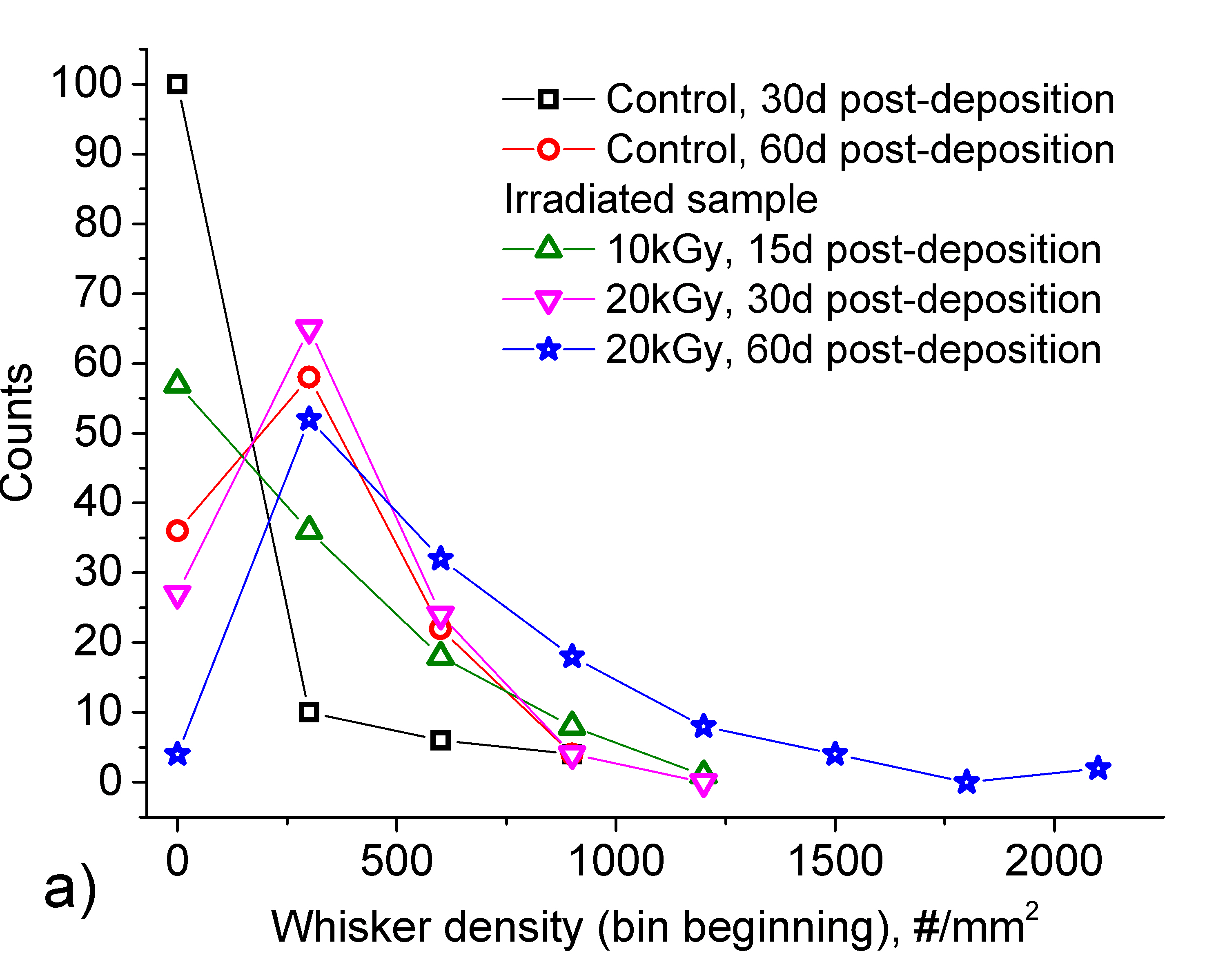
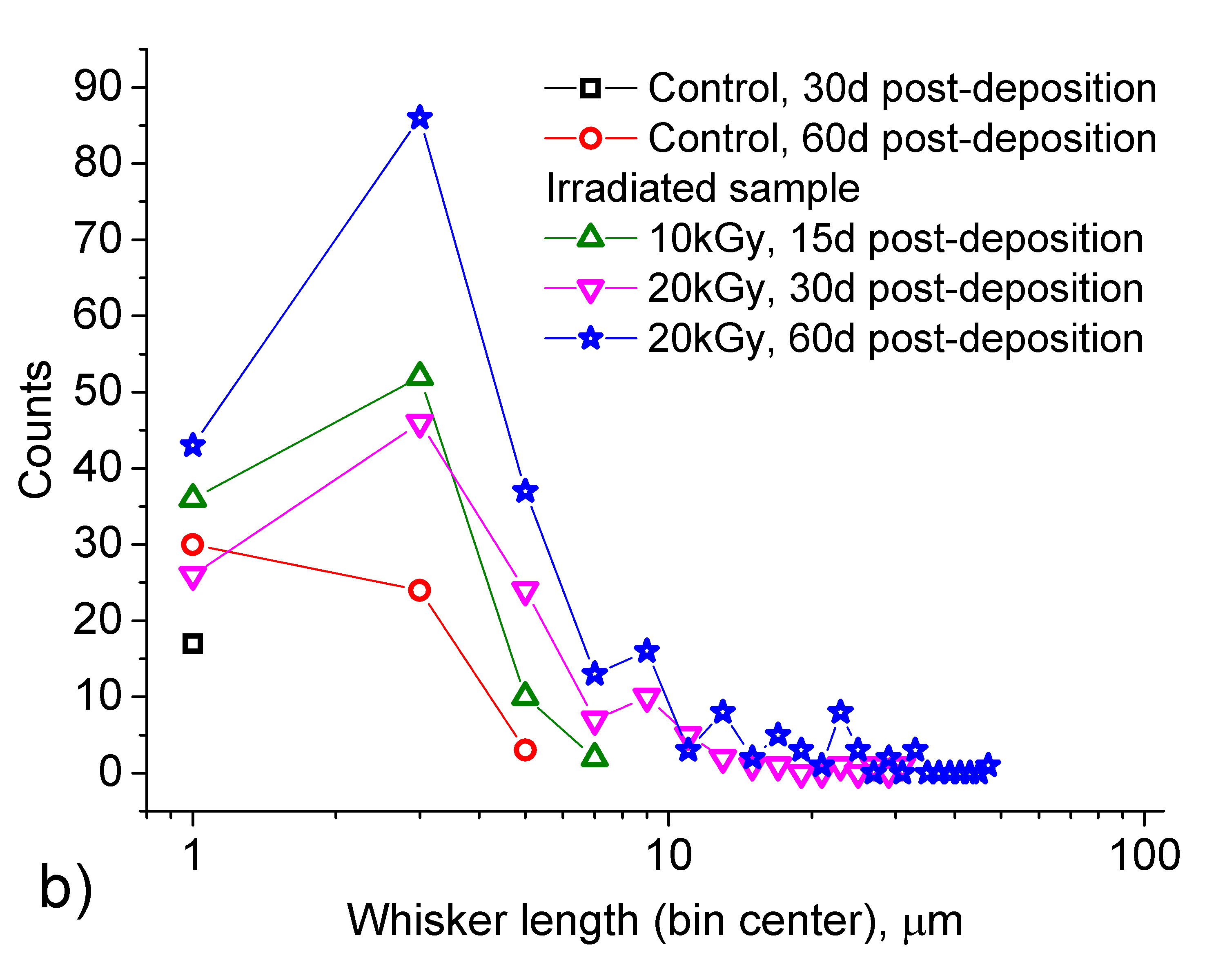
A whisker statistics summary is presented in Fig. 3 in a form of ”frequency counts”. Here the total number of counts in Fig. 3(a) is 120, corresponding to the number of SEM images processed per irradiation condition (the first point reflects the number of images with no identifiable whiskers). In Fig. 3(b) the total number of counts is defined by the total number of whiskers measured, leading to significantly higher count values for the irradiated sample, which developed substantial whiskering after 10kGy, greatly enhanced after the second round of irradiations to 20 kGy.
.
.
| Irradiated sample | Control sample | ||||
|---|---|---|---|---|---|
| Time, days | Dose, kGy | Whisker density, mm-2 | Whisker length, m | Whisker density, mm-2 | Whisker length,m |
| 15 | 10 | 282 32 | 2.57 0.12 | ||
| 30 | 20 | 378 25 | 4.98 0.42 | 101 33 | 0.75 0.02 |
| 60 | 20 | 696 41 | 6.50 0.47 | 339 36 | 1.99 0.12 |
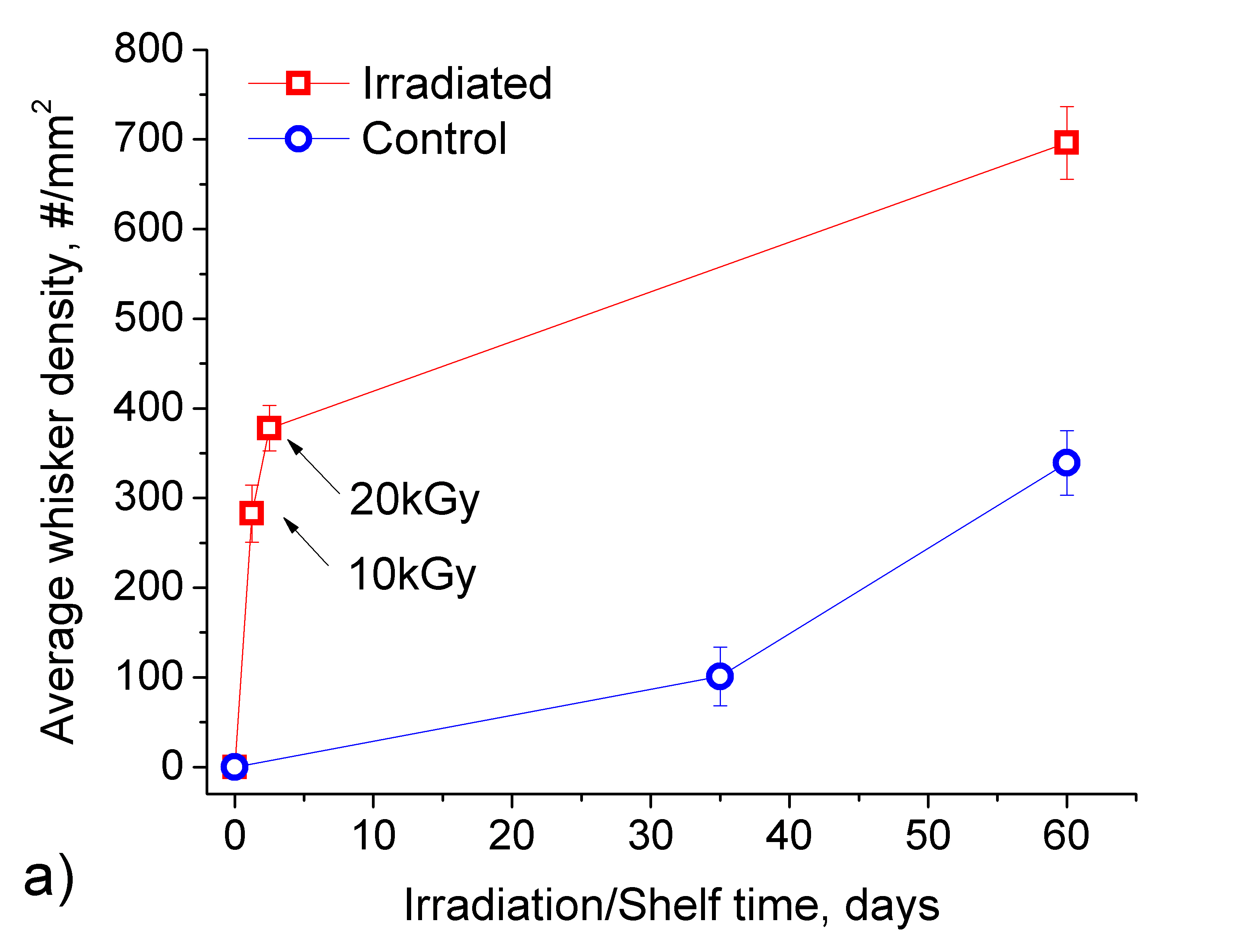
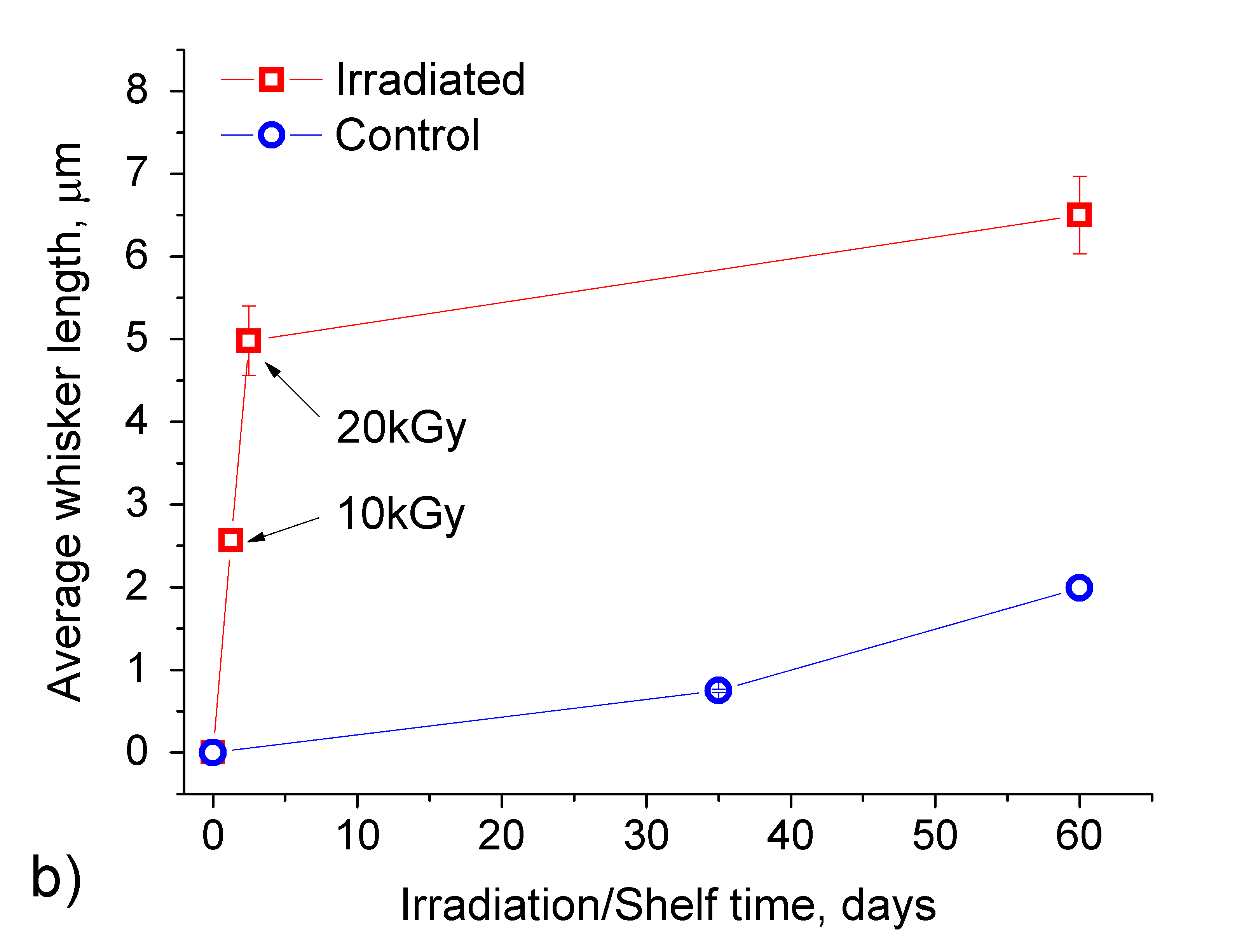
Temporal evolution of the average whisker densities and whisker lengths is presented in Fig. 4. The last point on each of ”Irradiated” curves represents the sample irradiated to 20 kGy, held on a shelf for additional 30 days. Error bars in this figure represent statistical uncertainties, calculated as corresponding standard deviations over , where N=120 is the number of SEM images taken for each sample/condition. While the data have large standard deviations, typical of whisker statistics, the number of processed images does reduce the uncertainty of the average value, making it a more relevant parameter to use for error bars. Numerical values for data summary in Fig. 4 are presented in Table 1; these are used to calculate whisker growth acceleration factor in what follows.
Discussion. X-ray and gamma-ray induced substrate charging have been studied in connection with various applications (x-ray lithography, degradation of optical devices, and degradation of MOS electronic devices), where such charging can be significant. The effect of radiation on the substrate atoms is that of ionization, knocking off the electrons from the substrates. The electric fields induced through resulting substrate charging fall in the range of MV/cm shaneyfelt ; schwank , corresponding to those predicted as accelerating whisker growth. karpov The field extends over the range of at least tenths of microns; at further distances from the film surface it is screened by ions of surrounding air and depends on the ambient pressure and humidity, as suggested by the Paschen law. lieberman
Here we utilized the phenomenon of substrate charging under irradiation to verify the effect of electric field on whisker growth. The observed concomitant darkening of the glass substrate under irradiation is due to a well-known phenomenon of color centers formation in glass, kreidl confirming radiation induced changes in the substrate.
As evident from Figures 3 and 4, whisker growth was significantly accelerated under irradiation, with both whisker average density and length affected, producing long whiskers, with some examples shown in Figs. 2 (c) and (e). While the control sample also grew some whiskers, they were order of magnitude shorter, almost at the level of ”nodules”, as presented in Figs. 2 (d) and (f).
A comment is in order regarding our observation of whisker density and length not correlating with the proximity to the radiation source, but dependent only on the total irradiation dose received. It is reasonable to assume that non-uniform irradiation results in the correspondingly nonuniform distribution of substrate charges, seemingly contradicting the observed uniform generation of whiskers. Although unanticipated, that contradiction is easily resolved by taking into effect the field screening due to the electrons in the TCO layer below Sn strip regions. The standard electrostatic calculation (to be published elsewhere along with the observations of nonuniform effects in samples without TCO) shows that free charge carriers in TCO rearrange themselves to fully screen the nonuniform field component while not affecting the average (uniform) field perpendicular to the substrate. We relate the latter to the observed significant acceleration of whisker growth.
To quantify the effect of electric field on whisker growth we use whisker creation rate
and distinguish between - spontaneous creation rate, with no external field applied, and - stimulated by applied external electric field, generated under gamma-ray irradiation. Next, we define the acceleration factor
Taking whisker growth time equal to the irradiation time hours vasko1 required to achieve 20kGy total dose for the irradiated sample, shelf time =35 days=840 hours for the control sample, and corresponding average whisker densities from Table 1, we arrive at acceleration ratio . Evaluating the acceleration factor in terms of relative increase in the average whisker length would result in even higher values for . Additional post-irradiation whisker growth appears to follow the rate of the shelf sample. From a practical standpoint that acceleration means growing whiskers under gamma-ray source should take one-two weeks rather than years.
Comparing the obtained results with those previously measured for tin and zinc samples under high-energy electron beams, vasko1 ; niraula we point out roughly 4 times lower values of acceleration factor observed under photon irradiation here. We attribute it to the lower dose rate of the gamma-ray source, resulting in longer irradiation times to achieve the same dose and charge density levels. More experiments with higher dose rate sources are being planned.
Conclusions. The experimentally observed effect of accelerated Sn whisker growth under gamma-ray irradiation presented here appears quite significant: both whisker densities and lengths are greatly enhanced in the irradiated sample. The effect is attributed to generation of electric charges in the insulating glass substrate supporting the Sn thin films. The charges create an electric field perpendicular to the film surface, thus providing conditions conducive to electrostatically driven whisker growth. The observed acceleration factors of about 50 make this approach promising as a non-destructive readily implementable accelerated life testing tool; the possibility of achieving even higher acceleration factors for higher intensity photon sources of radiation is under investigation.
Acknowledgements
We are grateful to G. Davy and A.D. Kostic for insightful discussions and critique of the manuscript.
References
- (1) NASA Goddard Space Flight Center Tin Whisker Homepage, website http://nepp.nasa.gov/whisker; accessed 12 June 2017.
- (2) J. Brusse, Metal Whisker Discussion (Tin and Zinc Whiskers), see http://www.calce.umd.edu/lead-free/other/BRUSSE_ACI.pdf for more info
- (3) Environmental Acceptance Requirements for Tin Whisker Susceptibility of Tin and Tin Alloy Surface Finishes, JEDEC Standard No. 201; Test Method for Measuring Whisker Growth on Tin and Tin Alloy Surface Finishes, JEDEC Standard No. 22-A121A, JEDEC Solid state technology association, available from Catalog of JEDEC Engineering Standards and Publications online at http://www.jedec.org/Catalog/catalog.cfm
- (4) NASA Goddard Space Flight Center Comments regarding JDEC ballot JC-14-04-226. Available at https://nepp.nasa.gov/whisker/reference/tech_papers/2004-GSFC-comments-JC-14-04-226.pdf
- (5) A.C. Vasko, G.R. Warrell, E.I. Parsai, V.G. Karpov, and Diana Shvydka, Electron beam induced growth of tin whiskers. J. Appl. Phys. 118,125301 (2015); doi:10.1063/1.4931426
- (6) V.G. Karpov , Electrostatic theory of metal whiskers, Phys. Rev. Applied, 1, 044001 (2014).
- (7) Diana Shvydka and V.G. Karpov, Surface parameters determining a metal propensity for whiskers, J. Appl. Phys. 119, 085301 (2016).
- (8) A.C. Vasko, C.R. Grice, A.D. Kostic, and V.G. Karpov, Evidence of electric-field-accelerated growth of tin whiskers, MRS Communications (Materials Research Society, 2015), doi: 10.1557/mrc.2015.64.
- (9) O.S. Oen and D.K. Holmes, Cross Sections for Atomic Displacements in Solids by Gamma Rays, J. Appl. Phys. 30, 1289 (1959); doi: 10.1063/1.1735307
- (10) V.Borra, D.G. Georgiev, C.R. Grice, Fabrication of optically smooth Sn thin films. Thin Solid Films. 2016; 616:311-315. doi:10.1016/j.tsf.2016.08.042.
- (11) J.Borg, D.W.O. Rogers, Spectra and air-kerma strength for encapsulated 192Ir sources, Med. Physics 26, 2441 (1999). doi: 10.1118/1.598763
- (12) H.E. Jons and J.R. Canningham, Physics of Radiology, 4th edition, Charles C. Thomas, Springfield, IL, 1983.
- (13) M.R. Shaneyfelt, D.M. Fleetwood, J.R. Schwank, and K.L. Hughes, Charge yield for 10-keV X-ray and Co-60 irradiation of MOS devices, IEEE Trans. Nucl. Sci., 38, 1187 (1991).
- (14) J.R. Schwank et. al., Correlation Between Co-60 and X-Ray Radiation-Induced Charge Buildup in Silicon-on-Insulator Buried Oxides, IEEE Trans. Nucl. Sci. 47, 2175-86, (2000).
- (15) M.A. Lieberman, A.J. Lichtenberg, (2005). Principles of plasma discharges and materials processing (2nd ed.). Hoboken, N.J.: Wiley-Interscience. 546. ISBN: 978-0-471-72001-0.
- (16) N.J. Kreidl , J.R. Hensler, Formation of color centers in glasses exposed to gamma radiation. J. Am. Ceram. Soc., 38, 423 (1955).
- (17) F.M. Ezz-Eldin, F. Abdel-Rehim, A.A. Abdel-Azim, A.A. Ahmed, Soda-lime-silica glass for radiation dosimetry, Med. Physics 21, 1085 (1994); doi: 10.1118/1.597357
- (18) D. Niraula, J. McCulloch, G. R. Warrell, R. Irving, V.G. Karpov, and Diana Shvydka, Electric Field Stimulated growth of Zn whiskers, AIP Advances, 6, 075201 (2016).