Comorbid CAD and Ventricular Hypertrophy Compromise The Perfusion of Myocardial Tissue at Subcritical Stenosis of Epicardial Coronaries
Background
Combined coronary artery disease and ventricular hypertrophy are not uncommon; they both share hypertension, which affects of the world population [1], as a risk factor. Accumulation of atheromatous plaques under tunica intima of the epicardial arteries restricts the blood flow to the supplied cardiac tissue. Chronic high-grade narrowing of the coronary arteries induces subendocardial ischemia during the escalation of the myocardial oxygen demand throughout exercise or stress [2]. The strained myocytes release mediators like adenosine and bradykinin [3][4] which in addition to stimulating coronary vasodilatation, irritate the nerve endings leading to anginal pain [5].
The treatment strategy for treating CAD aims to improve survival and/or relieve symptoms [6] including dyspnea and stable angina pectoris. Said strategy usually involves anti-anginal medications and/or PCI, or CABG in case of complex CAD and/or left main involvement, for achieving those aims. Trials show that revascularization by PCI or CABG is more effective than a strategy of medical therapy alone, in relieving symptoms like angina and dyspnea. Besides; it improves the quality of life by reducing the use of anti-angina drugs and increasing exercise capacity [7][8][9][10]. However; studies indicate that PCI, as an initial management strategy in patients with stable coronary artery disease, did not reduce the risk of complications as myocardial infarction or other major cardiovascular events when added to optimal medical therapy [11][12][13].
The guidelines recommend that ad hoc PCI should not automatically be applied after angiography [14]; and emphasize the usefulness of optimal medical treatment for selected patients, which can reduce angina and the risk of myocardial infarction and stroke substantially and prevent progression of atherosclerosis in the entire vasculature. Consequently; the clinical decisions for management of CAD generally need to balance adherence to guidelines against judgments based on specific patient, operator, social, economic, and cultural factors [15]. Generally; PCI and medical therapy should be viewed as complementary, rather than opposing, strategies [16]. Patients with stable coronary artery disease and functionally significant stenoses, benefit from the combination therapy of PCI plus optimal medical therapy by showing greater symptomatic improvement [17] and decreased the need for urgent revascularization. However; in patients without ischemia, the outcome appeared to be favorable with the optimal medical therapy alone [18].
For revascularization decisions and recommendations; said significant stenosis has been defined by most studies of CAD revascularization, which have been based on and reported according to angiographic criteria, as diameter narrowing, and/or for left main CAD [19]. Besides; angiophysiological criterion, such as an assessment of fractional flow reserve (FFR), has been used in deciding when revascularization is indicated. Thus, for recommendations about revascularization, coronary stenosis with FFR is also considered to be significant [20]. The standard values provided by both methods, and so the revascularization decision, don’t consider the relation between the resulting effective flow distal to the stenosis and the demand of a comorbid hypertrophied myocardial tissue.
Model
Hagen-Poiseuille law, which is an analytical solution to the Navier-Stokes equation [21], states that; the flow rate through a coronary vessel is directly proportional to the pressure gradient between the aortic root and the right atrium; and inversely proportional to the resistance within the vessel. Wherein; the resistance is inversely proportional to the radius of the vessel elevated to the fourth power, and is directly proportional to the blood viscosity and the vessel length . So, by considering a circular cross-section of the vessel:
when
so;
The blood flow, which is a non-Newtonian fluid, within the circulation doesn’t imitate precisely this law [22], because said relation is applied on a Newtonian fluid in the steady laminar flow moving through a long cylindrical pipe. Still, the law outlines the dominant determinants which influence the blood flow within the vasculature either in physiological or pathological conditions.
Atherosclerosis commonly affects the epicardial coronary vessels leading to narrowing of the vessel caliber and increase vascular resistance of the supplying vessel . While:
the corresponding supplied myocardial segment doesn’t actually suffer this severe blood flow reduction indicated in the above equation. The vasculature of the coronary circulation is arranged in-series, in addition to the in-parallel arrangement, so that the epicardial vascular resistance is a segmental resistance. The coronary circulation can be divided into two compartments, the large epicardial conduit vessels and the resistance vessels, which are typically less than in diameter [23]. Whereas the conduit vessels exert little if any resistance to flow, resistance to flow progressively rises as the vessel diameter of the resistance vessels declines from about in the small arteries to less than in the arteriolar vessels [24]. Therefore; the total resistance to blood flow comprises mainly the pre-capillary resistance , the resistance of microvasculature , and the negligible resistance of the epicardial or conductance vessels .
Narrowing of the radius of the epicardial vessel, due to atheromatous plaque, will increase the resistance in this vessel; but as:
the impact of mild to moderate increase of the epicardial resistance on the overall resistance of the coronary circulation is insignificant.
However; in case of combined coronary artery disease and ventricular hypertrophy, both and are increased. Microangiogenesis is activated during the pathogenesis of ventricular hypertrophy as a compensatory mechanism to maintain effective blood supply to the hypertrophied tissue. Accordingly; CAD causes an increase in due to epicardial arterial stenosis and ventricular hypertrophy increases due to neomicroangiogenesis, i.e. addition of a new microvascular segment.
consequently; the flow rate , and so the perfusion of myocardial tissue, diminish significantly upon subcritical stenosis of the supplying epicardial artery during the pathogenesis of CAD.
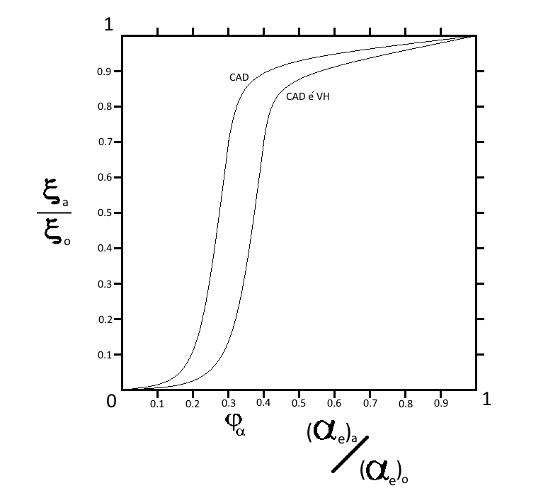
As mentioned; the identification of clinically-significant stenosis of an epicardial artery depends on an angiographic criterion, its radius , and/or an angiophysiologic criterion, the FFR, which is an absolute number result from the ratio of the pressure distal to the lesion, to the pressure proximal to the lesion during induced hyperemia [20]. The graphical representation of the relation between both these criteria of the supplying artery and the perfusion of the supplied myocardial tissue follows a direct proportional relationship represented by a sigmoid-shaped curve, due to the effect of segmental resistance. Myocardial perfusion describes the blood flow in ml/min per cubic centimeter of cardiac muscle volume .
According to the relation between the radius of an epicardial coronary artery, as an angiographic criterion, and the perfusion of the corresponding supplied myocardial tissue represented in Figure 1; the perfusion doesn’t decrease significantly with gradual stenosis in isolated CAD until a critical stenotic value is reached, wherein the perfusion collapses relatively. Clinically; said critical value is defined as radius reduction, significant stenosis [19]. However; in patients with comorbid CAD and ventricular hypertrophy; the curve is shifted to the right indicating an increase in the critical stenotic value , so that the perfusion of the corresponding supplied myocardial tissue collapses relatively at a clinically subsignificant stenosis. The right shift in said patients depends on the degree of ventricular hypertrophy.
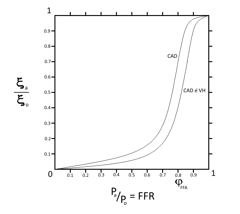
Additionally; the relation between another angiophysiologic criterion, the fractional flow reserve FFR, within a stenotic epicardial artery and the perfusion of the corresponding supplied myocardial tissue, as represented in Figure 2, indicates that the perfusion is not meaningfully reduced with the gradual decrease of FFR until a critical value is reached, wherein the perfusion collapses relatively. Clinical trials defined said critical value as a FFR [25][26]. Though; in patients with combined CAD and ventricular hypertrophy; the curve shows a right shift, which is directly proportional to the degree of ventricular hypertrophy, indicating an increase in the critical stenotic value , so that the perfusion of the corresponding supplied myocardial tissue collapses relatively at a clinically subsignificant reduction in the FFR.
The proposed model gives a more sensitive formula to detect the critical stenosis, which takes into account the demand of the supplied bulky myocardium. The isolated CAD curve is a logistic function; wherein represents the critical stenosis and is the curve slope:
in patients with comorbid CAD and ventricular hypertrophy; the curve is shifted to the right by yielding as a representation of the critical stenosis:
then;
wherein the curve shift is directly proportional to the difference in muscle bulk which obtained by Echocardiogram;
the value of the constant can be obtained experimentally. So; the percentage of the critical patency in patients with comorbid CAD and ventricular hypertrophy is:
Results
Individuals with pathological ventricular hypertrophy are more sensitive to haemodynamic changes of the coronary circulation or pathologies that reduce the coronary reserve. Ventricular hypertrophy stresses the subendocardial myotissue due to increase the structural resistance of the coronary circulation. Said stress is ameliorated by compensatory functional changes to sustain the normal coronary blood flow. Although during vigorous exercise, the compensatory capability of the coronary flow reserve is exhausted under the effect of demand upsurge and shortened diastolic period. Occasional hemodynamic disturbances or subclinical pathologies which lessen the maximum coronary reserve may lead to selective subendocardial hypoperfusion.
Comorbid CAD and ventricular hypertrophy cause the subendocardial tissue to suffer, during exercise or stress, from ischemia at an angiographically subsignificant stenosis in the supplying epicardial artery. CAD primes the structural resistance of the neomicrovasculature of the hypertrophied tissue. So; subcritical stenosis of the corresponding epicardial artery, mainly due to atherosclerosis, causes the total resistance to rise effectively to reduce the flow rate and exhaust the reactive compensatory mechanisms. The curve shift to the right in said patients doesn’t affect the risk of myocardial infarction, yet they are more susceptible to and usually presented by NSTEMI; with higher rates of transition from ischemia to necrosis in the affected hypertrophied endocardial tissue. Increased muscle bulk shifts the endocardium away from the main blood supply. Besides; subjection to higher extravascular pressure depletes the functional vasodilator reserve in long standing pathological hypertrophy.
Patients with combined CAD and ventricular hypertrophy have a higher risk to develop arrhythmias than their peers who suffer from isolated CAD. In pathological hypertrophy; the neomicroangiogenesis shows anatomical and architectural dysgenesis in relation to the hypertrophied tissue. Said dysgenesis leads to failure of the coronary bed to uniformly supply the cardiac muscle, rendering foci within the hypertrophied muscle bulk at greater risk of ischemic injury. These stressed foci can be arrhythmogenic upon increased cardiac demand leading to serious arrhythmia and sudden cardiac death.
Discussion
During cardiac catheterization; the main determinants of revascularization therapy in CAD patients, are either angiographic or angiophysiological criteria, measured during drug induced hyperemia, to identify the clinically-significant stenosis. Said determinants depend on the relation between the size of the insinuated plaque and the vascular diameter. A stenosis which reduces the radius of the epicardial vessel by is considered significant angiographically. In another determinant; a small sensor on the tip of the guidewire is used to measure the pressure, temperature and flow to determine the exact severity of the lesion. The ratio between the pressure distal to the lesion and the pressure proximal to the lesion, which is the fraction flow reserve FFR, measures the pressure drop caused by the stenosis. A fraction flow reserve value defines the stenosis to be significant. The FFR is a better method to detect the physiological significance of a stenosis [27], as it takes into account collateral flow, which can render an anatomical blockage functionally unimportant. Also, standard angiography can underestimate or overestimate narrowing, because it only visualizes contrast inside a vessel. Yet; the standard values provided by both methods to identify a stenotic lesion as significant, don’t consider the relation between the resulting effective flow distal to the stenosis and the demand of a hypertrophied myocardial tissue.
The pathogenesis of ventricular hypertrophy implicates an increase in the number of force-generating sarcomeres in the myocyte [28]. According to La Place law; an increase in pressure can be offset by an increase in wall thickness [29]. This mechanical input is transduced into a biochemical sequel that modifies gene transcription in the nucleus. The focal adhesion complex, in addition to the G-coupled neurohormonal augmentation [30], is proposed as the effector transducers of said mechanical input [31]. Integrins connect the internal cytoskeleton of the cell to the extracellular matrix, wherein multiple tyrosine-phosphorylated kinases and serine-threonine kinases that are implicated in the signaling of hypertrophy can be found in the ECM [32][33].
Angiogenesis is triggered during the pathogenesis of myocardial hypertrophy by increased cardiac work and oxygen demand; in an attempt to normalize maximal myocardial perfusion and capillary domains to sustain oxygen delivery. A limitation of capillary growth will increase diffusion distance for oxygen, while inadequate arteriolar growth will reduce maximal tissue perfusion. Pathogenesis of hypertrophy is categorized into pressure overload-induced, volume overload-induced, thyroxin-induced and exercise-induced models according to the stimulus for increasing muscle bulk. In exercise-induced and thyroxin-induced models; angiogenesis and arteriogenesis are well documented experimentally [34]. While in other models; there is a considerable variation in the reports of the literature about the extent and pattern of angiogenesis and the consequential coronary microvascular resistance. The reasons for the discrepancy between these studies are not evident, but the duration of the hypertrophy and the specificity of the stimulus may play a role.
Mathematically; angiogenesis increases the coronary microvascular resistance structurally, due to the addition of a new microvascular segment. However, in vivo; structural resistance can be modulated by functional changes, wherein autoregulatory adjustments involving the vasodilator reserve may ameliorate said structural resistance escalation. Well-trained athletes with physiological cardiac hypertrophy show a proportional increase of cardiac myocytes and coronary vasculature with no change in the proportion of extracellular collagen [35]. These structural modulations are accompanied by functional adaptations resulting in a compensatory exponential coronary reserve and vasodilator capacity. Functional adaptations can include changes in neurohumoral control and changes in local vascular control mechanisms [36][37]. In pathological severe hypertrophy; pathological features of the strained neoangiogenesis halt the functional compensation for the structural increase in the microvascular resistance. Endothelium-dependent vasodilation is markedly impaired in the coronary microvessels of patients with hypertension-induced ventricular hypertrophy [38]. Accordingly; severe ventricular hypertrophy is associated with a reduction in coronary vascular reserve [39].
Myocardial infarction is mainly caused by rupture of vulnerable fibroatheromatous plaque forming a thrombus that interferes with myocardial blood supply leading to excessive ischemia then necrosis [40]. Usually; soft non-stenotic plaques are more susceptible to rupture, causing major cardiovascular events [41]. The vulnerability of the plaque depends on lesion-specific characteristics like thin fibrous cap, large lipid-rich necrotic core, increased plaque inflammation, positive vascular remodeling, increased vasa-vasorum neovascularization, and intra-plaque hemorrhage [42]. Therefore; the comorbidity between CAD and ventricular hypertrophy doesn’t affect the risk of developing MI. However; patients with said comorbid diseases have higher rates of transition from ischemia to necrosis in the affected endocardial tissue, due to increase diffusion distance for oxygen and exhaustion of the functional compensation. They are also more susceptible to and usually presented by NSTEMI, due to the sensitivity of the endocardial myotissue to the equilibrium between the structural resistance of microvasculature and the reactive functional modulation.
Myocardial ischemia is characterized by ionic and biochemical alterations, creating an unstable electrical substrate capable of initiating and sustaining arrhythmias [43]. Theoretically; onerous angiogenesis in pathological hypertrophy shows patterns of anatomical and architectural dysgenesis rendering foci within the hypertrophied muscle bulk at greater risk of ischemic injury. Said stressed foci acquire different electrochemical properties, due to defective function of ATPase-dependent pumps, leading to tissue heterogeneity. Theses foci become arrhythmogenic, especially with increased cardiac demand during above-normal exercise or severe stressful conditions, leading to functional re-entry. Hence; presence of ventricular hypertrophy is associated with a greater risk of sustained arrhythmias [44].
Conclusion
The mathematical model establishes that ventricular hypertrophy increases the vascular structural resistance of the coronary circulation due to neomicroangiogenesis. Patients with comorbid CAD and ventricular hypertrophy suffer, due to exhaustion of functional compensation, from complications of myocardial hypoperfusion at angiographically sub-significant coronary artery stenosis. These patients are more susceptible to NSTEMI, serious arrhythmias and sudden cardiac death than patients with isolated CAD. Upon confirmation of such results by large investigational studies, said results should be taken into account during designing the treatment strategy of said patients.
Acknowledgement
The author states that there is no conflict of interest regarding this article.
References
- [1] Patricia M Kearney, Megan Whelton, Kristi Reynolds, Paul Muntner, Paul K Whelton, and Jiang He. Global burden of hypertension: analysis of worldwide data. The lancet, 365(9455):217–223, 2005.
- [2] Peter Libby and Pierre Theroux. Pathophysiology of coronary artery disease. Circulation, 111(25):3481–3488, 2005.
- [3] Noam Safran, Vladimir Shneyvays, Nissim Balas, Kenneth A Jacobson, Hermann Nawrath, and Asher Shainberg. Cardioprotective effects of adenosine a1 and a 3 receptor activation during hypoxia in isolated rat cardiac myocytes. Molecular and cellular biochemistry, 217(1):143–152, 2001.
- [4] James R Parratt, Agnes Vegh, I Jack Zeitlin, Maqsood Ahmad, Keith Oldroyd, Karoly Kaszala, and Julius Gy Papp. Bradykinin and endothelial-cardiac myocyte interactions in ischemic preconditioning. The American journal of cardiology, 80(3):124A–131A, 1997.
- [5] Filippo Crea, Giuseppe Pupita, Alfredo R Galassi, Hassan El-Tamimi, Juan Carlos Kaski, Graham Davies, and Attilio Maseri. Role of adenosine in pathogenesis of anginal pain. Circulation, 81(1):164–172, 1990.
- [6] William Wijns, Philippe Kolh, Nicolas Danchin, Carlo Di Mario, Volkmar Falk, Thierry Folliguet, Scot Garg, Kurt Huber, Stefan James, Juhani Knuuti, et al. Guidelines on myocardial revascularization: the task force on myocardial revascularization of the european society of cardiology (esc) and the european association for cardio-thoracic surgery (eacts). European heart journal, 31(20):2501–2555, 2010.
- [7] DA Chamberlain, KAA Fox, RA Henderson, DG Julian, et al. Coronary angioplasty versus medical therapy for angina: the second randomised intervention treatment of angina (rita-2) trial. The Lancet, 350(9076):461, 1997.
- [8] Time Investigators et al. Trial of invasive versus medical therapy in elderly patients with chronic symptomatic coronary-artery disease (time): a randomised trial. The Lancet, 358(9286):951–957, 2001.
- [9] William S Weintraub, John A Spertus, Paul Kolm, David J Maron, Zefeng Zhang, Claudine Jurkovitz, Wei Zhang, Pamela M Hartigan, Cheryl Lewis, Emir Veledar, et al. Effect of pci on quality of life in patients with stable coronary disease. New England Journal of Medicine, 359(7):677–687, 2008.
- [10] Paul Erne, Andreas W Schoenenberger, Dieter Burckhardt, Michel Zuber, Wolfgang Kiowski, Peter T Buser, Paul Dubach, Therese J Resink, and Matthias Pfisterer. Effects of percutaneous coronary interventions in silent ischemia after myocardial infarction: the swissi ii randomized controlled trial. Jama, 297(18):1985–1991, 2007.
- [11] William E Boden, Robert A O’rourke, Koon K Teo, Pamela M Hartigan, David J Maron, William J Kostuk, Merril Knudtson, Marcin Dada, Paul Casperson, Crystal L Harris, et al. Optimal medical therapy with or without pci for stable coronary disease. New England journal of medicine, 356(15):1503–1516, 2007.
- [12] Whady Hueb, Paulo R Soares, Bernard J Gersh, Luiz AM César, Protásio L Luz, Luiz B Puig, Eulógio M Martinez, Sergio A Oliveira, and José AF Ramires. The medicine, angioplasty, or surgery study (mass-ii): a randomized, controlled clinical trial of three therapeutic strategies for multivessel coronary artery disease: one-year results. Journal of the American College of Cardiology, 43(10):1743–1751, 2004.
- [13] Kathleen Stergiopoulos and David L Brown. Initial coronary stent implantation with medical therapy vs medical therapy alone for stable coronary artery disease: meta-analysis of randomized controlled trials. Archives of internal medicine, 172(4):312–319, 2012.
- [14] Stephan Windecker, Philippe Kolh, F Alfonso, JP Collet, J Cremer, V Falk, G Filippatos, C Hamm, SJ Head, P Jüni, et al. Authors/task force members. 2014 esc/eacts guidelines on myocardial revascularization: the task force on myocardial revascularization of the european society of cardiology (esc) and the european association for cardio-thoracic surgery (eacts) developed with the special contribution of the european association of percutaneous cardiovascular interventions (eapci). Eur Heart J, 35(37):2541–2619, 2014.
- [15] Andre Lamy, Madhu Natarajan, and Salim Yusuf. Medical treatment, pci, or cabg for coronary artery disease?, 2011.
- [16] Roger S Blumenthal, Gregory Cohn, and Steven P Schulman. Medical therapy versus coronary angioplasty in stable coronary artery disease: a critical review of the literature. Journal of the American College of Cardiology, 36(3):668–673, 2000.
- [17] Seema Pursnani, Frederick Korley, Ravindra Gopaul, Pushkar Kanade, Newry Chandra, Richard E Shaw, and Sripal Bangalore. Percutaneous coronary intervention versus optimal medical therapy in stable coronary artery disease. Circulation: Cardiovascular Interventions, pages CIRCINTERVENTIONS–112, 2012.
- [18] Bernard De Bruyne, Nico HJ Pijls, Bindu Kalesan, Emanuele Barbato, Pim AL Tonino, Zsolt Piroth, Nikola Jagic, Sven Möbius-Winkler, Gilles Rioufol, Nils Witt, et al. Fractional flow reserve–guided pci versus medical therapy in stable coronary disease. New England Journal of Medicine, 367(11):991–1001, 2012.
- [19] Glenn N Levine, Eric R Bates, James C Blankenship, Steven R Bailey, John A Bittl, Bojan Cercek, Charles E Chambers, Stephen G Ellis, Robert A Guyton, Steven M Hollenberg, et al. 2011 accf/aha/scai guideline for percutaneous coronary intervention: executive summary: a report of the american college of cardiology foundation/american heart association task force on practice guidelines and the society for cardiovascular angiography and interventions. Journal of the American College of Cardiology, 58(24):2550–2583, 2011.
- [20] Nico HJ Pijls, Bernard de Bruyne, Kathinka Peels, Pepijn H van der Voort, Hans JRM Bonnier, Jozef Bartunek, and Jacques J Koolen. Measurement of fractional flow reserve to assess the functional severity of coronary-artery stenoses. New England Journal of Medicine, 334(26):1703–1708, 1996.
- [21] Henrik Bruus. Theoretical microfluidics. Oxford university press Oxford, 2007.
- [22] Richard Klabunde. Cardiovascular physiology concepts. Lippincott Williams & Wilkins, 2011.
- [23] Heinrich R Schelbert. Anatomy and physiology of coronary blood flow. Journal of nuclear cardiology, 17(4):545–554, 2010.
- [24] Judy M Muller, Michael J Davis, and William M Chilian. Integrated regulation of pressure and flow in the coronary microcirculation. Cardiovascular research, 32(4):668–678, 1996.
- [25] Pim AL Tonino, Bernard De Bruyne, Nico HJ Pijls, Uwe Siebert, Fumiaki Ikeno, Marcel vant Veer, Volker Klauss, Ganesh Manoharan, Thomas Engstrøm, Keith G Oldroyd, et al. Fractional flow reserve versus angiography for guiding percutaneous coronary intervention. N Engl j Med, 2009(360):213–224, 2009.
- [26] Nico HJ Pijls, Pepijn van Schaardenburgh, Ganesh Manoharan, Eric Boersma, Jan-Willem Bech, Marcel van’t Veer, Frits Bär, Jan Hoorntje, Jacques Koolen, William Wijns, et al. Percutaneous coronary intervention of functionally nonsignificant stenosis: 5-year follow-up of the defer study. Journal of the American College of Cardiology, 49(21):2105–2111, 2007.
- [27] Nico HJ Pijls, William F Fearon, Pim AL Tonino, Uwe Siebert, Fumiaki Ikeno, Bernhard Bornschein, Marcel van’t Veer, Volker Klauss, Ganesh Manoharan, Thomas Engstrøm, et al. Fractional flow reserve versus angiography for guiding percutaneous coronary intervention in patients with multivessel coronary artery disease: 2-year follow-up of the fame (fractional flow reserve versus angiography for multivessel evaluation) study. Journal of the American College of Cardiology, 56(3):177–184, 2010.
- [28] Beverly H Lorell and Blase A Carabello. Left ventricular hypertrophy. Circulation, 102(4):470–479, 2000.
- [29] Stephen Gunther and William Grossman. Determinants of ventricular function in pressure-overload hypertrophy in man. Circulation, 59(4):679–688, 1979.
- [30] Peter H Sugden. Signaling in myocardial hypertrophy. Circulation Research, 84(6):633–646, 1999.
- [31] TK Borg and ML Burgess. Holding it all together: organization and function (s) of the extracellular matrix in the heart. Heart Failure, 8:230–230, 1992.
- [32] Dhandapani Kuppuswamy, Charlene Kerr, Takahiro Narishige, Vijaykumar S Kasi, Donald R Menick, and George Cooper. Association of tyrosine-phosphorylated c-src with the cytoskeleton of hypertrophying myocardium. Journal of Biological Chemistry, 272(7):4500–4508, 1997.
- [33] Louis Terracio, Kristofer Rubin, Donald Gullberg, ED Balog, Wayne Carver, Ron Jyring, and Thomas K Borg. Expression of collagen binding integrins during cardiac development and hypertrophy. Circulation research, 68(3):734–744, 1991.
- [34] Robert J Tomanek and Eduard I Dedkov. Angiogenesis and arteriogenesis in cardiac hypertrophy. Modern concepts in angiogenesis, pages 253–280, 2007.
- [35] Dirk J Duncker and Robert J Bache. Regulation of coronary blood flow during exercise. Physiological reviews, 88(3):1009–1086, 2008.
- [36] M HAROLD Laughlin. Effects of exercise training on coronary transport capacity. Journal of Applied Physiology, 58(2):468–476, 1985.
- [37] M Harold Laughlin. Physical activity in prevention and treatment of coronary disease: the battle line is in exercise vascular cell biology. Medicine & Science in Sports & Exercise, 36(3):352–362, 2004.
- [38] Charles B Treasure, J Larry Klein, Joseph A Vita, Steven V Manoukian, George H Renwick, Andrew P Selwyn, Peter Ganz, and R Wayne Alexander. Hypertension and left ventricular hypertrophy are associated with impaired endothelium-mediated relaxation in human coronary resistance vessels. Circulation, 87(1):86–93, 1993.
- [39] August D Pichard, Richard Gorlin, Harry Smith, John Ambrose, and Jose Meller. Coronary flow studies in patients with left ventricular hypertrophy of the hypertensive type: evidence for an impaired coronary vascular reserve. The American journal of cardiology, 47(3):547–554, 1981.
- [40] Grant W Reed, Jeffrey E Rossi, and Christopher P Cannon. Acute myocardial infarction. The Lancet, 389(10065):197–210, 2017.
- [41] Peter Libby. Mechanisms of acute coronary syndromes and their implications for therapy. New England Journal of Medicine, 368(21):2004–2013, 2013.
- [42] Pedro R Moreno. Vulnerable plaque: definition, diagnosis, and treatment. Cardiology clinics, 28(1):1–30, 2010.
- [43] AV Ghuran and AJ Camm. Ischaemic heart disease presenting as arrhythmias. British medical bulletin, 59(1):193–210, 2001.
- [44] Saurav Chatterjee, Chirag Bavishi, Partha Sardar, Vikram Agarwal, Parasuram Krishnamoorthy, Tomasz Grodzicki, and Franz H Messerli. Meta-analysis of left ventricular hypertrophy and sustained arrhythmias. The American journal of cardiology, 114(7):1049–1052, 2014.