11email: rajesh.ganai.physics@gmail.com 22institutetext: Homi Bhabha National Institute, Training School Complex, Anushakti Nagar, Mumbai - 400 085, India. 33institutetext: School of Physics and Material Sciences, Thapar University, Patiala, Punjab-147004, India 44institutetext: Sardar Vallabhbhai National Institute of Technology, Surat-395007, Gujarat, India.
A Proof-of-principle for Time-Of-Flight Positron Emission Tomography Imaging
Abstract
Time-Of-Flight (TOF) is a noble technique that is used in Positron Emission Tomography (PET) imaging worldwide. Scintillator based imaging system that is being used around the world for TOF-PET is very expensive. Multi-gap Resistive Plate Chambers (MRPCs) are gaseous detectors which are easy to fabricate, inexpensive and have excellent position and timing resolution. They can be used as a suitable alternative to highly expensive scintillators. For the sole purpose of TOF-PET, pair of 18 cm 18 cm, 5 gap, glass based MRPC modules have been fabricated. Our main aim was to determine the shift in the position of source (Na-22) with these fabricated MRPCs. In this document the details of the experimental results will be presented.
keywords:
Time of flight, Positron emission tomography, Multi-gap resistive plate chamber1 Introduction
Positron Emission Tomography PET)[1], is a radio-tracer, nuclear medicine imaging technique. PET is used to observe metabolic processes in the body. The basic principle of PET is detecting a pair of back to back 511 keV photons created by the annihilation of a positron with an electron. The positron emitter, Fludeoxyglucose (18F) (FDG) which is a radio-tracer, administered in the body annihilates into a pair of 511 keV photons, flying in opposite directions. PET is both a medical and research tool. It is used heavily in clinical oncology (medical imaging of tumors and the search for metastases), and for clinical diagnosis of certain diffuse brain diseases such as those causing various types of dementias. PET is also an important research tool to map normal human brain and heart function, and support drug development.
Time of flight (TOF) technique has found its application in PET imaging. The two gamma-ray interaction points define a so-called line-of-response (LOR) on which the annihilation must have taken place. A precise measurement of the arrival times of the coincident photons along with the time difference in flight-time of the two photons helps to localize the annihilation event on the LOR. Figure 1 illustrates the basic working and detection principle of any TOF-PET system.

2 Experimental set up and test results
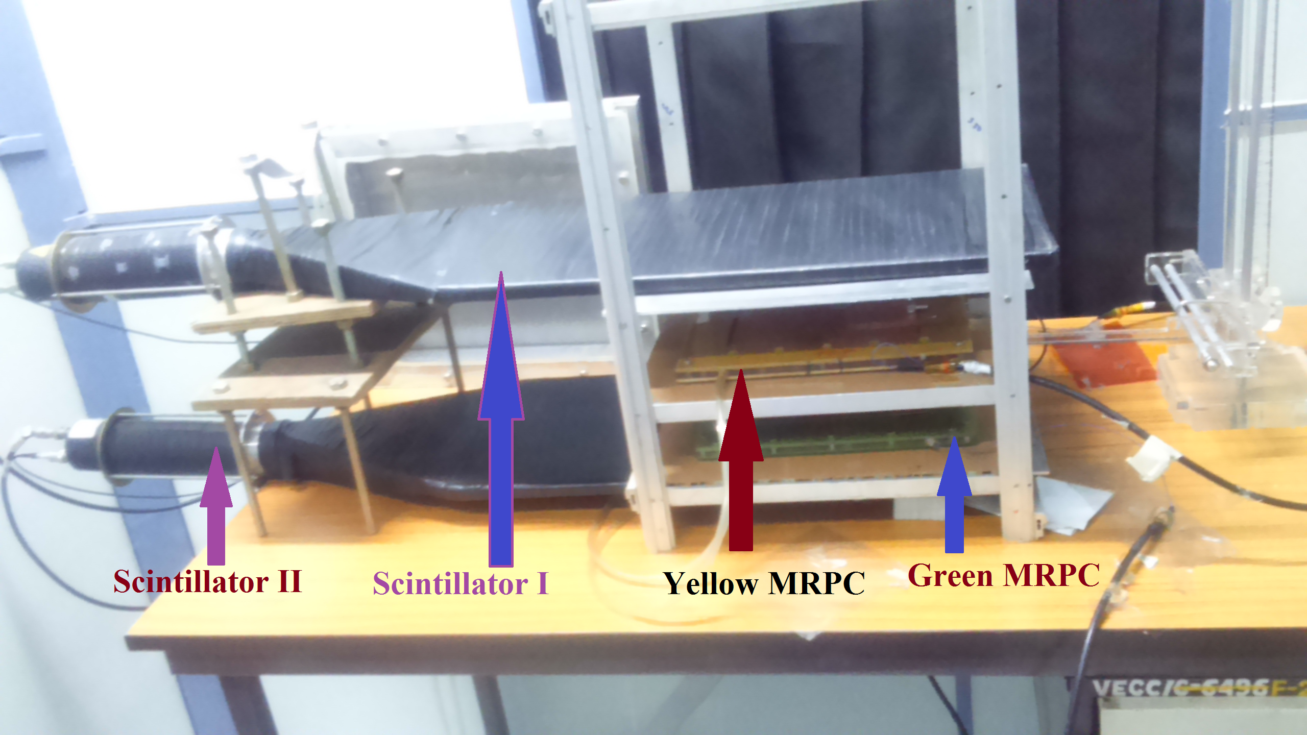
The aim of this work was to detect the two back to back gammas created by the annihilation of positron emitted from 22Na with an electron with the developed prototype 5-gap glass MRPCs[2],[3] and also to sense a change in the position of the 22Na source. In order to do so, the major challenge was to eliminate the cosmic muon background as MRPCs are known to have very good charged particle detection efficiency and was successfully achieved by veto method. The schematic of the experimental set up and the actual set up has been shown in figure 2. The two prototype MRPCs were kept horizontally and separated by a known distance of 17 cm. A source was kept in between the MRPCs. Two aluminium plates of dimensions 30 cm 30 cm 0.5 cm ensured that the back to back photons does not reach the scintillators. Two plastic scintillators each of dimension 50 cm 25 cm were used.
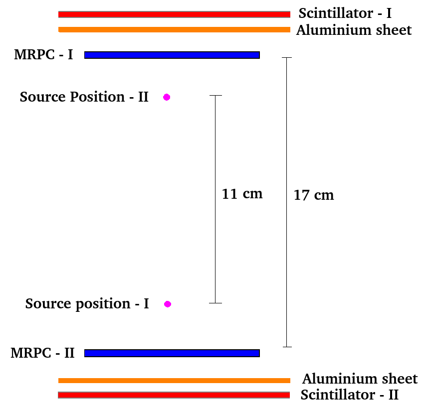
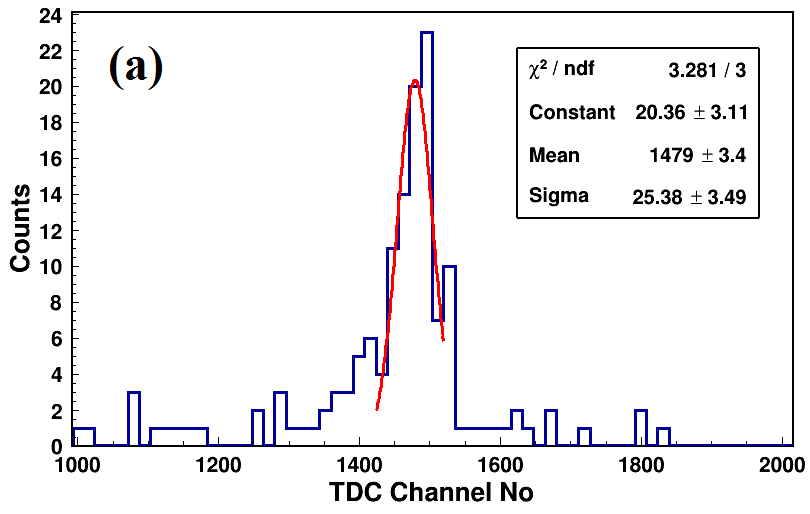
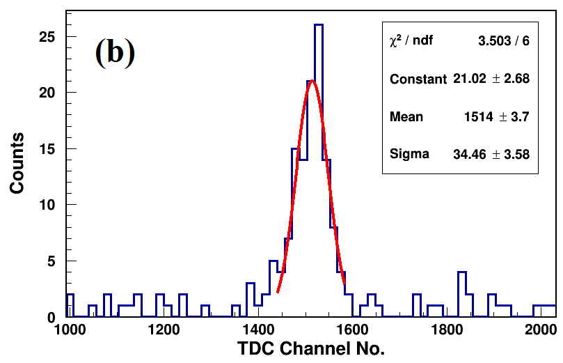
A suitable trigger ( MRPC - I) was also chosen which initiated the START of the TDC module. The first set of TDC spectra was taken when the source was placed 3 cm away from the bottom or MRPC - II and the second set was taken when the source was kept at 14 cm away from the bottom MRPC which have been shown in figure 3. The mean channel of the TDC spectra after a Gaussian fit was obtained to be 1479 for Figure 3(a) and 1514 for Figure 3(b). Clearly there is a shift in the mean of the TDC spectra, specially in the mean of the spectra by 35 TDC channels as the source was moved from position-I to position-II by 11 cm. Assuming the velocity of photons to be 30 cm/ns, a change in source position by 11 cm should give TDC channel difference of ∼30 channels which is close to to the obtained value of 35 channels. From another way of looking at it, a shift in the mean of the TDC spectra of 35 channels should yield a change in the source position by ∼ 12.8 cm which is close to the actual change in source position by 11 cm.
3 Summary
Excellent time resolution of MRPCs make them potential candidate to replace the scintillators in existing PET systems. If successful, the cost per scan of PET imaging will reduce drastically as MRPCs are relatively low cost detectors. As a first step towards this noble work, two prototype MRPCs have been tested in a two-MRPC coincidence set-up for the detection of back to back photons created by the annihilation of positron (emitted from Na22 source) with a nearby electron. The change in distance in the source position was successfully estimated from the time spectra obtained by using both the MRPCs.
References
- [1] Sorenson J. A, and Phelps M. E, Physics in Nuclear Medicine, 2nd Edition, Orlando, Grune and Stratton Inc., 1987. Ter-Pogossian M. M et. al., Positron Emission Tomography, Scientific American, 243, (1980), 170 - 181.
- [2] Rajesh Ganai, Proceedings of the DAE Symposium on Nuclear. Physics, 61, G18, (2016).
- [3] Rajesh Ganai, Proceedings of the DAE Symposium on Nuclear. Physics, 61, G23, (2016).