DNA capture into the ClyA nanopore: diffusion-limited versus reaction-limited processes
Abstract
The capture and translocation of biomolecules through nanometer-scale pores are processes with a potential large number of applications, and hence they have been intensively studied in the recent years. The aim of this paper is to review existing models of the capture process by a nanopore, together with some recent experimental data of short single- and double-stranded DNA captured by Cytolysin A (ClyA) nanopore. ClyA is a transmembrane protein of bacterial origin which has been recently engineered through site-specific mutations, to allow the translocation of double- and single-stranded DNA. A comparison between theoretical estimations and experiments suggests that for both cases the capture is a reaction-limited process. This is corroborated by the observed salt dependence of the capture rate, which we find to be in quantitative agreement with the theoretical predictions.
1 Introduction
Current nanopore technologies offer a large number of interesting applications for the analysis of DNA, proteins, peptides and other types of small molecules [1, 2, 3, 4, 5]. Such devices detect the presence of single molecules by measuring a drop in the ionic current passing through the pore. Two different types of nanopores are presently used; solid-state nanopores can be fabricated by various techniques that produce a small hole in a silicon [1] or graphene membrane [6]. The size and shape of these nanopores can be tuned during the fabrication process. Biological nanopores, on the other hand, are proteins, typically of bacterial origin, embedded within a lipid bilayer [2, 7]. Compared to solid-state nanopores the size of biological pore proteins cannot be tuned, but they can be engineered with atomic precision by site-specific mutations [8, 9]. The most studied biological nanopore is the alpha-hemolysin () protein, which is used in the first commercial nanopore DNA sequencer [10]. Owing to the narrow inner-pore constriction ( nm), translocation through is restricted to single-stranded DNA (ssDNA). While nanopore DNA sequencers are based on the translocation of ssDNA, for other applications it is desirable to consider pores also allowing the translocation of double-stranded DNA (dsDNA). A recent review about biological nanopore sensing and a discussion of commonly-utilized nanopores can be found in Ref. [7].
In this paper we analyze the capture of both ssDNA and dsDNA by Cytolysin A (ClyA), a biological nanopore which has been recently employed both for nucleic acid and protein analysis [3, 9, 4, 11]. In experiments, DNA molecules are initially placed in the cis-side of the membrane. An electric field is induced by applying a potential difference between two electrodes placed at the two opposite sides of the membrane (see Fig. 1a). As a result, negatively-charged DNA molecules diffusing in the vicinity of the nanopore are attracted to the pore entry. After their eventual capture they either translocate to the trans-side, or are released back to the cis-side. Here we review the theory of the DNA capture and discuss two possible mechanisms of diffusion-limited and reaction-limited capture [12, 13]. We compare the two mechanisms with experiments for short ssDNA and dsDNA captured by a ClyA nanopore. We show that the dependence of these rates on the ionic strength of the solution suggests that for both molecules the capture is a reaction-limited process.
2 The ClyA nanopore
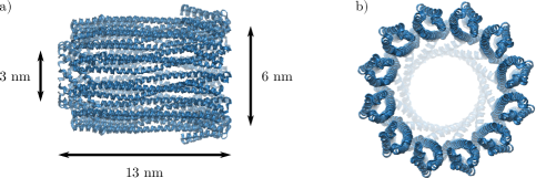
Cytolysin A (ClyA) is a toxin synthesized by several bacteria, and is employed to disrupt cellular membranes of other organisms. It is initially synthesized as a monomer, and then it spontaneously assembles into a 12-mer, cylindrically-shaped pore (Fig. 2). The internal diameter is about nm at the narrower side and nm at the wider side, while its length is nm. Although the diameter of ClyA can, in principle, fit both ssDNA and dsDNA, owing to the negative charges present in the pore lumen, translocation in the wild type ClyA can only occur in solutions of high ionic strength. For this reason ClyA mutants were recently engineered [11], that contain additional positive charges in the lumen and the wide entrance of the pore, allowing DNA translocation at physiological salt concentrations ( mM NaCl).
3 Modeling the DNA capture
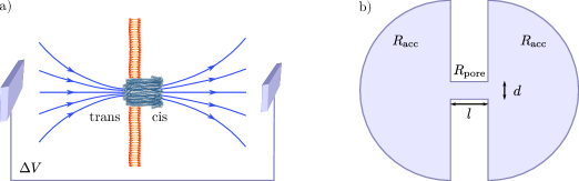
Figure 1a shows a typical experimental setup, in which a single nanopore is inserted in the lipid bilayer membrane, in contact with a NaCl solution. When a potential difference is applied between two electrodes, and in absence of blockages at the pore, a steady electric current pore is generated, with oppositely-charged ions flowing through the pore in opposite directions. The current can be calculated by first decomposing the system into two semi-infinite spherical shells (cis and trans side), connected with each other through a cylinder of diameter and length (nanopore). Then, treating the three regions as resistors in series (Fig. 1b) and using Ohm’s law yields [12]
| (1) |
where is the conductivity. We have denoted by the resistance of each semi-infinite half sphere, known as the access resistance (the derivation can be found in Ref. [15]), and by the electric resistance of the pore (Fig. 1b). The contribution of becomes dominant for wider solid-state nanopores, as confirmed by experiments with nanopores of varying [16]. In the case of ClyA, which has dimensions nm and nm, one finds . Assuming that the equipotential surfaces are semi-spherical outside the pore, one obtains the electrostatic potential [12]
| (2) |
where we set the potential to zero at the electrode and defined the characteristic length
| (3) |
which depends only on the geometry of the pore (for the case of ClyA one finds nm). The DNA molecule performs a drift-diffusive motion in the potential (2) until it reaches the close vicinity of the pore. There it is either directly translocated to the other side of the membrane, corresponding to a diffusion-limited case, or it encounters an additional free energy barrier that needs to overcome for a successful translocation. If the barrier is large compared to the thermal energy and the attractive electrostatic potential, it will dominate the capture kinetics, and the process becomes reaction-limited. We will discuss these two cases separately, following closely the theory developed in [12].
3.1 Diffusion-limited capture
In spite of its high complexity, far from the pore the problem becomes spherically symmetric [see Eq. (3)], and DNA can be treated as a charged point particle. Let us consider a collection of such diffusing particles characterized by a concentration and subject to an electrophoretic force, given by a radial potential . The continuity equation in spherical coordinates reads
| (4) |
where the radial current density contains the contribution from diffusion and electrophoretic drift
| (5) |
Combining Eqs. (4) and (5) one obtains the drift-diffusion equation, with and the diffusion coefficient and the electric mobility, respectively. Note that the two terms in Eq. (5) enter with a different sign because DNA is negatively charged, and by convention . It should be stressed that the Einstein relation does not hold for free electrophoresis of DNA [17], i.e. . This arises from the fact that a free DNA in solution is accompanied by a collection of counterions, while the application of an electric field pushes the two in opposite directions. This leads to different typical molecular configurations, hence the Einstein relation breaks down.
The stationary solution of Eq. (4) is obtained by setting , corresponding to constant . For the potential of Eq. (3) one finds [12]
| (6) |
where is the bulk concentration and a characteristic length given by
| (7) |
For the derivation of Eq. (6) we used as boundary conditions and , with a microscopic distance of the order of the pore size. From Eq. (6) one can estimate the capture rate, which is equal to the number of particles per unit time reaching the absorbing boundary at . This is obtained by integrating the current density on a half-spherical shell of radius [12]
| (8) |
where we have used , which is a valid approximation for typical systems [11]. Here can be interpreted as the distance at which the DNA is irreversibly captured by the pore [12], and increases with the applied potential and the electrophoretic mobility [see Eq. (7)]. Equation (8) is identical to the Smoluchowski diffusion-limited reaction rate for a free diffusing particle absorbed by a sphere of radius , with instead of due to the semi-infinite geometry [12].
3.2 Reaction-limited capture
In a reaction-limited capture the actual translocation takes place once DNA overcomes an additional barrier at the pore entry. Ref. [13] discussed this type of process, which we review here. Let us consider a drift-diffusion model with an additional short-range repulsive potential , i.e. nonvanishing only in the close vicinity of the pore. The radial current density is then given by
| (9) |
where we distinguish between the electrophoretic mobility and a mobility connected to other external forces [18]. Although the former does not satisfy the Einstein relation, the latter does [19], i.e. . Thus, we one can rewrite the current as
| (10) |
with
| (11) |
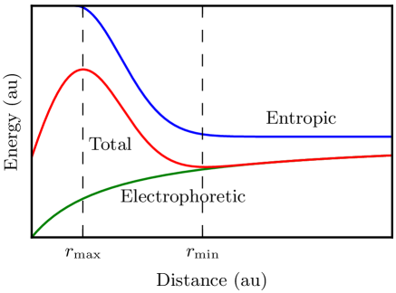
Thus, the dynamics is described by a drift-diffusion equation in the effective potential . The electrophoretic contribution to the force is attractive, while is short-range and repulsive and expected to originate from the steric hindrance of the DNA threading into the pore [12, 20] (Fig. 3). To initiate translocation the molecules have to overcome a barrier , where and are the positions of the maximum and minimum of , respectively, located in the vicinity of the pore (Fig. 3). If the capture process is reaction-limited (i.e. ), we expect that the rate will be given by
| (12) |
where is a characteristic rate constant.
4 Experiments
Having reviewed the existing theory of DNA capture by nanopore, we will now apply it to the experimental data of Ref. [11]. In that study the capture rates of both ssDNA and dsDNA by the ClyA nanopore were measured as a function of the ionic strength of the solution. In what follows we will show that the experimental data in both cases (shown in Fig. 4) are not in line with the theory of diffusion-limited capture. Instead, they seem to exhibit the exponential dependence predicted by Eq. (12), suggesting that it is more likely a reaction-limited process. Finally, we will show that the fitted exponent is in line, at least in the order of magnitude, with the theoretical predictions, further reinforcing our argument.
Since both theories involve the electrophoretic mobility of DNA, we will first estimate its value. Assuming that DNA is a cylinder of diameter with an effective charge per length equal to (with the separation between successive phosphate groups), Ref. [12] estimates the electrophoretic mobility as
| (13) |
where kg m-1s-1 is the water viscosity, and is a numerical factor. The latter takes into account that counterions are bound to the phosphate charges, rendering the effective charge of DNA smaller than the bare one. Finally, one should use and for ssDNA and dsDNA, respectively. Since this quantity enters in both theories, we will calculate for both ssDNA and dsDNA. Using nm and mV we obtain 111For dsDNA it is nm and nm, while for ssDNA one has nm and nm [21].
| (14) |
where for simplicity we have taken . Thus, the electrophoretic mobilities of ssDNA and dsDNA are found to be quite similar.
We will first test whether the data can be described by the theory of diffusion-limited capture. Combining Eqs. (7) and (8) yields , so using the experimental concentration M, the characteristic length nm [see discussion below Eq. (3)] and Eq. (14) yields
| (15) |
where we have used a representative value nm for the Debye length. A comparison with the experimental data of Fig. 4 indicates that this result overestimates the capture rates by two orders of magnitude. Note that some of the phosphate DNA charges can be bound to counterions, leading to . However, to reconcile the data with diffusion-limited capture, one would need a very small value of , which is unlikely. In addition, the data are not consistent with a linear dependence on , as expected from a diffusion-limited process (). We, thus, conclude that the capture of both ssDNA and dsDNA is not diffusion-limited. 222Ref. [11] suggested for ssDNA a reaction-limited capture and for dsDNA a diffusion-limited capture. The latter conclusion was based on an erroneous estimate of . This is in agreement with measurements for dsDNA of comparable size captured by solid-state nanopores [22].

Having excluded a diffusion-limited capture, let us now test the other limiting case, that of a reaction-limited process. Combining Eqs. (11) and (12) one obtains
| (16) |
where we have defined and the maximum and minimum values of the electrophoretic potential. In combination with Eq. (13), this relation implies an exponential dependence of the capture rate on the Debye length. This is indeed the observed trend of the experimental data, as seen in Fig. 4. For a more quantitative comparison, we get from Eq. (2)
| (17) |
where is a characteristic length, and is expected to be comparable to the pore diameter, i.e. nm. Combining this with Eq. (16) yields
| (18) |
The only missing element is the determination of the mechanical mobility . For this purpose one may use Stokes’ law, which gives
| (19) |
where is the hydrodynamic radius. Combining this with Eqs. (14) and (18), and using once more nm and nm, gives , where
| (20) |
is a parameter that can be fitted to the experimental data (see Fig. 4). Since the contour length nm of ssDNA is much larger than its persistence length nm, and if we neglect excluded-volume effects, we can approximate it as a sphere of radius (radius of gyration). Using this for the estimation of its hydrodynamic radius gives nm, from which we find nm. In the case of dsDNA, the contour length nm is lower than its persistence length nm, suggesting it behaves more like a rigid rod. If we, once more, approximate it as a cylinder of diameter , and use the results of Ref. [23], we obtain nm. Finally, plugging this in Eq. (20) yields nm. These results are in a good agreement with fits of the experimental data (Fig. 4), which yielded the values nm and nm for ssDNA and dsDNA, respectively, despite the simplicity of the theory. The agreement further corroborates the validity of the reaction-limited capture scenario.
5 Conclusion
We have reviewed two basic mechanisms of DNA capture by a nanopore: the diffusion-limited and the reaction-limited capture. The theoretical description of these mechanisms was developed in Refs. [22, 12, 20], and these ideas were tested in translocation experiments through solid-state nanopores with dsDNA sequences ranging from 800 to 50,000 base pairs [22]. The shortest lengths dsDNA (up to 8,000 base pairs) showed a reaction-limited capture, characterized by an exponential growth of the capture rate with the sequence length. A second regime, for sequences longer than 10,000 base pairs, was found to be consistent with a diffusion-limited capture, in which is independent of the sequence length [22]. Overall, solid-state nanopore experiments [22] were found to be in agreement with the theoretical framework of dsDNA capture.
Here we tested the theory in a set of experiments with ClyA, a biological nanopore recently engineered to allow translocation of both ssDNA and dsDNA at physiological salt concentrations [11]. The experiments involved short ssDNA and dsDNA sequences ( nucleotides and base pairs, respectively), and were performed at varying salt concentration [11]. Diffusion-limited capture rates estimated for a nanopore with the ClyA size were shown to be much higher than experimental measurements for both ssDNA and dsDNA, suggesting for both a reaction-limited capture (this corrects the erroneous conclusion in Ref. [11]) Our analysis showed that the experiments are in quantitative agreement with the theory, which predicts an exponential dependence on the Debye length , with the prefactors determined by the local properties of the barrier. The theoretical estimates for the characteristic length for both ssDNA and dsDNA are in agreement with fits to the experimental data, confirming the validity of the modeling approach. A consistent picture thus emerges for the capture mechanism of DNA from ClyA nanopore. A reaction-limited capture was also found to be in agreement with the results of Ref. [22], and with other studies of ssDNA capture into HL nanopores [24, 25, 26]. Still, it would be desirable to have more insight on the nature of the barrier. A question, which could be addressed by additional experiments or computer simulations of the capture mechanism, similar to those of Refs. [27, 28].
Acknowledgement – SN acknowledges financial support from the Research Funds Flanders (FWO Vlaanderen) grant VITO-FWO 11.59.71.7.
References
References
- [1] Dekker C 2007 Nature Nanotechnology 2 209
- [2] Venkatesan B M and Bashir R 2011 Nature Nanotechnology 6 615
- [3] Soskine M, Biesemans A, Moeyaert B, Cheley S, Bayley H and Maglia G 2012 Nano letters 12 4895–4900
- [4] Soskine M, Biesemans A and Maglia G 2015 J. Am. Chem. Soc. 137 5793–5797
- [5] Huang G, Willems K, Soskine M, Wloka C and Maglia G 2017 Nature Comm. 8 935
- [6] Heerema S J and Dekker C 2016 Nature Nanotechnology 11 127
- [7] Shi W, Friedman A K and Baker L A 2016 Anal. Chem. 89 157–188
- [8] Ayub M and Bayley H 2016 Curr. Opin. Chem. Biol. 34 117–126
- [9] Soskine M, Biesemans A, De Maeyer M and Maglia G 2013 J. Am. Chem. Soc. 135 13456–13463
- [10] Bayley H 2015 Clinical Chem. 61 25–31
- [11] Franceschini L, Brouns T, Willems K, Carlon E and Maglia G 2016 ACS Nano 10 8394–8402
- [12] Grosberg A Y and Rabin Y 2010 J. Chem. Phys. 133 165102
- [13] Rowghanian P and Grosberg A Y 2013 Phys. Rev. E 87 042722
- [14] Humphrey W, Dalke A and Schulten K 1996 J. Mol. Graphics 14 33–38
- [15] Hall J E 1975 J. Gen. Physiol. 66 531–532
- [16] Kowalczyk S W, Grosberg A Y, Rabin Y and Dekker C 2011 Nanotechnology 22 315101
- [17] Nkodo A E, Garnier J M, Tinland B, Ren H, Desruisseaux C, McCormick L C, Drouin G and Slater G W 2001 Electrophoresis 22 2424–2432
- [18] Long D, Viovy J L and Ajdari A 1996 Phys. Rev. Lett. 76 3858–3861
- [19] Rowghanian P and Grosberg A Y 2013 Phys. Rev. E 87 042723
- [20] Muthukumar M 2010 J. Chem. Phys. 132 05B605
- [21] Chi Q, Wang G and Jiang J 2013 Physica A 392 1072–1079
- [22] Wanunu M, Morrison W, Rabin Y, Grosberg A Y and Meller A 2010 Nature Nanotechnology 5 160
- [23] Hansen S 2004 J. Chem. Phys. 121 9111–9115
- [24] Henrickson S E, Misakian M, Robertson B and Kasianowicz J J 2000 Phys. Rev. Lett. 85 3057
- [25] Meller A, Branton D et al. 2002 Electrophoresis 23 2583–2591
- [26] Meller A 2003 J. Phys.: Cond. Matt. 15 R581
- [27] Farahpour F, Maleknejad A, Varnik F and Ejtehadi M R 2013 Soft Matter 9 2750–2759
- [28] Farahpour F, Ejtehadi M R and Varnik F 2014 Int. J. Mod. Phys. C 25 1441010