Clustering of Janus Particles in Optical Potential Driven by Hydrodynamic Fluxes
I Abstract
Self-organisation is driven by the interactions between the individual components of a system mediated by the environment, and is one of the most important strategies used by many biological systems to develop complex and functional structures. Furthermore, biologically-inspired self-organisation offers opportunities to develop the next generation of materials and devices for electronics, photonics and nanotechnology. In this work, we demonstrate experimentally that a system of Janus particles (silica microspheres half-coated with gold) aggregates into clusters in the presence of a Gaussian optical potential and disaggregates when the optical potential is switched off. We show that the underlying mechanism is the existence of a hydrodynamic flow induced by a temperature gradient generated by the light absorption at the metallic patches on the Janus particles. We also perform simulations, which agree well with the experiments and whose results permit us to clarify the underlying mechanism. The possibility of hydrodynamic-flux-induced reversible clustering may have applications in the fields of drug delivery, cargo transport, bioremediation and biopatterning.
II 1 Introduction
Self-organisation entails the emergence of complex patterns and structures from relatively simple constituting building blocks Zhang and Glotzer [2004], van Blaaderen [2004], Jiang et al. [2010], Walther and Müller [2013], Velu et al. [2013], Mijalkov et al. [2016]. Phenomena such as flocking of birds and growth of bacterial colonies are examples of self-organisation in nature. Also artificial microscopic systems feature similar forms of organisation with the emergence of clusters, sometimes referred to as “living crystals” Palacci et al. [2013], Buttinoni et al. [2013], Gao et al. [2013], Stenhammar et al. [2015], Schmidt et al. [2018]. In the past two decades, studies on self-organisation focused on systems made of complex colloids with anisotropic surface Perro et al. [2005], Pawar and Kretzschmar [2010], such as Janus particles Walther and Müller [2013], de Gennes [1992]. Depending on their surface material properties, Janus particles have been used in different fields for various applications such as self-assembly, microrheology and emulsion stabilisation Jiang et al. [2010], Walther and Müller [2013]. Under certain conditions, Janus particles have the ability of self-propelling and behave as active Brownian particles Howse et al. [2007], Gangwal et al. [2008], Volpe et al. [2011], Buttinoni et al. [2012], Illien et al. [2017]; these active Janus particles might be used in future biomedical nano-devices for diagnostics, drug delivery and microsurgery Wang and Gao [2012], Baraban et al. [2012].
Studies on clustering of Janus particles have been performed by Palacci et al. Palacci et al. [2013], who have shown the formation of living crystals in systems of light-activated Janus particles (Fe2O3-TPM) in hydrogen peroxide solution. Similarly, Buttinoni et al. Buttinoni et al. [2013] demonstrated the clustering of light-activated Janus particles (carbon-SiO2) in a water-lutidine binary mixture. Other research groups have shown self-assembly and controlled crystal formations in a mixed system of light-activated Janus particles and passive colloids Gao et al. [2013], Stenhammar et al. [2015]. In all these studies, a necessary ingredient for the clustering is the active nature of the particles. In systems of passive colloidal particles, crystallisation was observed at the bottom of an attractive optical potential Pinçe et al. [2016], close to the hard boundary during electrophoretic deposition Solomentsev et al. [1997], and in the presence of an external temperature gradient Weinert and Braun [2008], Di Leonardo et al. [2009].
Here, we investigate the behaviour of a system composed of Janus particles (silica microspheres half-coated with gold) close to a planar surface in the presence of an optical potential, and we experimentally demonstrate reversible clustering triggered by the presence of the optical field. Experimental results are compared and validated by numerical simulations, where the key ingredient for clustering is the presence of an attractive potential of hydrodynamic nature. Such results are confirmed also in mixtures of Janus particles and passive colloids (silica microspheres), where the hydrodynamic flux due to the Janus particles causes the clustering of the particles in the hybrid system and the formation of living crystals. As a further confirmation that the presence of Janus particles in the optical potential is crucial for the clustering, we show that a system with only non-Janus particles does not give rise to any clustering.
III 2 Experiments
The experiments are performed on a homemade inverted microscope, as schematically shown in Fig. 1a. A laser beam (wavelength ; power ) is focused by a convex lens (L, focal length ) onto the sample chamber (S) in order to generate a broad Gaussian optical potential (beam waist ) Pesce et al. [2015]. The height of the sample chamber is . The particles are tracked by digital video microscopy using the image projected by a microscope objective (, ) on a monochrome CCD camera with an acquisition rate of .
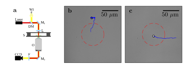
In Fig. 1(b), we show the typical motion of a Janus particle in the optical potential generated by the Gaussian laser beam. The Janus particle does not stay for a long time within the region of maximum intensity, but it is driven outwards by a combination of optical forces and optical torques: the presence of the reflecting thin gold layer results in an optical force directed towards the region of lower light intensity. On the contrary, in Fig. 1(c), we show that a silica particle is driven by the optical force towards the region of maximum intensity, as expected in the presence of optical forces Jones et al. [2015].
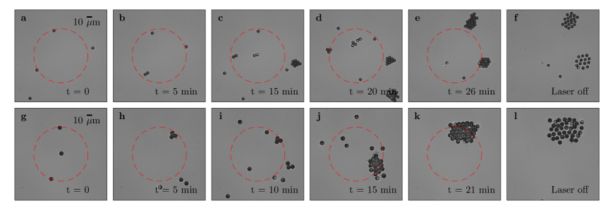
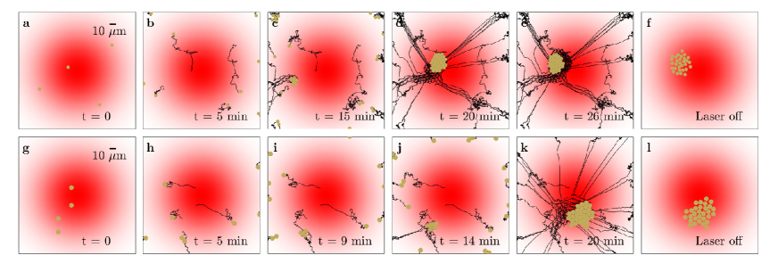
In Fig. 2, we show the behaviour of multiple Janus particles in a Gaussian optical potential. Figs. 2(a-f) show a time sequence for a solution of Janus particles of diameter, and Figs. 2(g-l) a time sequence for a solution of Janus particles of diameter. In both cases, when the optical potential is turned on, the Janus particles cluster together and the centres of the clusters lay outside the centre of the optical potential. The process is slow at the beginning but accelerates as the size of the clusters increases. When the optical potential is switched off, the clusters immediately start to disassemble because of the attractive force between the particles disappears.
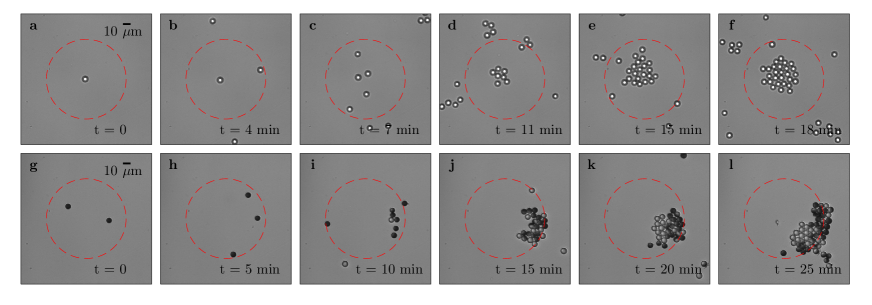
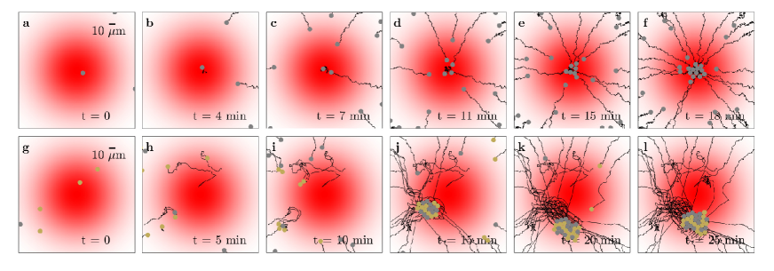
We do not observe this clustering behaviour in the case of a colloidal suspension composed only of silica particles with the same dilution as for the solution containing the Janus particles. In fact, when employing silica microspheres, aggregation and formation of a colloidal crystal are observed only with a significantly higher concentration (as observed, for example, in Ref. Pinçe et al., 2016). In this case, the aggregation is not due to an effective attraction between the particles, but to the interplay between the optical forces pushing each particle towards the centre of the potential and the steric repulsion between the particles. The combined effect of these two interactions determines the formation of a regular close-packed lattice structure. However, this mechanism is ineffective at low concentrations, as shown in the time sequence presented in Figs. 3(a-f): while the silica particles are attracted towards the centre of the potential, they do not form a cluster. If we add some Janus particles to this system of silica particles, the clustering of the particles away from the centre of the beam is recovered, as shown in Figs. 3(g-l).
IV 3 Model
In order to understand the physical mechanism underlying the clustering of the Janus particles, we developed a numerical model of this system. In this model, we take into account the optical forces and torques acting on the Janus particles Callegari et al. [2015], Jones et al. [2015], their Brownian motion Fernandes and de la Torre [2002], Volpe and Volpe [2013], Jones et al. [2015], and thermophoretic forces and torques Bickel et al. [2013].
We model the optical forces and torques acting on a spherical Janus particle using the geometric optics approximation, because the size of the particle is significantly larger than the wavelength of the incident field and, in these conditions, the geometrical optics approximation reproduces well the features of the dynamics observed experimentally Callegari et al. [2015], Jones et al. [2015]. We model a Janus particle as a spherical dielectric microsphere plus a surface layer shaped as the hemispherical gold cap with a given thickness, mass density and refractive index. When the Janus particle is suspended in a solution and subject to an optical potential, there are three elements influencing its motion: (i) optical forces and torques due to the scattering of the light between media with different refractive indices Callegari et al. [2015], Jones et al. [2015]; (ii) Brownian forces due to the presence of a thermal noise Fernandes and de la Torre [2002], Volpe and Volpe [2013], Jones et al. [2015]; and (iii) thermophoretic forces and torques, which are due to the partial light absorption by the golden cap determining a temperature gradient around the particle and, therefore, a self-propelled motion Bickel et al. [2013]. In addition, one should also take into account (iv) the combined effect of gravity and buoyancy, which keep the particles hovering just above the sample chamber bottom surface and; and (v) the gravitational torque due to the inhomogeneity of the mass distribution of the Janus particle due to the gold coating, which, in the absence of any optical field, always results in a preferential downwards orientation of the golden cap.
In order to calculate the scattering and absorption of the golden cap, we use the thin film approximation for an absorbing layer on a transparent substrate Heavens [1991]. This permits us to obtain the reflectance, transmittance and absorbance of the metallic cap, and therefore to calculate the scattering of the light on the Janus particle. From the scattered rays, we obtain the optical force and torque according to the procedure in Refs. Callegari et al., 2015, Jones et al., 2015.
In order to simulate the Brownian motion of a Janus particle, we have to take into account the asymmetry due to the presence of the metal cap, even though the shape of the Janus particle is accurately represented by a sphere. This entails that we need to take into account not only the translational motion, but also the rotational motion. Therefore, we use the diffusion matrix, as in Ref. Fernandes and de la Torre, 2002. Furthermore, since the Janus particle are not in bulk but near a planar wall, we need to correct the translational and rotational diffusion for the effects of the close proximity to the boundary Happel and Brenner [2012], Lee et al. [1979].
The self-propelled motion originates from the presence of a local temperature gradient around the particle due to the light absorption by the metal-coated side of the Janus particle. A non-spherically symmetric temperature profile is induced around the particle due to the non-spherically symmetric shape of the absorbing layer. Such configuration induces a local force field tangential to the surface of the Janus particle. This interfacial force leads to a slip velocity at the interface, i.e., a jump in the tangential fluid velocity component. This slip velocity drives the particle in the opposite direction along the temperature gradient axis Bickel et al. [2013], inducing the particle to self-propel. The velocity of this self-propulsion (i.e. the thermophoretic velocity) depends linearly on the temperature gradient, i.e. , where is the thermophoretic mobility (or thermal diffusion coefficient) Weinert and Braun [2008]. However, there is no certain law for the amplitude and sign of the thermophoretic mobility, which strongly depends on the microscopic nature of the particle-solvent interactions at the boundary layer of thickness (Debye length) Dhont et al. [2007], Ruckenstein [1981], Würger [2007], Piazza [2008]. Depending on the sign of , the particle moves either towards the cold or the hot region Weinert and Braun [2008], Piazza [2008].
In the case of two Janus particles close to a planar wall, a further effect of hydrodynamic nature has to be considered. This hydrodynamic effect creates an effective attraction among the particles. To explain this behaviour, one can use the same approach proposed for two immobile colloidal particles close to a wall Weinert and Braun [2008], Morthomas and Würger [2010], Di Leonardo et al. [2009]. Indeed, a particle moves toward the horizontal bottom surface of the sample cell due to gravity, radiation force and interfacial driving force. Eventually, this particle would be fixed at a certain distance from the wall, due to the repulsive interaction with the wall and the viscous stress. The particle is then immobile Weinert and Braun [2008], Morthomas and Würger [2010], Di Leonardo et al. [2009] and the surrounding velocity field is squeezed by the boundary. Due to the temperature gradient surrounding the particles, the fluid continues to move along its surface. This creates a flow with a horizontal incoming radial component (parallel to the planar boundary) and outgoing vertical components, directed upwards from the wall. The thermophoretically-induced flow field affects the motion of other neighbouring particles, so that a second nearby particle experiences an attractive hydrodynamic drag force toward the first particle. Following Ref. Morthomas and Würger, 2010, the radial component of the flow velocity at a horizontal distance from the centre of the immobile particle, , is:
| (1) |
where , , is the absorbed power by the gold cap with the total outward heat flow, ( and are the thermal conductivity of fluid and particle, respectively), , , , is the distance of the centre of the particle with radius from the wall, and . From the flow velocity, , one can obtain the effective hydrodynamic force on nearby particle as
| (2) |
where is a dimensionless factor which accounts for the effect of the presence of a planar wall on the effective friction coefficient in the direction parallel to the wall Happel and Brenner [2012], Di Leonardo et al. [2009]. When this lateral flow is strong enough, the attractive hydrodynamic force can be larger than other repulsive contributions and than the thermal fluctuations, leading to a stable aggregation of the particles. The strength of the attractive interaction can be regulated by the light intensity since the power absorbed by the gold cap determines the temperature gradient around the immobile particle and therefore the entity of the hydrodynamic lateral flow.
Using this model we investigate in the next section the motion of a single and multiple Janus particles suspended in a water solution and in presence of a broad Gaussian optical potential.
V 4 Numerical results
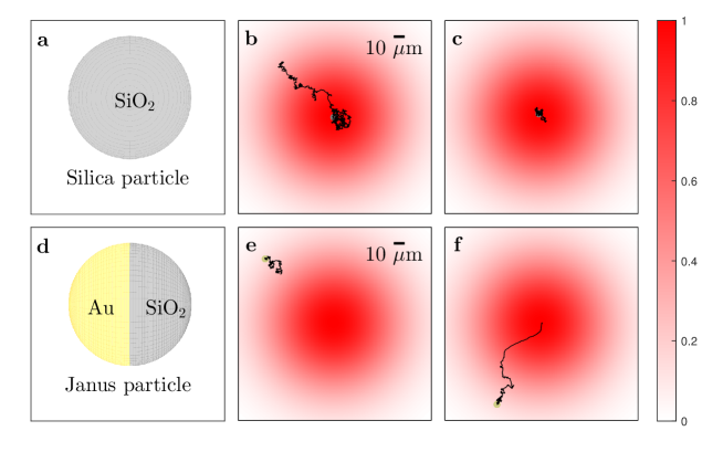
In Fig. 4, we show the behaviour of a silica particle (Fig. 4(a-c)) and of a Janus particle (Fig. 4(d-f)) in the presence of a Gaussian optical potential (radius , wavelength , power ). The particles’ trajectories are shown by the black lines. The particles are made of silica and have a diameter of , and the Janus particle is half-coated with a gold layer (at the wavelength , the refractive indices of silica and gold film are and , respectively Johnson. and Christy [1972]). The silica particle (Fig. 4(a)) moves toward the high intensity area and tends to remain in the region of higher optical intensity because of the presence of gradient optical forces Ashkin et al. [1986], Jones et al. [2015], both when it is initially placed outside the potential (Fig. 4(b)) and at its centre (Fig. 4(c)). On the contrary, the Janus particle (Fig. 4(d)) tends to move towards a circular region at a fixed distance from the centre of the potential, independently from whether it is initially placed outside the trapping potential (Fig. 4(e)) or at its centre (Fig. 4(f)). These results are in agreement with the experiments shown in Figs. 1(b) and 1(c).
In Fig. 5, we show the time sequences corresponding to the clustering of Janus particles in a Gaussian optical potential. As in the experiments shown in Fig. 2, the diameter of the Janus particles is in Figs. 5(a-f) and in Figs. 5(g-l). Both kinds of particles form clusters at a certain distance from the centre of the beam, which is in good agreement with the experiments shown in Fig. 2. Furthermore, the clustering speed depends on the Janus particles size: larger particles aggregate more rapidly, as observed in experiments.
In Fig. 6(a-f), we show the simulated collective behaviour of a system of silica particles (diameter ). In agreement with the experimental results shown in Figs. 3(a-f), the silica particles go toward the centre of the Gaussian optical potential because of optical gradient forces, but do not form a colloidal crystal. In Fig. 6(g-l), we show the simulated collective behaviour of a mixture of silica particles and Janus particles (diameter ). Again in agreement with the experiments presented in Figs. 3(g-l), the Janus particles generate a hydrodynamic flow that is sufficient to induce the clustering of all particles away from the optical potential center.
VI 5 Conclusions
We have shown with experiments and numerical simulations that the presence of Janus particles triggers the formation of clusters in a Gaussian optical potential. This is due to the presence of attractive hydrodynamic interactions among the particles. The presence of Janus particles is crucial for the cluster formation, since the attractive interaction is generated by the presence of a temperature gradient around the Janus particles: When a Janus particle is close to a boundary, this temperature gradient induces a hydrodynamic flow that drags other particles towards the Janus particle. In the absence of Janus particles, this hydrodynamic flow is absent and thus no clusters form. We have shown experimentally that the clustering process is reversible, since the cluster starts to disassemble as soon as the optical potential is switched off. Beyond their fundamental interest, the reported results are potentially relevant for various applications in the fields of self-assembly, targeted drug-delivery and bioremediation. For example, the possibility of forming clusters at a controllable distance from the minimum of a potential well offers a new route towards self-assembly near a target. Future work will be devoted to understanding how the clustering behaviour can be controlled or altered by using more complex optical potentials.
VII Acknowledgment
SMM acknowledges a Tubitak 2216 fellowship and Tubitak project 114F207. SKPV acknowledges Tubitak projects 114F207 and 116F068. AC acknowledges Tubitak projects 115F401 and 116F111.
References
- Zhang and Glotzer [2004] Z. Zhang and S. C. Glotzer. Self-assembly of patchy particles. Nanolett., 4(8):1407–1413, 2004.
- van Blaaderen [2004] A. van Blaaderen. Colloids under external control. MRS Bull., 29(2):85–90, 2004.
- Jiang et al. [2010] S. Jiang, Q. Chen, M. Tripathy, E. Luijten, K. S. Schweizer, and S. Granick. Janus particle synthesis and assembly. Adv. Mat., 22(10):1060–1071, 2010.
- Walther and Müller [2013] A. Walther and A. H. E. Müller. Janus particles: Synthesis, self-assembly, physical properties, and applications. Chem. Rev., 113(7):5194–5261, 2013.
- Velu et al. [2013] S. K. P. Velu, M. Yan, K.-P. Tseng, K.-T. Wong, D. M Bassani, and P. Terech. Spontaneous formation of artificial vesicles in organic media through hydrogen-bonding interactions. Macromolecules, 46(4):1591–1598, 2013.
- Mijalkov et al. [2016] M. Mijalkov, A. McDaniel, J. Wehr, and G. Volpe. Engineering sensorial delay to control phototaxis and emergent collective behaviors. Phys. Rev. X, 6(1):011008, 2016.
- Palacci et al. [2013] J. Palacci, S. Sacanna, A. P. Steinberg, D. J. Pine, and P. M. Chaikin. Living crystals of light-activated colloidal surfers. Science, 339(6122):936–940, 2013.
- Buttinoni et al. [2013] Ivo Buttinoni, J. Bialké, F. Kümmel, H. Löwen, C. Bechinger, and T. Speck. Dynamical clustering and phase separation in suspensions of self-propelled colloidal particles. Phys. Rev. Lett., 110(23):238301, 2013.
- Gao et al. [2013] W. Gao, A. Pei, X. Feng, C. Hennessy, and J. Wang. Organized self-assembly of janus micromotors with hydrophobic hemispheres. J. Am. Chem. Soc., 135(3):998–1001, 2013.
- Stenhammar et al. [2015] J. Stenhammar, R. Wittkowski, D. Marenduzzo, and M. E. Cates. Activity-induced phase separation and self-assembly in mixtures of active and passive particles. Phys. Rev. Lett., 114(1):018301, 2015.
- Schmidt et al. [2018] F. Schmidt, B. Liebchen, H. Löwen, and G. Volpe. Light-controlled assembly of active colloidal molecules. arXiv, page 1801.06868, 2018.
- Perro et al. [2005] A. Perro, S. Reculusa, S. Ravaine, E. Bourgeat-Lami, and E. Duguet. Design and synthesis of janus micro-and nanoparticles. J. Mat. Chem., 15(35-36):3745–3760, 2005.
- Pawar and Kretzschmar [2010] A. B. Pawar and I. Kretzschmar. Fabrication, assembly, and application of patchy particles. Macromol. Rap. Commun., 31(2):150–168, 2010.
- de Gennes [1992] P. G. de Gennes. Soft matter. Science, 256(5056):495–497, 1992.
- Howse et al. [2007] J. R. Howse, R. A. L. Jones, A. J. Ryan, T. Gough, R. Vafabakhsh, and R. Golestanian. Self-motile colloidal particles: From directed propulsion to random walk. Phys. Rev. Lett., 99(4):048102, 2007.
- Gangwal et al. [2008] S. Gangwal, O. J. Cayre, M. Z. Bazant, and O. D. Velev. Induced-charge electrophoresis of metallodielectric particles. Phys. Rev. Lett., 100(5):058302, 2008.
- Volpe et al. [2011] G. Volpe, I. Buttinoni, D. Vogt, H.-J. Kümmerer, and C. Bechinger. Microswimmers in patterned environments. Soft Matter, 7(19):8810–8815, 2011.
- Buttinoni et al. [2012] I. Buttinoni, G. Volpe, F. Kümmel, G. Volpe, and C. Bechinger. Active Brownian motion tunable by light. J. Phys.: Condens. Matter, 24:284129, 2012.
- Illien et al. [2017] P. Illien, R. Golestanian, and A. Sen. ‘Fuelled’ motion: Phoretic motility and collective behaviour of active colloids. Chem. Soc. Rev., 46:5508, 2017.
- Wang and Gao [2012] J. Wang and W. Gao. Nano/microscale motors: biomedical opportunities and challenges. ACS Nano, 6(7):5745–5751, 2012.
- Baraban et al. [2012] L. Baraban, D. Makarov, R. Streubel, I. Mönch, D. Grimm, S. Sanchez, and O. G. Schmidt. Catalytic janus motors on microfluidic chip: deterministic motion for targeted cargo delivery. ACS Nano, 6(4):3383–3389, 2012.
- Pinçe et al. [2016] E. Pinçe, S. K. P. Velu, A. Callegari, P. Elahi, S. Gigan, G. Volpe, and G. Volpe. Disorder-mediated crowd control in an active matter system. Nature Commun., 7:10907, 2016.
- Solomentsev et al. [1997] Y. Solomentsev, M. Böhmer, and J. L. Anderson. Particle clustering and pattern formation during electrophoretic deposition: A hydrodynamic model. Langmuir, 13(23):6058–6068, 1997.
- Weinert and Braun [2008] F. M. Weinert and D. Braun. Observation of slip flow in thermophoresis. Phys. Rev. Lett., 101(16):168301, 2008.
- Di Leonardo et al. [2009] R. Di Leonardo, F. Ianni, and G. Ruocco. Colloidal attraction induced by a temperature gradient. Langmuir, 25(8):4247–4250, 2009.
- Pesce et al. [2015] G. Pesce, G. Volpe, O. M. Maragò, P. H. Jones, S. Gigan, A. Sasso, and G. Volpe. Step-by-step guide to the realization of advanced optical tweezers. J. Opt. Soc. Am. B, 32:B84–B98, 2015.
- Jones et al. [2015] P. H. Jones, O. M. Maragò, and G. Volpe. Optical tweezers: Principles and applications. Cambridge University Press, 2015.
- Callegari et al. [2015] A. Callegari, M. Mijalkov, A. B. Gököz, and G. Volpe. Computational toolbox for optical tweezers in geometrical optics. J. Opt. Soc. Am. B, 32(5):B11–B19, 2015.
- Fernandes and de la Torre [2002] M. X. Fernandes and J. G. de la Torre. Brownian dynamics simulation of rigid particles of arbitrary shape in external fields. Biophys. J., 83(6):3039–3048, 2002.
- Volpe and Volpe [2013] G. Volpe and G. Volpe. Simulation of a Brownian particle in an optical trap. Am. J. Phys., 81(3):224–230, 2013.
- Bickel et al. [2013] T. Bickel, A. Majee, and A. Würger. Flow pattern in the vicinity of self-propelling hot janus particles. Phys. Rev. E, 88(1):012301, 2013.
- Heavens [1991] O. S. Heavens. Optical properties of thin solid films. Courier Corporation, 1991.
- Happel and Brenner [2012] J. Happel and H. Brenner. Low Reynolds number hydrodynamics: with special applications to particulate media, volume 1. Springer Science & Business Media, 2012.
- Lee et al. [1979] S. H. Lee, R. S. Chadwick, and L. G. Leal. Motion of a sphere in the presence of a plane interface. part 1. an approximate solution by generalization of the method of Lorentz. J. Fluid Mech., 93(4):705–726, 1979.
- Dhont et al. [2007] J. K. G. Dhont, S. Wiegand, S. Duhr, and D. Braun. Thermodiffusion of charged colloids: Single-particle diffusion. Langmuir, 23(4):1674–1683, 2007.
- Ruckenstein [1981] E. Ruckenstein. Can phoretic motions be treated as interfacial tension gradient driven phenomena? J. Colloid Interfac. Sci., 83(1):77–81, 1981.
- Würger [2007] A. Würger. Thermophoresis in colloidal suspensions driven by Marangoni forces. Phys. Rev. Lett., 98(13):138301, 2007.
- Piazza [2008] R. Piazza. Thermophoresis: moving particles with thermal gradients. Soft Matter, 4(9):1740–1744, 2008.
- Morthomas and Würger [2010] J. Morthomas and A. Würger. Hydrodynamic attraction of immobile particles due to interfacial forces. Phys. Rev. E, 81(5):051405, 2010.
- Johnson. and Christy [1972] Peter B. Johnson. and R.-W. Christy. Optical constants of the noble metals. Phys. Rev. B, 6(12):4370, 1972.
- Ashkin et al. [1986] A. Ashkin, J. M. Dziedzic, J. E. Bjorkholm, and S. Chu. Observation of a single-beam gradient force optical trap for dielectric particles. Opt. Lett., 11(5):288–290, 1986.