Fluorescence coherence with anodic aluminum oxide hybrid photonic-plasmonic structure
Abstract
A class of hybrid photonic-plasmonic structures (HPPS) with vertical cylindrical cavities is proposed and its performance in providing a coherent light from spontaneous emission is investigated. It is shown that the proposed easy-to-fabricate and robust anodic aluminum oxide structure dramatically enhances the temporal and spatial coherence compared to the previously examined HPPS. The physical mechanism of achieving the temporal and spatial coherence is explained based on the special optical properties of the proposed HPPS and it is discussed how this structure enables one adjusting hybrid photonic-plasmonic optical modes to obtain coherence for emitters of different frequencies.
![[Uncaptioned image]](/html/1811.10465/assets/TOCfig.png)
Fluorescence is a vastly used optical method for chemical and biological detection Shcheslavskiy et al. (2018); Giljohann and Mirkin (2009); however, its performance and efficiency shall be enhanced especially in sensing and imaging applications Ribeiro, Baleizão, and Farinha (2017); Moerland, Eguiluz, and Kaivola (2013); Zhao et al. (2014); Wang et al. (2015). Plasmonics appears to be the best method for tailoring and enhancing the fluorescence emission Stockman et al. (2018); Bauch et al. (2014). It is known that light can be confined in close vicinity of metallic surfaces or nano-particles due to the coupling between electromagnetic (EM) waves and oscillations of electrical charges at the surface. The idea is to employ This interaction, which is referred to as surface plasmon resonance (SPR) in a way that a large enhancement in the optical density of states can be obtained in the neighborhood of fluorophores. This will be an effective means for elevating the excitation rate and raising the quantum yield as well as controlling the angular distribution of the fluorescence emission Kwon et al. (2008); Kinkhabwala et al. (2009); Aouani et al. (2011); Lozano et al. (2013); Langguth et al. (2013).
Although the presence of fluorophore in the vicinity of metallic structures leads to the above mentioned appealing features, this adjacency may also increase the probability of quenching and non-radiative energy loss for the excited fluorophore Anger, Bharadwaj, and Novotny (2006); Pons et al. (2007); Li et al. (2009); Reineck et al. (2013). This inadequacy can be resolved by means of a hybrid photonic-plasmonic structure (HPPS), that is created by attaching a photonic crystal (PC) to the metal surface (plasmonic structure). In other words, coupling between the surface plasmon polaritons (SPPs), i.e. the EM waves which are confined along the metal-dielectric interface, and the guided or trapped modes of the photonic crystal leads to the striking features of increasing the propagation length of SPPs, confining light in a deep subwavelength scale, and highly guided modes and cavity resonances in the PC structure Romanov et al. (2011); Yang et al. (2011); Zhang et al. (2012); Schokker et al. (2017). Moreover, by using an HPPS, the effective length of the evanescent normal component of SPPs can extend to tens of nanometers above the metal surface, therefore, the enhancement and directionality are obtained without any significant quenching Zhu et al. (2012); López-García et al. (2010); Ding et al. (2013).
On the other hand, due to the weak correlation between the spontaneous emissions of fluorophores, the resulted light is isotropic in space and broad in spectrum, which in turn lower the detectability. Therefore, it is potentially advantageous to utilize a technique to create a coherent light from such spontaneous emitters. It must be noted that a desirable technique shall also prevent any significant loss in the emission intensity Raghunathan, Schouten, and Visser (2012); Greffet et al. (2002); De Zoysa et al. (2012). In order to address these requirements, Shi and his coworkers Shi et al. (2014a, b) have proposed using a HPPS whose optical attributions was previously investigated by the group Shi et al. (2010). It is shown that the interaction between the leaky modes (that can scape from propagating along the metal-dielectric interface) of HPPS and the fluorescent molecules successfully provides both the temporal and spatial coherence for the fluorescent emission. Such a technique can play an important role in different fluorescence applications. However, further investigations are needed to obtain a more efficient structure with an inherent capability to be adjusted for different spontaneous emitters. This is the aim of the present work.
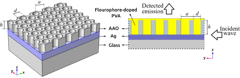
Among different options for the photonic-crystal part of an HPPS, anodic aluminium oxide (AAO) presents a special feature; the vertical cylindrical cavities in the structure facilitates one’s control over the direction of the propagation of the leaky modes. This provides the directionality of the emitted light and therefore can enhance the spatial coherence. Moreover, the frequency of the leaky modes can be adjusted by adapting the geometry of the cavities. In the literature, the capability of AAO in enhancing the intensity of fluorescent emission has also been addressed Li et al. (2012). Nevertheless, AAO has not yet been used in forming an HPPS to achieve coherent fluorescent emission. In this work, a robust and easy to fabricate HPPS using the AAO structure is proposed that substantially enhances the coherence, while brings the required flexibility to be adjusted for spontaneous emitters with different excitation/emission frequencies.
Here, the previously proposed method ARXIV which empowers finite difference time domain to simulate fluorescent molecules is applied to a novel HPPS that is constructed by placing an inverse photonic crystal, made by pore-openedBruschi et al. (2015) anodic aluminium oxide (AAO), on top of a 200 nm thick silver (Ag) layer (Fig.1). The cylindrical holes of PC are filled with S101-doped PVA, which also forms a layer of 50 nm thickness on top of the AAO. The structural parameters, i.e., thickness of AAO layer nm, diameter of cylindrical holes nm, and the center-to-center distance the neighboring cylinders nm, have been set in a way that not only the structure can be easily fabricated, but also, it significantly enhances the coherence as seen in the rest of this paper.
Similar to the previous test-case, the proposed HPPS experiences an incident continuous EM wave with a wavelength of 575 nm from its side that is perpendicular to the -axis (Fig. 1) and the resulting vertical emission (along -axis) is recorded. In this way, the detected wave would not be masked by the incident wave. Moreover, this is a wise choice of the excitation and detection, which is appropriate for imaging and LED applications. In Fig. 2, the recorded field is shown in frequency domain. The graph hits its peak at nm with a FWHM of nm. On the other hand, using TCF, a coherence time of sec or equivalently nm is calculated. It is observed that the presence of the proposed HPPS leads to an almost eight times greater coherence length for the fluorescence emission.
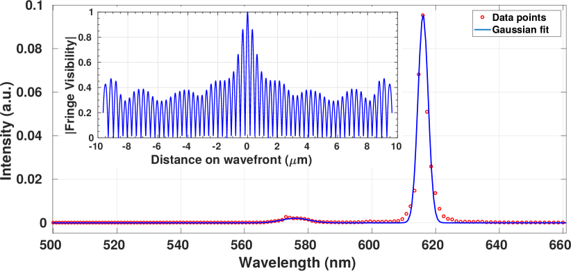
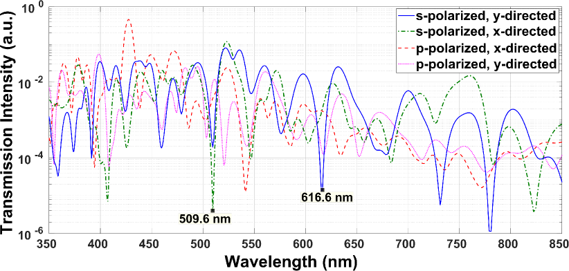
In order to identify the underlying cause of the emergence of such a narrow emission bandwidth, all modes propagated horizontally (along the plane) in PC of the proposed HPPS are also calculated and shown in Fig. 3. It is known that there is no possibility for p-polarized modes to be vertically propagated since the associated electric field is aligned with z-axis. On the other hand the electrical field is directed horizontally for s-polarized modes and thus, for each wavelength that the horizontal propagation is prevented by HPPS, a vertical reflection is possible. Therefore, considering that the excitation input wave is aligned with y-axis, one should focus on the y-directed s-polarized propagation graph in Fig. 3, which reveals a trough at nm. In this sense, the portion of the emission band of S101 molecules that coincides with this trough is expected to reflect vertically while the rest propagates along the metal surface. Since for S101 molecules, the excitation band overlaps partially with the emission band, the horizontally propagated modes can excite the neighboring fluorophores. This synchronizes the transitions of the molecules and therefore, a spatial coherence is also expected. The visibilities obtained as results of the double-slits tests are shown in Fig. 2 (inset), which clearly shows a spatial coherence width of greater than m. Anyway, this coherence width is proportional to the propagation length of the horizontally propagated modes, which is determined by the imaginary part of the corresponding wave-vectors. It can state that in the near-field, the plasmonic modes are responsible for coherence while the s-polarized modes are capable to transfer this coherence to the far-field. It must also be noted that the conversion between the s- and p-polarized modes is possible due to the random orientation of the dipole moment of the molecules and internal reflections inside cavities. It is worth noting that the wider the spectral range of the horizontally propagated modes, the larger the portion of energy absorbed by structure. This is the case for the proposed HPPS as seen in Fig. 3, where only a small portion of modes (corresponding to the troughs) is prevented from being horizontally propagated, and thus, a large portion of the incident energy is responsible for exciting the fluorophores (see Fig. 2). This is also among desirable features of the proposed HPPS compared to the previously proposed structures.
References
- Shcheslavskiy et al. (2018) V. I. Shcheslavskiy, M. V. Shirmanova, V. V. Dudenkova, K. A. Lukyanov, A. I. Gavrina, A. V. Shumilova, E. Zagaynova, and W. Becker, “Fluorescence time-resolved macroimaging,” Opt. Lett. 43, 3152–3155 (2018).
- Giljohann and Mirkin (2009) D. A. Giljohann and C. A. Mirkin, “Drivers of biodiagnostic development,” Nature 462, 461–464 (2009).
- Ribeiro, Baleizão, and Farinha (2017) T. Ribeiro, C. Baleizão, and J. P. S. Farinha, “Artefact-free evaluation of metal enhanced fluorescence in silica coated gold nanoparticles,” Scientific Reports 7 (2017).
- Moerland, Eguiluz, and Kaivola (2013) R. J. Moerland, L. Eguiluz, and M. Kaivola, “Shaping single emitter emission with metallic hole arrays: strong focusing of dipolar radiation,” Optics express 21, 4578–4590 (2013).
- Zhao et al. (2014) T. Zhao, K. Yu, L. Li, T. Zhang, Z. Guan, N. Gao, P. Yuan, S. Li, S. Q. Yao, Q.-H. Xu, et al., “Gold nanorod enhanced two-photon excitation fluorescence of photosensitizers for two-photon imaging and photodynamic therapy,” ACS applied materials & interfaces 6, 2700–2708 (2014).
- Wang et al. (2015) L. Wang, Q. Song, Q. Liu, D. He, and J. Ouyang, “Plasmon-enhanced fluorescence-based core–shell gold nanorods as a near-ir fluorescent turn-on sensor for the highly sensitive detection of pyrophosphate in aqueous solution,” Advanced Functional Materials 25, 7017–7027 (2015).
- Stockman et al. (2018) M. I. Stockman, K. Kneipp, S. I. Bozhevolnyi, S. Saha, A. Dutta, J. Ndukaife, N. Kinsey, H. Reddy, U. Guler, V. M. Shalaev, A. Boltasseva, B. Gholipour, H. N. S. Krishnamoorthy, K. F. MacDonald, C. Soci, N. I. Zheludev, V. Savinov, R. Singh, P. Groß, C. Lienau, M. Vadai, M. L. Solomon, D. R. Barton, M. Lawrence, J. A. Dionne, S. V. Boriskina, R. Esteban, J. Aizpurua, X. Zhang, S. Yang, D. Wang, W. Wang, T. W. Odom, N. Accanto, P. M. de Roque, I. M. Hancu, L. Piatkowski, N. F. van Hulst, and M. F. Kling, “Roadmap on plasmonics,” Journal of Optics 20, 043001 (2018).
- Bauch et al. (2014) M. Bauch, K. Toma, M. Toma, Q. Zhang, and J. Dostalek, “Plasmon-enhanced fluorescence biosensors: a review,” Plasmonics 9, 781–799 (2014).
- Kwon et al. (2008) M.-K. Kwon, J.-Y. Kim, B.-H. Kim, I.-K. Park, C.-Y. Cho, C. C. Byeon, and S.-J. Park, “Surface-plasmon-enhanced light-emitting diodes,” Advanced Materials 20, 1253–1257 (2008).
- Kinkhabwala et al. (2009) A. Kinkhabwala, Z. Yu, S. Fan, Y. Avlasevich, K. Müllen, and W. Moerner, “Large single-molecule fluorescence enhancements produced by a bowtie nanoantenna,” Nature Photonics 3, 654–657 (2009).
- Aouani et al. (2011) H. Aouani, O. Mahboub, E. Devaux, H. Rigneault, T. W. Ebbesen, and J. Wenger, “Plasmonic antennas for directional sorting of fluorescence emission,” Nano letters 11, 2400–2406 (2011).
- Lozano et al. (2013) G. Lozano, D. J. Louwers, S. R. Rodríguez, S. Murai, O. T. Jansen, M. A. Verschuuren, and J. G. Rivas, “Plasmonics for solid-state lighting: enhanced excitation and directional emission of highly efficient light sources,” Light: Science & Applications 2, e66 (2013).
- Langguth et al. (2013) L. Langguth, D. Punj, J. Wenger, and A. F. Koenderink, “Plasmonic band structure controls single-molecule fluorescence,” ACS nano 7, 8840–8848 (2013).
- Anger, Bharadwaj, and Novotny (2006) P. Anger, P. Bharadwaj, and L. Novotny, “Enhancement and quenching of single-molecule fluorescence,” Physical review letters 96, 113002 (2006).
- Pons et al. (2007) T. Pons, I. L. Medintz, K. E. Sapsford, S. Higashiya, A. F. Grimes, D. S. English, and H. Mattoussi, “On the quenching of semiconductor quantum dot photoluminescence by proximal gold nanoparticles,” Nano letters 7, 3157–3164 (2007).
- Li et al. (2009) X. Li, J. Qian, L. Jiang, and S. He, “Fluorescence quenching of quantum dots by gold nanorods and its application to dna detection,” Applied Physics Letters 94, 063111 (2009).
- Reineck et al. (2013) P. Reineck, D. Gómez, S. H. Ng, M. Karg, T. Bell, P. Mulvaney, and U. Bach, “Distance and wavelength dependent quenching of molecular fluorescence by au@ sio2 core–shell nanoparticles,” ACS nano 7, 6636–6648 (2013).
- Romanov et al. (2011) S. G. Romanov, A. V. Korovin, A. Regensburger, and U. Peschel, “Hybrid colloidal plasmonic-photonic crystals,” Advanced Materials 23, 2515–2533 (2011).
- Yang et al. (2011) X. Yang, A. Ishikawa, X. Yin, and X. Zhang, “Hybrid photonic- plasmonic crystal nanocavities,” Acs Nano 5, 2831–2838 (2011).
- Zhang et al. (2012) Z. Zhang, L. Zhang, M. N. Hedhili, H. Zhang, and P. Wang, “Plasmonic gold nanocrystals coupled with photonic crystal seamlessly on tio2 nanotube photoelectrodes for efficient visible light photoelectrochemical water splitting,” Nano letters 13, 14–20 (2012).
- Schokker et al. (2017) A. H. Schokker, F. van Riggelen, Y. Hadad, A. Alù, and A. F. Koenderink, “Systematic study of the hybrid plasmonic-photonic band structure underlying lasing action of diffractive plasmon particle lattices,” Physical Review B 95, 085409 (2017).
- Zhu et al. (2012) X. Zhu, F. Xie, L. Shi, X. Liu, N. A. Mortensen, S. Xiao, J. Zi, and W. Choy, “Broadband enhancement of spontaneous emission in a photonic-plasmonic structure,” Optics letters 37, 2037–2039 (2012).
- López-García et al. (2010) M. López-García, J. F. Galisteo-López, A. Blanco, J. Sánchez-Marcos, C. López, and A. García-Martín, “Enhancement and directionality of spontaneous emission in hybrid self-assembled photonic–plasmonic crystals,” small 6, 1757–1761 (2010).
- Ding et al. (2013) B. Ding, C. Hrelescu, N. Arnold, G. Isic, and T. A. Klar, “Spectral and directional reshaping of fluorescence in large area self-assembled plasmonic–photonic crystals,” Nano letters 13, 378–386 (2013).
- Raghunathan, Schouten, and Visser (2012) S. B. Raghunathan, H. F. Schouten, and T. D. Visser, “Correlation singularities in partially coherent electromagnetic beams,” Optics letters 37, 4179–4181 (2012).
- Greffet et al. (2002) J.-J. Greffet, R. Carminati, K. Joulain, J.-P. Mulet, S. Mainguy, and Y. Chen, “Coherent emission of light by thermal sources,” Nature 416, 61–64 (2002).
- De Zoysa et al. (2012) M. De Zoysa, T. Asano, K. Mochizuki, A. Oskooi, T. Inoue, and S. Noda, “Conversion of broadband to narrowband thermal emission through energy recycling,” Nature Photonics 6, 535–539 (2012).
- Shi et al. (2014a) L. Shi, T. Hakala, H. Rekola, J.-P. Martikainen, R. Moerland, and P. Törmä, “Spatial coherence properties of organic molecules coupled to plasmonic surface lattice resonances in the weak and strong coupling regimes,” Physical review letters 112, 153002 (2014a).
- Shi et al. (2014b) L. Shi, X. Yuan, Y. Zhang, T. Hakala, S. Yin, D. Han, X. Zhu, B. Zhang, X. Liu, P. Törmä, et al., “Coherent fluorescence emission by using hybrid photonic–plasmonic crystals,” Laser & Photonics Reviews (2014b).
- Shi et al. (2010) L. Shi, X. Liu, H. Yin, and J. Zi, “Optical response of a flat metallic surface coated with a monolayer array of latex spheres,” Physics Letters A 374, 1059–1062 (2010).
- Li et al. (2012) X. Li, Y. He, T. Zhang, and L. Que, “Aluminum oxide nanostructure-based substrates for fluorescence enhancement,” Optics express 20, 21272–21277 (2012).
- Bruschi et al. (2015) L. Bruschi, G. Mistura, P. T. M. Nguyen, D. D. Do, D. Nicholson, S.-J. Park, and W. Lee, “Adsorption in alumina pores open at one and at both ends,” Nanoscale 7, 2587–2596 (2015).