Automatic lesion boundary detection indermoscopy
Automatic lesion boundary detection in dermoscopy
1 Abstract
This manuscript addresses the problem of the automatic lesion boundary detection in dermoscopy, using deep neural networks.
My approach is based on the adaptation of the U-net convolutional neural network with skip connections for lesion boundary segmentation task. Network was trained with the dataset provided by ISIC [8].
I hope this paper could serve, to some extent, as an experiment of using deep convolutional networks in biomedical segmentation task and as a guideline of the boundary detection benchmark, inspiring further attempts and researches.
2 Introduction
Melanoma is the most dangerous type of skin cancer. Visual inspection is the most common diagnostic technique. Moles that are irregular in color or shape are typically treated as a candidates thus total body photography is widely used in the follow-up of high-risk patients and can be coupled with digital dermoscopy or video-dermoscopy.
Semantic segmentation is an active area of research in medical image analysis. Segmentation is performed manually by pathologists, and it is time-consuming and tedious. Growing quantity of medical images and significant increase of neural networks popularity makes biomedical experiments more common across the independent researchers and scientists. Nowadays, automatic biomedical image segmentation is a hot topic. The task still remains very challenging, because of high variability in medical images due to a big variation of objects and structures, contrast and image quality.
With the introduction of Convolutional Neural Networks (CNN), significant improvements in performance have been achieved in many standard datasets. [6, 3]. Automatic skin lesion segmentation in dermoscopy becomes one of the hottest and active areas of research in biomedical image analysis. With the usage of deep convolutional networks the quality of detection arises each year following with the increasing level of survival rates.
3 Dataset

[5]
Training set consists of 2594 images with the corresponding ground truth masks. The validation data has 100 and the test data has 1000 RGB dermoscopic images with their corresponding ground truth labels. Resolutions are ranging from 576x768 to 6748x4499. The participants can use validation set to submit their masks and receive immediate scores to evaluate their algorithms. Test set submission is used for final evaluation of the challenge according to the thresholded jaccard score of each participant.

4 Methods
In recent years, variety of methods have been suggested to solve the problem of pixel-wise semantic segmentation. Deep CNN architectures have achieved the state-of-the-art results in variety of computer vision challenges. Most popular solutions for such kind of challenges are region based convolutional neural networks [4] and U-net like encoder-decoder architectures and these solutions are proved to be ones of the best in such kind computer vision tasks.
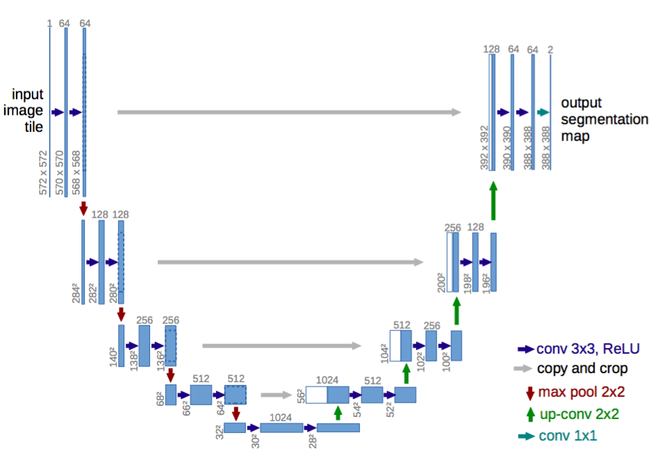
In this particular competition, U-net [7] style network was used as the main model to segment the boundaries of the skin. The method based on ensembling deep convolutional networks with different architectures and snapshots for better generalization.
Deep encoders were used for better catching complex patterns. Wide ResNet38 and DPN backbone networks were used as encoders (same padding everywhere) with FPN based decoder, pretrained on ImageNet.
As an improvement over the standard U-Net architecture convolution with a 7x7 kernel and stride 2 was used as an initial block followed by max-pooling with stride 2. Each decoder block includeded 1 x 1 convolution operation to reduce the filters. Besides, in-place activated batch normalization was used to reduce the memory and speed up the training process. In-Place Activated BatchNorm (InPlace-ABN) [11] is a memory efficient replacement for BatchNorm + Activation step, this approach saves up to 25 percent of memory consumption which helps to increase batch size and speed up the training process.
To enhance the quality of the predictions - cycle learning rate, snapshot ensembling and hypercolumn[1] were used as well.
Data was preprocesed by resizing the image to 224x224, then normalizing it with the mean and standard deviation estimated from the training set. Then, training set was separated into 5 stratified folds to preserve class distribution. For each image, around 14 random augmentations were used with different random coefficients. [2] (e.g. MotionBlur, MedianBlur, RandomContrast, RandomBrightness, ShiftScaleRotate, CLAHE, IAASharpen, Distort, HueSaturation, ToGray).
4.1 Loss function
Loss-function - w1*BCE + w2*(1 - dice) with corresponding weights: 0.5, 0.5. Binary cross-entropy - for more certain body of the object and dice to make the boundary more precise. Dice coefficient is different from Jaccard Similarity Index in the way of counting pixels which are presented in both correct and identified regions.
4.2 Post-processing
Marker based watershed was used in post-processing stage where the connected component was used as a marker with some dilation and erosion. Afterall, test time augmented predictions, 7 augmentations for each model, were ensembled. For averaging i used mean of the predicted masks.
5 Results
The best model achieved results with 0.752. Other experiments with different architectures and single models achieved results between 0.700 and 0.750.
6 Conclusion
In this paper, U-net architecture was proposed as a solver of the current lesion boundary segmentation task. For this challenge, encoder based, pre-trained Wide-ResNet[9] and DPN[10] networks were used with U-net style decoders. The model could be optimized by reducing and selecting only optimal augmentations and, of course, by using deeper architectures following with the ensembles.
References
- [1] Ross Girshick Jitendra Malik Bharath Hariharan, Pablo Arbeláez. Hypercolumns for object segmentation and fine-grained localization. Arxiv, 2015.
- [2] Alexander V. Buslaev, Alex Parinov, Eugene Khvedchenya, Vladimir I. Iglovikov, and Alexandr A. Kalinin. Albumentations: fast and flexible image augmentations. CoRR, abs/1809.06839, 2018.
- [3] Alexander V. Buslaev, Selim S. Seferbekov, Vladimir I. Iglovikov, and Alexey A. Shvets. Fully convolutional network for automatic road extraction from satellite imagery. CoRR, abs/1806.05182, 2018.
- [4] Kaiming He, Georgia Gkioxari, Piotr Dollár, and Ross B. Girshick. Mask r-cnn. 2017 IEEE International Conference on Computer Vision (ICCV), pages 2980–2988, 2017.
- [5] Xiaojuan Qi Xiaogang Wang Hengshuang Zhao, Jianping Shi and Jiaya Jia. Pyramid scene parsing network. Arxiv, 2016.
- [6] Glib Kechyn, Svyatoslav Kechyn, Lucius Yu, and Yangguang Zang. Sales forecasting using wavenet within the framework of the kaggle competition. arXiv, 2018.
- [7] P. Fischer O. Ronneberger and T. Brox. U-net: Convolutional networks for biomedical image segmentation. In International Conference on Medical image computing and computerassisted intervention, Springer 2015.
- [8] Harald Kittler Philipp Tschandl, Cliff Rosendahl. The ham10000 dataset: A large collection of multi-source dermatoscopic images of common pigmented skin lesions. Arxiv, 2018.
- [9] N Komodakis S Zagoruyko. Wide residual networks. Arxiv, 2016.
- [10] Huaxin Xiao Xiaojie Jin Shuicheng Yan Jiashi Feng Yunpeng Chen, Jianan Li. Dual path networks. Arxiv, 2017.
- [11] Chiyuan Zhang, Samy Bengio, Moritz Hardt, Benjamin Recht, and Oriol Vinyals. Understanding deep learning requires rethinking generalization. CoRR, abs/1611.03530, 2016.