THE POSSIBILITY OF BLOCKING THE PROCESS OF DNA BASE PAIRS OPENING BY HYDROGEN PEROXIDE
Abstract
One of the most progressive methods of cancer treatment is ion beam therapy. The simulations of water radiolysis show that in the cell medium the most long-living species are hydrogen peroxide () molecules. But up to the present time the role of molecules in the deactivation of cancer cells has not been determined yet. To understand the possible role of in ion beam therapy, the competitive interaction of and molecules with nucleic bases in a pair on the different stages of genetic information transfer is studied in the present work. Atom-atomic potential functions method is used for the calculations. It is shown, that some configurations of A·T and G·C complementary pairs are stabilized much better by molecule compared to water molecule. Formation of such interaction complexes can terminate the processes of DNA unzipping by enzymes and consequently block genetic information transfer processes in cancer cells during ion beam therapy treatment. Experimental verification method of hydrogen peroxide interaction with nucleic base pairs is proposed.
1 Intorduction
Ion beam therapy is one of the most progressive methods for cancer treatment. Nowadays ion cancer facilities are built throughout the Europe where patients undergo treatment on special accelerators. In the basis of ion therapy lies the so-called Bragg effect [1], which means that the maximum amount of energy is transferred to the medium by heavy ions at the end of their track in a certain localized area of space. This effect makes ion therapy especially effective for the treatment of tumors that are deep enough in the body.
In radiotherapy it is considered that to destroy the cancer cells, it is necessary in some way to deactivate their DNA [2]. However, the definite mechanisms of action of high-energy ions on DNA of cancer cells have not been determined yet [3].
It is known that DNA macromolecule is situated in a cell in water-ionic solution. This solution stabilizes the double helix, determines its shape, and, accordingly, affects its functioning in a living cell. Due to water radiolysis, which takes place during ion beam therapy treatment, this environment changes significantly. A large number of species occurs in solution: free radicals, secondary electrons, ions, as well as molecular products such as and . Until recently, in the literature [4, 2] the most attention was paid to the DNA strand breaks, which occur due to the action of secondary electrons and free radicals. However, it is known that there are DNA reparation mechanisms, which can eliminate DNA strand breaks [5].
Experimental studies as well as the Monte Carlo simulation of water radiolysis [6, 7, 8] showed that on the biological time scales one of the most long-living species are hydrogen peroxide molecules. But their role in ion beam therapy has not been discussed in the previous works properly.
In our paper [9], the new hypothesis was proposed. According to this hypothesis, a hydrogen peroxide molecule can form stable complexes with active DNA sites, thus blocking the processes of genetic information transfer in living cells. To prove the hypothesis, in our works the interaction of molecules with nonspecific DNA recognition sites - phosphate groups () [9, 10], as well as with specific DNA recognition sites - nucleic bases [11] was investigated. Methods of atom-atom potential functions and density functional theory were used for the calculations. Results of both methods revealed that the hydrogen peroxide molecule can form a complex with DNA phosphate groups that is no less stable than the same complex with water molecule. Also the definite sites of Adenine (A), Thymine (T), Guanine (G) and Cytosine (C) nucleic bases, where the molecule can interact much stronger than the water molecule, are determined. These interactions can block processes of DNA recognition by the enzyme.
It should be noted, that in the DNA double helix, the nucleic bases form complementary pairs - A·T and G·C. During genetic information transfer the complementary base pairs become to be open for the interaction with the surrounding molecules, i.e. expose their atomic groups to the solvent. The base pair opening can be blocked by molecules during ion beam therapy treatment and can serve as an evidence of our proposed mechanism of DNA deactivation. It is sufficient that now base pairs opening can be investigated experimentally with the help of single-molecule manipulation technique [12, 13, 14], therefore can be used for the direct observation of the interaction of molecules with DNA.
In Sec. 2 the methodology of our calculations is described. In Sec. 3, stability of the complexes consisting of ‘preopened’ and ‘stretched’ A·T and G·C base pairs together with hydrogen peroxide and water molecule is studied. The possibility of blocking the process of the nucleic base pairs opening by molecules is considered. In Sec 4, the experimental observation method of the interaction of hydrogen peroxide molecules with DNA nucleic bases is discussed.
2 Calculation methods
In the present work we will perform our calculations with the help of atom-atom potential function method. This method is now widely used in such force fields as CHARMM and AMBER [15, 16, 17] for studying the structure of molecular complexes. Calculations are made using GNU Octave software package [18]. In the framework of this method, the energy of intermolecular interaction consists of van der Waals interactions, hydrogen bonds and Coulomb interactions:
| (1) |
In the framework of the present method we consider all the covalent bonds and angles as rigid.
Van der Waals’s interaction is described by Lennard-Jones’s ‘6-12’ potential:
The energy of the hydrogen bond between atoms i and j is modeled by the modified Lenard-Jones potential ‘10 -12’:
| (3) |
where - the distance between the atoms and , - the angle of the hydrogen bond. For example, when the hydrogen bond is , then is an angle between the lines of covalent bond () and the hydrogen bond ().
Coulomb interaction is described by the electrostatic potential:
| (4) |
where and are the charges of the atoms and located at a distance , 0 is the vacuum permittivity, and (r) is the dielectric permittivity of the medium.
The charges , for nucleic bases were taken from the works [19, 20]. Charges of and molecules were calculated from the condition that the dipole moment of water molecule should be equal to [21], and of hydrogen peroxide molecule [22]. Hence, for the molecule we obtain the charges , , and, accordingly, for , . The values of charges on the atoms of molecule are in good agreement with charges obtained by quantum-chemical calculations in the work [23]. Also the same charge values are used in recently developed force field for hydrogen peroxide molecule [24].
Since DNA in the living cell is situated in a water-ion solution, the interacting atoms are screened by water molecules. This leads to a weakening of the Coulomb interaction. Thus, more effective accounting of Coulomb interactions can be achieved using the dependence of the dielectric permittivity upon distance ((r)), developed by Hingerty et al. [25] in the explicit form:
| (5) |
where .
3 Calculation results
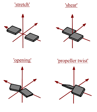
It is known that the nucleic bases in the complementary pair have many degrees of freedom, which are defined by the standard nomenclature [26]. Since the base pairs are situated in a double helix, their structure is stabilized by the stacking interaction between adjacent pairs. This essentially limits the degrees of freedom that remove the bases beyond the plane of the pair. Consequently, in this paper, only the degrees of freedom of the bases in the plane of the pair (‘stretch’, ‘opening’, ‘shear’) are considered. In addition, the degree of freedom ‘propeller twist’ was considered (Appendix, Fig. A1), which is essential to be taken into account due to the spatial structure of molecule.
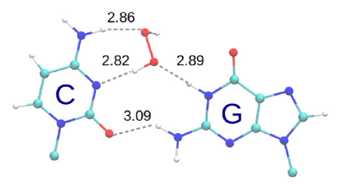
Firstly, in order to verify the correctness of the parameters chosen for our calculations, the stable Watson-Crick configuration of the complementary pairs of A·T and G·C were calculated (Fig. 2). As can be seen from Tabl. 1, the spatial structures of these configurations are close to those obtained from the X-ray structural analysis and the experiment on nuclear magnetic resonance. The main differences occur in the parameter ‘propeller twist’ due to the fact that the experiment measures pairs which are not isolated, but which constitute a structure of a double helix. These parameters are calculated using the 3DNA software package [27]. Visualization is made by VMD [28]. Also Tabl. 1 shows that the spatial structure of the pairs does not differ much, depending on whether the formula (5) is used in the calculations. At the same time, the difference in the interaction energy for G·C pair is significant, which is due to the anomalous contribution of the Coulomb interaction. More details of the necessity of using the dependency (5) are discussed in [29]. It was shown in [11] that the calculation results of the water-water and peroxide-water complexes with the use of the dependence (5) are much closer to the results of quantum-chemical calculations than without this dependence. Therefore, all further calculations in this paper will be carried out taking into account formula (5).
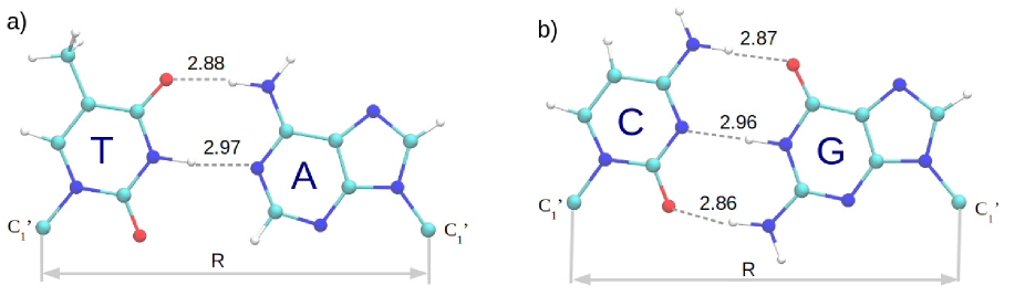
[t] Parameter A·T G·C X-ray1 NMR2 Our calculations X-ray1 NMR2 Our calculations vacuum using (5) vacuum using (5) ‘stretch’ ‘shear’ ‘opening’ ‘propeller twist’
-
1
calculated from the spatial structures of the Dickerson-Drew dodecamer (files 1bna.pdb, 7bna.pdb, 9bna.pdb, 436d.pdb) obtained by X-ray analysis. The values are averaged separately by the A·T and the G·C base pairs and the standard errors are calculated.
-
2
calculated from the spatial structures of the Dickerson-Drew dodecamer (files 1duf.pdb, 1gip.pdb, 1naj.pdb, 2dau.pdb) obtained by the method of nuclear magnetic resonance. The values are averaged separately by the A·T and the G·C base pairs and the standard errors are calculated.
The interaction of nucleic bases with water molecules was considered in a series of papers [30, 31, 32]. In the paper [33] hydration shells of A·T and G·C base pairs were calculated, that is, simultaneous interaction with a large number of water molecules was considered. However, in the literature it was not paid essential attention to the interaction of nucleic bases with hydrogen peroxide molecules (which, as described in Sec. 1, appears in the cell as a result of ion beam treatment). Only the work [34] is known, where the interaction of hydrogen peroxide molecules with the Adenine base was considered.
In the present paper calculations of interaction energy of complexes consisting of A·T and G·C nucleic base pairs with hydrogen peroxide and water molecules are carried out. In the work [35] it was shown that structures stabilized by water molecules occur on the pathway of base pairs opening during DNA unzipping. For the purposes of the present work, it is important to analyze the possibility of forming the simplest ‘non-Watson-Crick’ configurations of A·T and G·C base pairs, stabilized by hydrogen peroxide and water molecules, and to establish whether the formation of these complexes can block the genetic information transfer processes. Consequently, the complexes that include only one hydrogen peroxide or water molecule are taken into account.
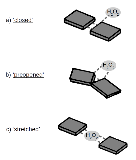
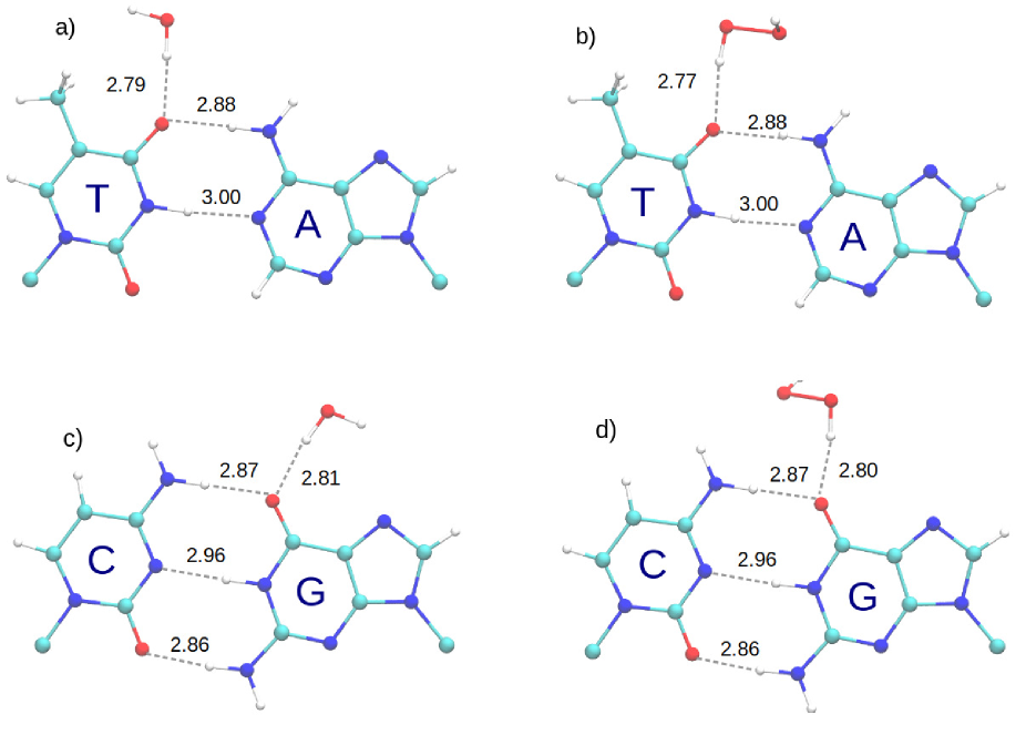
In the present work we consider three configurations of nucleic base pairs with hydrogen peroxide molecules and water molecules. The complexes consisting of complementary A·T and G·C base pairs and the hydrogen peroxide or water molecule that interacts with the base from the side of the major groove we denote as ‘closed’ pairs (Fig. 1 a). The configuration of the pair where the ‘opening’ pathway dominates, we denote ‘preopened’ (Fig. 1 b), and those in which the ‘stretch’ pathway dominates will be denoted as ‘stretched’ pair (Fig. 1 c).
Firstly, let us calculate ‘closed’ configurations. It can be seen, that the interaction with these molecules almost does not change the geometry of the Watson-Crick pairs (Tabl. 2, Appendix, Fig. A3).
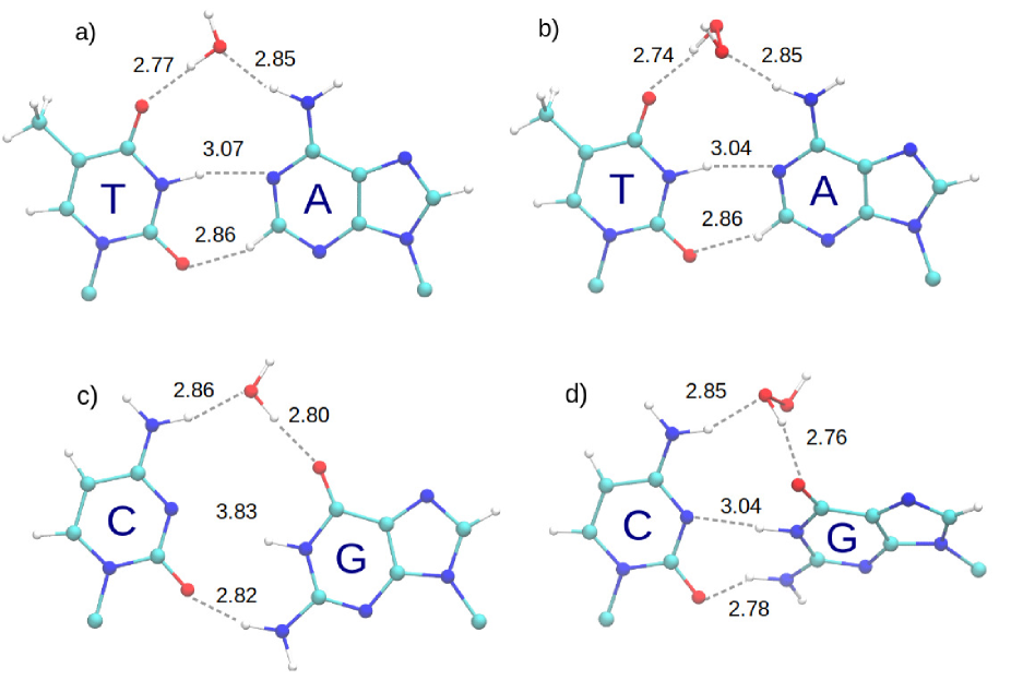
On Fig. A4 (Appendix) stable configurations of the ‘preopened’ A·T and G·C base pairs with water and hydrogen peroxide molecules are shown. For the convenience of the analysis, in Tabl. 2 only structural parameters (difference in distances (Fig. 2 a) in the corresponding and Watson-Crick pair), ‘opening’ and ‘propeller’, as well as interaction energies are shown. From Tabl. 2 it can be seen that the configurations of ‘preopened’ A·T pairs with water and hydrogen peroxide molecules have almost identical parameters. The ‘opening’ parameter has the maximum value, the rest of the parameters do not change significantly. It also should be noted that the ‘propeller twist’ parameter almost does not take the bases out of the plane of the pair. Moreover, configurations of ‘preopened’ G·C pairs with and molecules are significantly different, since the spatial structure of peroxide increases the parameter ‘propeller twist’, taking the bases out of the plane of the pair.
It also should be noted that in the case of G·C pair, there is still a configuration with the molecule (Appendix, Fig. A2), which, on the one hand, is similar to the ‘preopened’ state because the parameter is almost unchanged, and the ‘opening’ parameter is significantly larger than in the Watson-Crick ones, but since the peroxide molecule is ‘embedded’ to the internal () hydrogen bond in this case, we call this state ‘opened’. Note, that due to the geometry of the molecules, the ‘opened’ state occurs only for G·C pair and only with molecules.
[t] State ‘opening’ ‘propeller twist’ ‘closed’ A·T G·C ‘preopened’ A·T G·C ‘opened’ G·C ‘stretched’ A·T G·C
-
∗ All the values are rounded to the first decimal due to the accuracy of the parameters that are used for the calculations of the corresponding structures.
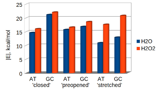
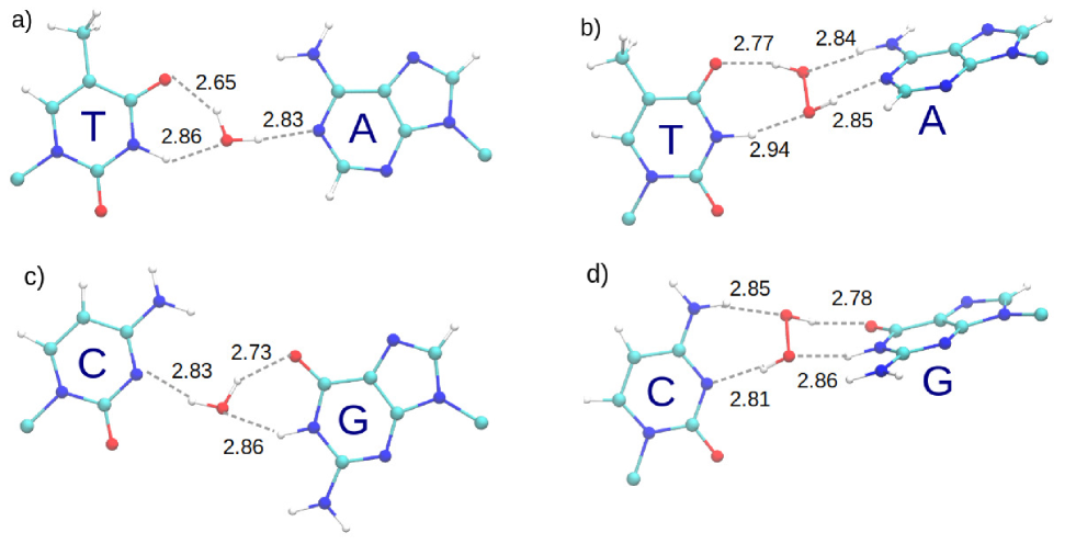
Configurations of ‘stretched’ pairs with water and hydrogen peroxide differ significantly by their parameters (Appendix, Fig. A5). It should be noted that for both the A·T and the G·C base pairs, the interaction energy of the ‘stretched’ pair for the complex with water molecule is lower than the corresponding value for the ‘preopened’ configuration, and for complexes with hydrogen peroxide molecule, on the contrary, it is substantially higher (Tabl. 2).
4 About the possibility of an experimental observation of the formation of complexes of hydrogen peroxide molecules with the DNA base pairs
During last decades the technique that allows to study features of single molecules was improved significantly. With the help of single-molecule micromanipulation methods important properties of a DNA macromolecule, such as stretching, bending, twisting [12], and the consequent opening of nucleic base pairs under the action of external force (unzipping) [13, 14] can be investigated. The experiment is carried out at a constant opening velocity, and the dependence of the opening force on the displacement value is measured. At the beginning, the force is not enough to open a double helix, so it sharply increases, and then reaches the plateau, which corresponds to the beginning of the opening of the base pairs.
In the work [29], it has been shown that during the unzipping of a double helix, depending on the opening velocity, base pairs can open as along the ‘stretch’ pathway, as well as along the ‘opening’ pathway. And in the work [35] it was shown that during unzipping process configurations of base pairs that are stabilized by water bridges occur. In this regard, if a certain concentration of hydrogen peroxide is added to the solution, ‘preopened’, and ‘stretched’ pairs stabilized by molecules can appear in the conditions of this experiment. As it follows from our calculations, the binding energy of these complexes is significantly higher than the energy of the same complexes with the water molecule. Therefore, the opening force of a double helix in the experiment carried out in the presence of a certain concentration of molecules should be higher as a result of the interaction of the base pairs with molecules. The observation of these force growing in the unzipping experiment can serve as the proof of our hypothesis about the blocking of the DNA base pairs by hydrogen peroxide molecules.
Also the complexes of hydrogen peroxide with DNA base pairs can be observed by the methods of Raman spectroscopy. It is known that all DNA atomic groups have their own vibrational frequency. As known, the vibration frequencies of atomic groups of DNA nucleic bases are in the range of [36]. Interaction of base pairs with water molecules can slightly lower frequency and amplitude of the vibrations [37]. Therefore, the interaction of hydrogen peroxide molecules with DNA base pairs must manifest itself as the shifting of the absorption peak to the less-frequency range and lower its height with comparison to the same complexes with water molecules.
5 Discussion and conclusions
In the present paper, the spatial configurations of complexes consisting of nucleic base pairs with hydrogen peroxide and water molecules are investigated. The comparison of the interaction energy values is schematically shown on the diagram (Fig. 3). As it can be seen from the results, there are configurations of A·T and G·C base pairs (‘preopened’ and ‘stretched’), which are stabilized by hydrogen peroxide molecules much better than by water molecules.
Interaction of hydrogen peroxide molecules with DNA base pairs can manifest itself in living cell. Namely, in the process of DNA replication, when two DNA macromolecules are formed from one double helix. At the initial stage of this process, an enzyme passes along a double helix [38] and consequently opens its base pairs one after another. DNA bases interact with water molecules all the time. But, as it was mentioned above, significant amount of molecules are introduced to the medium during ion beam therapy. If the interaction energy of nucleic bases of the pair with hydrogen peroxide molecule is large enough compared to the same interaction energy with water molecule, the DNA unzipping by enzyme can be terminated.
As can be seen from the obtained results, the most significant difference in interaction energies is for ‘stretched’ configurations of the pairs. Therefore, the possibility of blocking ‘stretched’ pairs by hydrogen peroxide molecules is significantly more probable. In this case the difference between the opening energies is (Tabl. 2). This is due to the fact that the hydrogen peroxide molecule, because of its spatial structure, forms four hydrogen bonds with nucleic bases (Appendix, Fig. A5 b,d). At the same time, water molecule forms three hydrogen bonds, and two of them are substantially curved, that is, their energy is weakened (Appendix, Figures A5 a,c). It should also be noted that, since in ‘stretched’ configurations the parameter ‘propeller twist’ is significant, the formation of such configurations is possible only in the unzipping fork, where there is no stacking interaction from one side of the pair.
It should be noted, that the energy values obtained in the present work are only the enthalpy values, entropy is not taken into account. However, due to the similar structure of and molecules, the entropy contribution to the interaction of this molecules with nucleic bases, should also be similar. Therefore, our approach allows us to obtain a qualitative picture of the formation of complexes of hydrogen peroxide molecules with base pairs, and to see a essential energy advantage compared to the same complexes with a water molecule.
Formation of complexes of molecules with DNA can completely block the DNA transcription of cancer cells, and can be a key factor of the action of high-energy ions on cancerous tumors in the process of ion beam therapy.
Acknowledgement. The present work was partially supported by the Program of Fundamental Research of the Department of Physics and Astronomy of the National Academy of Sciences of Ukraine (project number 0116U003192).
APPENDIX
References
- [1] W.H. Bragg, R. Kleeman. LXXIV. On the ionization curves of radium. The London, Edinburgh, and Dublin Philosophical Magazine and Journal of Science 48, 726 (1904).
- [2] E. Surdutovich, A.V. Yakubovich, A.V. Solov’yov. Multiscale approach to radiation damage induced by ion beams: complex DNA damage and effects of thermal spikes. The European Physical Journal D 60, 101 (2010).
- [3] M. Krämer, M. Durante. Ion beam transport calculations and treatment plans in particle therapy. The European Physical Journal D 60, 195 (2010).
- [4] P.L. Olive. The role of DNA single- and double-strand breaks in cell killing by ionizing radiation. Radiation Research 150, 11 (1998).
- [5] C.M. Gustafsson. Mechanistic studies of DNA repair. Royal Swedish Academy of Sciences (2015).
- [6] M.S. Kreipl, W. Friedland, H.G. Paretzke. Time- and space-resolved monte carlo study of water radiolysis for photon, electron and ion irradiation. Radiation and Environmental Biophysics 48, 11 (2008).
- [7] S. Uehara and H. Nikjoo. Monte carlo simulation of water radiolysis for low-energy charged particles. Journal of Radiation Research 47, 69 (2006).
- [8] D. Boscolo, M. Krämer, M. Durante, M.C. Fuss, and E. Scifoni. Trax-chem: A pre-chemical and chemical stage extension of the particle track structure code trax in water targets. Chemical Physics Letters 698, 11 (2018).
- [9] D.V. Piatnytskyi, O.O. Zdorevskyi, S.M. Perepelytsya, S.N. Volkov. Understanding the mechanism of dna deactivation in ion therapy of cancer cells: hydrogen peroxide action. The European Physical Journal D 69, 255 (2015).
- [10] D.V. Piatnytskyi, S.N. Volkov. Complexes of hydrogen peroxide and dna phosphate group in quantum-chemical calculations. Biophysical bulletin 39, 5 (2018).
- [11] O. Zdorevskyi, D. Piatnytskyi, S.N. Volkov. Blocking of DNA specific recognition sites by hydrogen peroxide molecules in the process of ion beam therapy of cancer cells. arXiv preprint , arXiv:1811.11026 (2018).
- [12] C. Bustamante, Z. Bryant, S.B. Smith. Ten years of tension: single-molecule DNA mechanics. Nature 6921, 423 (2003).
- [13] U. Bockelmann, Ph. Thomen, B. Essevaz-Roulet, V. Viasnoff, F. Heslot. Unzipping DNA with optical tweezers: high sequence sensitivity and force flips. Biophysical journal 82, 1537 (2002).
- [14] J.M. Huguet, C.V. Bizarro, N. Forns, S.B. Smith, C. Bustamante, F. Ritort. Single-molecule derivation of salt dependent base-pair free energies in DNA. Proceedings of the National Academy of Sciences 107, 15431 (2010).
- [15] K. Vanommeslaeghe, E. Hatcher, C. Acharya, S. Kundu, S. Zhong, J. Shim, E. Darian, O. Guvench, P. Lopes, I. Vorobyov, and A. D. Mackerell. Charmm general force field: A force field for drug-like molecules compatible with the charmm all-atom additive biological force fields. Journal of Computational Chemistry 31, 671 (2010).
- [16] T.E. Cheatham, D.A. Case. Twenty-five years of nucleic acid simulations. Biopolymers 99, 969 (2013).
- [17] R Lavery. Modeling nucleic acids: fine structure, flexibility and conformational transitions. Adv. Comput. Biol. 1, 69 (1994).
- [18] J.W. Eaton, D. Bateman, S. Hauberg, R. Wehbring. GNU Octave version 4.2.1 manual: a high-level interactive language for numerical computations (2017).
- [19] V.I. Poltev, N.V. Shulyupina. Simulation of interactions between nucleic acid bases by refined atom-atom potential functions. Journal of Biomolecular Structure and Dynamics 4, 739 (1986).
- [20] V.B. Zhurkin, V.I. Poltev, and V.L. Florent’ev. Atom–atomic potential functions for conformational calculations of nucleic acids. Molekuliarnaia biologiia 14, 1116 (1980).
- [21] S.A. Clough, Y. Beers, G.P. Klein, L.S. Rothman. Dipole moment of water from Stark measurements of H2O, HDO, and D2O. The Journal of Chemical Physics 59, 2254 (1973).
- [22] J.T. Massey , D.R. Bianco. The Microwave Spectrum of Hydrogen Peroxide. The Journal of Chemical Physics 22, 442 (1954).
- [23] S.T. Moin, T.S. Hofer, B.R. Randolf, B.M. Rode. An ab initio quantum mechanical charge field molecular dynamics simulation of hydrogen peroxide in water. Computational and Theoretical Chemistry 980, 15 (2012).
- [24] E. A. Orabi, A. M. English. A Simple Additive Potential Model for Simulating Hydrogen Peroxide in Chemical and Biological Systems. Journal of chemical theory and computation . 14, 2808 (2018).
- [25] B. E. Hingerty, R. H. Ritchie, T. L. Ferrell, and J. E. Turner. Dielectric effects in biopolymers: The theory of ionic saturation revisited. Biopolymers 24, 427 (1985).
- [26] S. Diekmann. Definitions and nomenclature of nucleic acid structure parameters. Journal of molecular biology 205, 787 (1989).
- [27] X.-J. Lu, W.K. Olson. 3DNA: a software package for the analysis, rebuilding and visualization of three-dimensional nucleic acid structures. Nucleic acids research 31, 5108 (2003).
- [28] W. Humphrey, A. Dalke, K. Schulten. VMD: visual molecular dynamics.Journal of molecular graphics 14, 33 (1996).
- [29] O. Zdorevskyi, S.N. Volkov. Possible scenarios of DNA double-helix unzipping process in single-molecule manipulation experiments. European Biophysics Journal 47, 917 (2018).
- [30] E.S. Kryachko, S.N. Volkov. Preopening of the DNA base pairs. International Journal of Quantum Chemistry 82, 193 (2001).
- [31] E. Giudice, P. Várnai, R. Lavery. Base pair opening within B-DNA: free energy pathways for GC and AT pairs from umbrella sampling simulations. Nucleic acids research 31, 1434 (2003).
- [32] V.I. Poltev, E.H. Gonzalez, A.V. Teplukhin. Possible role of rare tautomers of nucleic bases in mutagenesis: Effect of hydration on tautomer equilibrium. Molecular Biology 29, 213 (1995).
- [33] L. Gorb, Y. Podolyan, P. Dziekonski, W.A. Sokalski, J. Leszczynski. Double-proton transfer in adenine- thymine and guanine- cytosine base pairs. A post-hartree-fock ab initio study. Journal of the American Chemical Society 126, 10119 (2004).
- [34] J.A. Dobado, J. Molina. Adenine-hydrogen peroxide system: DFT and MP2 investigation. The Journal of Physical Chemistry A 103, 4755 (1999).
- [35] S.N. Volkov, E.V. Paramonova, A.V. Yakubovich, A.V. Solov’yov. Micromechanics of base pair unzipping in the DNA duplex. Journal of Physics: Condensed Matter, 24, 035104 (2011).
- [36] B. Prescott, W. Steinmetz, G.J. Thomas Jr. Characterization of DNA structures by laser raman spectroscopy. Biopolymers: Original Research on Biomolecules 23, 235 (1984).
- [37] V.Y. Maleev, M.A. Semenov, A.I. Gasan, V.A. Kashpur. Physical properties of the DNA-water system. Biofizika 38, 768 (1993).
- [38] R. Galletto, M.J. Jezewska, W. Bujalowski. Unzipping mechanism of the double-stranded DNA unwinding by a hexameric helicase: quantitative analysis of the rate of the dsDNA unwinding, processivity and kinetic step-size of the escherichia coli DNAb helicase using rapid quench-flow method. Journal of molecular biology 343, 83 (2004).