MIDLMedical Imaging with Deep Learning
\jmlrpages
\jmlryear2019
\jmlrworkshopMIDL 2019 – Extended Abstract Track
\midlauthor\NameMickael Tardy\nametag1,2 \Emailmickael.tardy@ec-nantes.fr
\addr1 LS2N UMR6004, Ecole Centrale Nantes, France,
\addr2 Hera-MI, SAS, Nantes, France and \NameBruno Scheffer\nametag2,3 \Emailbruno.scheffer@hera-mi.com
\addr3 Institut de cancérologie de l’Ouest, Saint-Herblain, France and \NameDiana Mateus\nametag1 \Emaildiana.mateus@ec-nantes.fr
A closer look onto breast density with weakly supervised dense-tissue masks
Abstract
This work focuses on the automatic quantification of the breast density from digital mammography imaging. Using only categorical image-wise labels we train a model capable of predicting continuous density percentage as well as providing a pixel wise support frit for the dense region. In particular we propose a weakly supervised loss linking the density percentage to the mask size.
keywords:
Deep Learning, Weakly supervised segmentation, Regression, Breast density1 Purpose
Breast density is a biomarker for breast cancer development risk that suggests that the risk of cancer development increases with denser breasts. Moreover, the detection of cancer in dense tissues and, more generally, in dense breasts is often considered more challenging due to the similar visual aspects of normal and abnormal tissues, which complicates the interpretation of mammographic images. For the above reasons, we argue that computer-aided decision systems for early breast cancer detection should both, quantitatively evaluate the breast density, and evaluate the spatial distribution of the dense tissues.
2 Methods
In clinical practice, breast density is usually assessed image-wise using a classification grid like the BI-RADS (Breast imaging-reporting and data system) [Irshad et al.(2016)Irshad, Leddy, Ackerman, Cluver, Pavic, Abid, and Lewis]. In the present work, we propose to estimate breast density at the pixel level while using only image-wise ground truth from the BI-RADS scale. Our goal is to generate a breast density mask, identifying pixels associated with the tissue that contributed to the density class. To achieve our goal, we propose a novel loss linking the sought breast density mask to the globally estimated breast density (fig. LABEL:fig:architecture). We formulate the problem as a weakly supervised binary semantic segmentation. Our approach is related to recent efforts to reduce the amount of supervision [Carneiro et al.(2017)Carneiro, Peng, Bayer, and Navab, Dubost et al.(2017)Dubost, Bortsova, Adams, Ikram, Niessen, Vernooij, and De Bruijne].
In practice, we rely on a modified U-Net architecture [Ronneberger et al.(2015)Ronneberger, Fischer, and Brox] and on an extended 12-class density grid that improves the density resolution compared to traditional BI-RADS classification (4th edition). Compared to the state-of-the-art, our classification and segmentation scheme does not rely on the modelfls attention but uses a loss function efficiently correlating a tissue mask with the target breast density values. Moreover, the output is constrained with the breast binary mask removing useless activations.
3 Results
The database for training and tests consists of 1232 and 370 images respectively. We got promising results with a mean absolute error (MAE) of 6.7% for the density regression estimate (see tab. LABEL:tab:performance_overview_rgr) and an accuracy of 78% for 4-class BI-RADS density classification (tab. LABEL:tab:performance_overview_cls). Our comparison baseline is a VGG-like regression model trained on the same dataset.
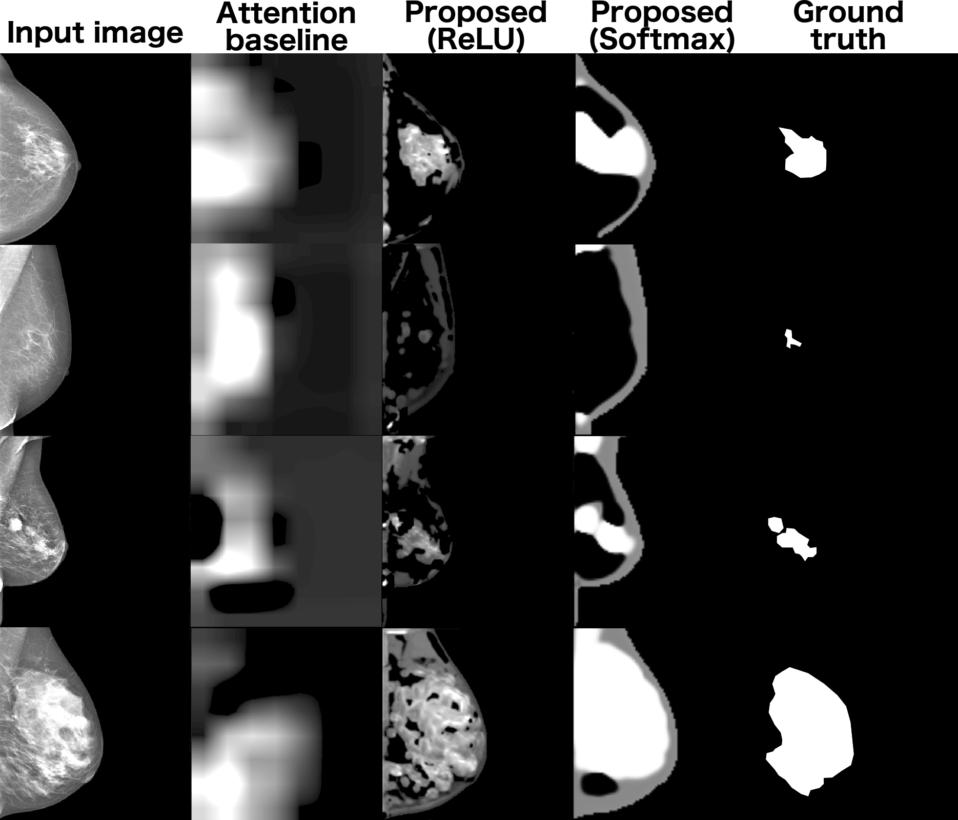
To validate the segmentation performance, we collected regions of interest on several images (16) and calculated the . Overall we obtain clinically meaningful segmentation masks offering valuable insights into the spatial distribution of the dense tissues (fig. LABEL:fig:dense_masks). In comparison, we demonstrate the inefficiency of the attention-based techniques for the breast density mask generation.
In addition we validate our approach on the INBreast [Moreira et al.(2012)Moreira, Amaral, Domingues, Cardoso, Cardoso, and Cardoso] database. Without any additional training and a simple preprocessing we obtained accuracy and . We note that our results are comparable to other works on the same dataset (, [Schebesch et al.()Schebesch, Unberath, Andersen, and Maier], [Angelo et al.(2015)Angelo, Carneiro, Granado, and Patrocinio].
4 Conclusions
Our approach to link breast density classification to the spatial distribution of dense tissue has a positive effect on classification scores while providing an additional output mask of the dense regions. These results are interesting given the considerably low requirements on ground truth (just a class instead of an image mask) and the size of the training dataset.
tab:performance_overview_cls Metrics Model Accuracy Precision Recall -score Cohen kappa VGG+Sigmoid+MSE 0.764 0.782 0.764 0.766 0.891 U-NET+Softmax+ 0.684 0.729 0.684 0.679 0.838 U-NET+ReLU+ 0.779 0.809 0.779 0.781 0.891
tab:performance_overview_rgr Metrics Dataset MAE () MxAE () C-index VGG+Sigmoid+MSE 6.545 31.964 0.820 U-NET+Softmax+ 8.303 34.404 0.789 U-NET+ReLU+ 6.661 32.156 0.839
fig:architecture
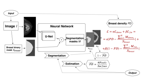
fig:dense_masks
Research funding is provided by Hera-MI, SAS and Association Nationale de la Recherche et de la Technologie via CIFRE grant no. 2018/0308.
References
- [Angelo et al.(2015)Angelo, Carneiro, Granado, and Patrocinio] Michele Fúlvia Angelo, P. C. Carneiro, T. C. Granado, and A. C. Patrocinio. Influence of contrast enhancement to breast density classification by using sigmoid function. In IFMBE Proceedings, volume 51, pages 33–36. Springer, Cham, 2015. ISBN 9783319193878. 10.1007/978-3-319-19387-8_9.
- [Carneiro et al.(2017)Carneiro, Peng, Bayer, and Navab] Gustavo Carneiro, Tingying Peng, Christine Bayer, and Nassir Navab. Automatic Quantification of Tumour Hypoxia from Multi-Modal Microscopy Images Using Weakly-Supervised Learning Methods. IEEE Transactions on Medical Imaging, 36(7):1405–1417, jul 2017. ISSN 1558254X. 10.1109/TMI.2017.2677479.
- [Dubost et al.(2017)Dubost, Bortsova, Adams, Ikram, Niessen, Vernooij, and De Bruijne] Florian Dubost, Gerda Bortsova, Hieab Adams, Arfan Ikram, Wiro J Niessen, Meike Vernooij, and Marleen De Bruijne. GP-Unet: Lesion Detection from Weak Labels with a 3D Regression Network. In Maxime Descoteaux, Lena Maier-Hein, Alfred Franz, Pierre Jannin, D Louis Collins, and Simon Duchesne, editors, Medical Image Computing and Computer-Assisted Intervention MICCAI 2017, pages 214–221. Springer International Publishing, 2017. ISBN 978-3-319-66179-7.
- [Irshad et al.(2016)Irshad, Leddy, Ackerman, Cluver, Pavic, Abid, and Lewis] Abid Irshad, Rebecca Leddy, Susan Ackerman, Abbie Cluver, Dag Pavic, Ahad Abid, and Madelene C. Lewis. Effects of changes in BI-RADS density assessment guidelines (fourth versus fifth edition) on breast density assessment: Intra-and interreader agreements and density distribution. American Journal of Roentgenology, 207(6):1366–1371, dec 2016. ISSN 15463141. 10.2214/AJR.16.16561.
- [Moreira et al.(2012)Moreira, Amaral, Domingues, Cardoso, Cardoso, and Cardoso] Inês C. Moreira, Igor Amaral, Inês Domingues, António Cardoso, Maria João Cardoso, and Jaime S. Cardoso. INbreast: Toward a Full-field Digital Mammographic Database. Academic Radiology, 19(2):236–248, 2012. ISSN 10766332. 10.1016/j.acra.2011.09.014.
- [Ronneberger et al.(2015)Ronneberger, Fischer, and Brox] Olaf Ronneberger, Philipp Fischer, and Thomas Brox. U-net: Convolutional networks for biomedical image segmentation. Technical report, 2015.
- [Schebesch et al.()Schebesch, Unberath, Andersen, and Maier] Frank Schebesch, Mathias Unberath, Ingwer Andersen, and Andreas Maier. Breast density assessment using wavelet features on mammograms. In Informatik aktuell, pages 38–43. Springer Vieweg, Berlin, Heidelberg. ISBN 9783662494646. 10.1007/978-3-662-49465-3_9.