Direct observation of bimolecular reactions of ultracold KRb molecules
Abstract
Femtochemistry techniques have been instrumental in accessing the short time scales necessary to probe transient intermediates in chemical reactions. Here we take the contrasting approach of prolonging the lifetime of an intermediate by preparing reactant molecules in their lowest ro-vibronic quantum state at ultralow temperatures, thereby drastically reducing the number of exit channels accessible upon their mutual collision. Using ionization spectroscopy and velocity-map imaging of a trapped gas of potassium-rubidium molecules at a temperature of 500 nK, we directly observe reactants, intermediates, and products of the reaction 40K87Rb + 40K87Rb K2Rb K2 + Rb2. Beyond observation of a long-lived energy-rich intermediate complex, this technique opens the door to further studies of quantum-state resolved reaction dynamics in the ultracold regime.
The creation of ensembles of molecules at ultralow temperatures enables a variety of high resolution spectroscopic studies, allows broader exploration of reaction phase space, and promises quantum state control over the outcome of chemical reactions. Already, investigations of single partial wave collisions have provided detailed benchmarks of short-range molecular potentials Henson et al. (2012); Yang et al. (2019), exotic conditions at low temperatures have facilitated the synthesis of new chemical species Puri et al. (2017), and highly sensitive and precise methods of detection have traced state-to-state reactions between atoms and weakly-bound Feshbach molecules Hoffmann et al. (2018). Further, chemical reaction rates for barrierless reactions can be altered Ospelkaus et al. (2010); Kilaj et al. (2018), in some case by orders of magnitude, merely by changing the nuclear spins of the reactants and entering quantum degeneracy De Marco et al. (2019). These studies all rely on the extraordinary control attainable over the quantum states of the ultracold molecules.
Despite recent advances in ultracold molecule studies, a key capability has been missing: namely, characterization of transient reaction intermediates and final products. Previous experiments have shown evidence of ultracold reactions between bialkali molecules through the quantum-state-specific detection of loss of reactants Ospelkaus et al. (2010), similar to that shown in the inset to Fig. 1, giving insights into how long-range forces determine the kinetic collision rates of the reactants. These reactions have been observed to occur with a high probability after just a single collision, approaching unity in certain cases Ospelkaus et al. (2010); Ye et al. (2018); Gregory et al. (2019). Despite tour-de-force levels of control over the precise ro-vibrational quantum state of the reactants to open up additional energetically allowed reaction channels, no significant differences based on the reactant species or initial quantum state have yet been observed Ye et al. (2018); Gregory et al. (2019), and the nature of the molecular loss is still under debate Christianen et al. (2019a).
When two molecules approach one another, they initially form an energy-rich intermediate collision complex, the dynamics of which could hold the key for understanding the details of ensuing these ultracold, barrierless, bimolecular reactions. In higher-temperature reactions, this transient complex only persists for one or two vibrational periods, and at most on the order of a rotational period ( ps) Bauer (1979); Miller et al. (1972). Studying the dynamics or kinetics of such complexes in the gas phase has typically required ultrafast Brooks (1988); Zewail (2000); Gruebele et al. (1991); Sims et al. (1992); Polanyi and Zewail (1995) or stabilizing collisional Womack et al. (2015); Bjork et al. (2016); Bui et al. (2018) techniques. Structural investigations of these complexes have been previously obtained by photodetachment Garand et al. (2008); Continetti and Guo (2017), photoabsorption Su et al. (2013), or photodissociation Sato (2001). Based on the Rice-Ramsperger-Kassel-Marcus (RRKM) theory, the lifetime of an intermediate complex is given by , where denotes the density of states of the intermediate complex near the incident energy, , and is the number of energetically allowed exit channels (Fig. 1). Preparing reactant KRb molecules in the pure ro-vibronic ground state in the ultralow temperature regime tightly constrains the number of energetically allowed exit channels, greatly extending the lifetime of the intermediate complex. For reactions between bialkali molecules, depending on the species, has been estimated to be on the order of hundreds of nanoseconds to microseconds Mayle et al. (2013); Christianen et al. (2019b), which makes direct observation of the complex a possible goal. However, no such observations have been made because all previous work has been based on the observation of loss of reactants. Direct multi-species detection methods are necessary in order to fully describe the details of these ultracold reactions Nesbitt (2012).
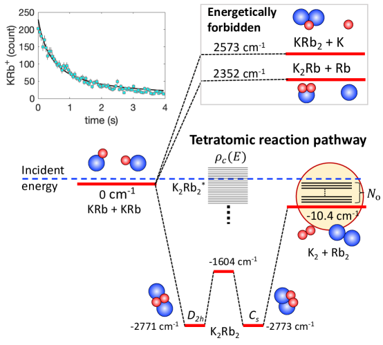
Here, we report the direct detection of a predicted intermediate as well as products in the ultracold chemical metathesis reaction 40K87Rb + 40K87Rb K2Rb K2 + Rb2 (see Fig. 1) Mayle et al. (2013); Christianen et al. (2019b). We combine precise quantum-state preparation of the ultracold reactants with an ionization-based detection method that allows for direct and simultaneous detection of reactants (KRb), intermediates (K2Rb), and final products (K2, Rb2).
We began by implementing an established protocol Ni et al. (2008) to create an optically trapped gas of , ground-state KRb molecules. Here and are the vibrational and rotational quantum number of the molecules, respectively. In brief, ultracold K and Rb atoms are first converted to weakly-bound molecules with 20% efficiency by a magnetic field sweep (1.4 ms) through a Feshbach resonance at 546.62 G Cumby et al. (2013). Then a pair of Raman beams are applied in a stimulated Raman adiabatic passage (STIRAP) Vitanov et al. (2001) pulse sequence (4 s) to coherently transfer the weakly-bound molecules into a single hyperfine state of the ro-vibronic ground state with 85% efficiency. 8 s after the STIRAP pulse, we remove residual Rb and K atoms. Because the atom-to-molecule transfer is mostly coherent, we can reverse the transfer with high efficiency. To detect the ground-state KRb molecules, a reversed STIRAP sequence is applied followed by absorption imaging on an atomic transition (see example image shown in Fig. 2A). Typically, KRb molecules are created at 500 nK with a peak density of cm-3 and are trapped by a crossed optical dipole trap (ODT) at a laser wavelength of 1064 nm.
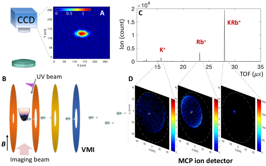
Because the absorption imaging detection is tied directly to the quantum-state-specific STIRAP transfer, it is only sensitive to the KRb molecules in the STIRAP populated quantum state. To probe chemical reaction products and the intermediate complex, we chose a more general detection method that entailed photoionization of neutral reaction species into bound molecular ions, acceleration of the ions in an electric field, and measurement of their arrival time and position on a multi-channel plate (MCP) (Fig. 2C). By combining mass spectrometry and velocity-map imaging (VMI) Eppink and Parker (1997) in our ultracold molecule apparatus, we could thereby identify reacting species and study reaction dynamics.
We performed three separate experiments to probe the reactants, intermediate complex, and products of the ultracold reaction. The detection procedure worked as follows: After KRb creation but before the ionization pulse, we ramped the magnetic field down to 30 G within 15 ms to reduce subsequent Lorentz forces that might deflect ions away from the detector, housed 1 m downstream. We then applied an ultraviolet (UV) ionization pulse while simultaneously triggering the MCP to record ion signals. For the detection of reactants and products, we chose a photoionization wavelength of 285 nm, which is above the ionization threshold of KRb, K, Rb, as well as any species that consist of combinations of multiple K and Rb atoms (table S1). For the detection of the intermediate complex, the wavelength was varied over the 285 to 356 nm range. To avoid space-charge effects, the laser power was kept low enough to ensure at most one ion was generated per UV pulse. The ODT was switched off for a variable time period during and before the ionization pulse to preclude its influence on the chemical reaction, the lifetime of the intermediate collisional complex, and the photoionization process. We repeated this detection procedure at 1 kHz for the reactant and product detection (see timing diagram in fig. S1), and at 7 kHz for the intermediate complex detection (see inset of Fig. 4D). The mass, and thereby elemental composition, of each detected ion could be inferred from its time-of-flight (TOF), whereas the momentum of the ion was mapped through its location on the VM image SM .
To demonstrate the ionization detection capability in our ultracold molecular apparatus and to gain information beyond absorption imaging, we first probed the trapped KRb molecules in the ODT (Fig. 2C,D). As expected, the dominant signal results from KRb+. The VM image for the KRb+ signal has a width limited by the detector resolution, consistent with the negligible translational energy in the ultracold regime. Measurable amounts of Rb+ and K+ were also detected. The VM images for K+ and Rb+ both show two distinct components: an isotropic central peak and an anisotropic ring. The ions forming the central peak originate from residual ultracold atoms from the molecule creation process after the cleanup pulses. Based on the known ionization cross sections and estimated ion detection efficiencies (table S1), we put an upper bound of 250 atoms of each species in the trap. These populations are small compared to KRb, ensuring that the dominant reaction in the subsequent study is the desired bimolecular reaction. The sensitivity of ionization detection allowed us to quantify the small number of residual atoms in the ODT, which are not seen using absorption imaging. To analyze the Rb+ ions forming the ring pattern, we extracted the translational energy release (TER) from the diameter of the ring to obtain a TER of cm-1. By comparing this TER to the calculated molecular potentials of KRb and KRb+ Korek et al. (2003) we identified a two-photon dissociative ionization pathway that contributes to this atomic ion signal. The same analysis also applies to the ring pattern of the K+ ions (fig. S3).
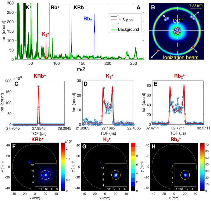
After KRb molecules are created, the bimolecular reaction occurs continuously with a measured decay rate coefficient of cm3/s until the reactants are depleted (Fig. 1 inset), consistent with previous studies Ospelkaus et al. (2010). To probe the products of the bimolecular reaction while reducing the perturbation to the reactants during ionization, we shaped our ionization beam into a “hollow-bottle” (Fig. 3B) with the laser intensity concentrated in a ring outside of the ODT to keep the reactants in the dark; the measured intensity contrast between the peak and center of the beam was 500 SM . To further reduce the hollow volume for higher efficiency ionization, we crossed two hollow-bottle beams at a 40∘ angle centered on the ODT SM . To observe the bimolecular reaction without the possible influence of the ODT light, we shut off the ODT for 170 s prior to each ionization pulse, thereby precluding any role of the ODT in the formation of all but those products with translational energy less than 0.0127 foo .
The dominant peaks in the mass spectrum (Fig. 3A) are again K+, Rb+, and KRb+, primarily from photoionization of trapped KRb molecules by the residual intensity at the centers of the hollow-bottle beams. Aside from these dominant peaks, we can clearly identify ions corresponding to the masses of K and Rb. All peaks aside from these five species appear with comparable intensities in a background spectrum (green trace) taken in the absence of ultracold atoms and molecules.
We postulate that K and Rb come from direct ionization of reaction products, K2 and Rb2 (Fig. 1). To support such an assignment, we draw evidence from the TOF lineshapes and the VM images. The TOF lineshapes characterize the spatial origin of the ions in the ionization beam. The KRb+ lineshape (Fig. 3C) is sharp and described well by the time resolution limited Gaussian for ions that come from the central part of the hollow ionization beams, which coincides with the position of the ODT. K and Rb share similar TOF lineshapes, as shown in Figs. 3D & E, which are much wider than that of KRb+. The simulated lineshape (with only total amplitude as a free parameter, see Sec. S3 in SM) based on the beam geometry for particles ionized by the ring portion of the hollow ionization beams matches well to the data, which supports the assignment that these signals is from reaction products escaping the central KRb cloud. The presence of a center peak in Fig. 3E that is not captured by the simulated curve is likely due to the product ionization at the center of the hollow beams, where the beams are not perfectly dark. We also rule out the role of ion-neutral reactions due to their negligible estimated rates (Sec. S4 in SM).
In addition to the mass spectrometry of the K and Rb ions, we simultaneously recorded the momentum distribution of the K and Rb ions with VMI (Figs. 3G & H). To characterize the radius of the distribution, we performed Bayesian fits (Sec. S5 in SM) to the images, assuming a circular Gaussian density on a flat background with uninformative priors. The radius of K (Rb) corresponds to a translational energy of 0.59 cm-1 (0.29 cm-1), well above the MCP resolution of 0.02 cm-1. The ionization process of K2 (Rb2) would impart to the resulting ion a photon recoil energy of 0.0159 cm-1 (0.0112 cm-1), too small to significantly impact the momentum distribution of the ions. Therefore, the measured K and Rb translational energies closely resemble that of their parent neutrals. The sum of measured translational energies is smaller than the exothermicity, cm-1, of the bimolecular KRb reaction (Fig. 1). Further, their translational energy ratio, , is consistent with the expected ratio, 0.46, originating from two different mass products flying apart with zero center-of-mass momentum. This provides further evidence that supports the identification of K and Rb ions as arising from ionization of the products of the KRb + KRb chemical reaction.
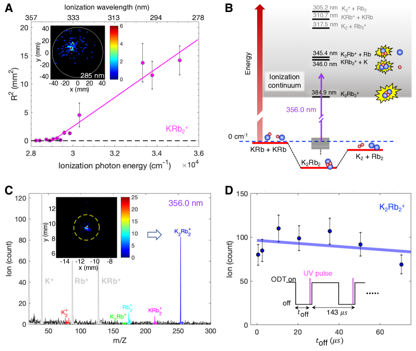
Next, we focused on the transient intermediate collision complex, K2Rb. In order to observe the complexes which by conservation of momentum should only exist in the vicinity of the reactants, we shaped the UV ionization beam into a Gaussian beam profile. After data accumulation, we observed signals consistent with the masses of K2Rb+ and KRb (fig. S2). Based on their VM images, which show large translational energies (Fig. 4A inset), we hypothesize that these ions are from dissociative ionization of K2Rb. To substantiate this idea, we varied the wavelength of the ionization beam in order to determine the relationship between the translational energy of the triatomic ions and the energy of the photon. We found that the characteristic translational energy associated with the KRb ion decreases as the ionization energy decreases. The ionization energy where the translational energy becomes zero (at nm) agrees with our theoretical predictions ( nm) of the dissociative ionization threshold for the transient intermediate, K2Rb + h KRb + K(4s) + .
These theoretical calculations of ionization threshold energies of diatomic, triatomic, and tetratomic K- and Rb-containing molecules (shown in Fig. 4B) are based on the same methodology used in Vexiau et al. (2017) and references therein. Briefly, each alkali-metal atom was modeled as a one-electron system in the field of an ionic core (K+ or Rb+). We used a semi-empirical effective core potential plus a core polarization potential to represent the correlation between the valence electron and the core electrons SM . The K2Rb+ and KRb triatomic ions were modeled as two-valence-electron systems, and the K2Rb ion as a three-valence-electron molecule. In the framework of such a simplification, the ground-state potential energy surface (PES) can be obtained with good accuracy via the diagonalization of the full electronic Hamiltonian (i.e. full configuration interaction) expressed on a large Gaussian basis set. For all molecular and atomic species, the energies were computed with respect to the same origin, namely the energy of the four cores (K+ + K+ + Rb+ + Rb+). This allowed for the determination of transition energies between different species.
To directly observe the transient intermediate complex K2Rb, we tuned the wavelength of our ionization laser to 356 nm, with energy well below the lowest dissociative ionization channel. Figure 4C displays a mass spectrum obtained with ionization at 356 nm, and a strong signal of K2Rb is evident. We emphasize that the ionization process transforms the transient intermediate into a bound molecular ion that has no energetically allowed dissociation channel (Fig. 4B) and can therefore survive its flight to the MCP. Although we have not yet directly measured the lifetime of the complex due to the technical challenges of precisely establishing a zero of time, the signal strength of our direct observation puts an estimate of a lifetime of 350 ns (or 3 s), assuming the ionization cross-section of the K2Rb2 intermediate complex is 10 Mb (or 1 Mb). This cross-section has not been reported in the literature.
The origin of the observed intermediate complex has been the subject of previous debate Mayle et al. (2013); Christianen et al. (2019a). The long-lived transient complex could potentially collide with another KRb, causing the prior’s decay into a deeply-bound K2Rb2 molecule and leading to the conversion of its internal energy into a large, observable TER Mayle et al. (2013); Christianen et al. (2019b). In contrast, we observe a detector resolution-limited small momentum distribution of the K2Rb ions (Fig. 4C inset), consistent with the zero-momentum transient intermediate.
Moreover, because the reactants are trapped in the ODT, a light-assisted process could be a competing, confounding factor, as suggested by Christianen et al. Christianen et al. (2019a). To examine the role of ODT on the detected intermediate complex, we varied the length of time that the ODT was switched off prior to ionization, from 1 s to 70 s. If the ODT contributed to the formation of deeply-bound K2Rb2 molecules, which have no radiative decay pathway and only potentially leave the probed volume on a millisecond time scale if they are untrapped, the K2Rb2 would steadily build up in concentration in the presence of the ODT. As a result, the concentration of K2Rb2 should decrease monotonically as we increase the ODT off duration. Instead, we find the yield of K2Rb ions has no monotonic trend with the ODT off duration (see Fig. 4D). This result is evidence that the intermediates we observe are formed upon collision of two KRb molecules, with no significant effect from the ODT, on or off.
The direct observation of 2 KRb K2Rb K2 + Rb2 opens numerous possibilities of exploring the detailed role of quantum mechanics in ultracold chemical reaction dynamics by measuring the lifetime of the intermediate complex Mayle et al. (2013); Christianen et al. (2019b), testing the transition from quantum to semiclassical reactions Gao (2010), and resolving the quantum states of the reaction products Croft et al. (2017) and the intermediate.
Acknowledgments: We thank D. Herschbach, L. Zhu, T. Karman, and J. Ye for discussion, K. Liu for introducing us to the VMI techniques, T. Pfau, E. Narevicius and M. Greiner for discussions on apparatus design, J. Doyle for loaning laser equipment, and W. Stwalley, P. Gould and the late E. Eyler for sharing KRb spectroscopy literature. The 40K isotope used in this research was supplied by the United States Department of Energy Office of Science by the Isotope Program in the Office of Nuclear Physics. Funding: This work is supported by the DOE Young Investigator Program, the David and Lucile Packard Foundation, and the NSF through Harvard-MIT CUA. Author contributions: The experimental work and data analysis were carried out by M.-G.H., Y.L., D.D.G., Y.-W.L., A.H.G., T.R., and K.-K.N.. Theoretical calculations were done by R.V., N.B., and O.D.. All authors contributed to interpreting the results and writing the manuscript. Competing interests: The authors declare that they have no competing financial interests. Data and materials availability: Data from the main text and supplementary materials are available through the Harvard Dataverse at Hu et al. (2019).
References
- Henson et al. (2012) A. B. Henson, S. Gersten, Y. Shagam, J. Narevicius, and E. Narevicius, Science 338, 234 (2012).
- Yang et al. (2019) H. Yang, D.-C. Zhang, L. Liu, Y.-X. Liu, J. Nan, B. Zhao, and J.-W. Pan, Science 363, 261 (2019).
- Puri et al. (2017) P. Puri, M. Mills, C. Schneider, I. Simbotin, J. A. Montgomery, R. Côté, A. G. Suits, and E. R. Hudson, Science 357, 1370 (2017).
- Hoffmann et al. (2018) D. K. Hoffmann, T. Paintner, W. Limmer, D. S. Petrov, and J. H. Denschlag, Nature communications 9, 5244 (2018).
- Ospelkaus et al. (2010) S. Ospelkaus, K.-K. Ni, D. Wang, M. De Miranda, B. Neyenhuis, G. Quéméner, P. Julienne, J. Bohn, D. Jin, and J. Ye, Science 327, 853 (2010).
- Kilaj et al. (2018) A. Kilaj, H. Gao, D. Rösch, U. Rivero, J. Küpper, and S. Willitsch, Nature communications 9 (2018).
- De Marco et al. (2019) L. De Marco, G. Valtolina, K. Matsuda, W. G. Tobias, J. P. Covey, and J. Ye, Science 363, 853 (2019).
- Ye et al. (2018) X. Ye, M. Guo, M. L. González-Martínez, G. Quéméner, and D. Wang, Science advances 4, eaaq0083 (2018).
- Gregory et al. (2019) P. D. Gregory, M. D. Frye, J. A. Blackmore, E. M. Bridge, R. Sawant, J. M. Hutson, and S. L. Cornish, Nature communications 10, 3104 (2019).
- Christianen et al. (2019a) A. Christianen, M. W. Zwierlein, G. C. Groenenboom, and T. Karman, Physical Review Letters 123, 123402 (2019a).
- Bauer (1979) S. Bauer, Annual Review of Physical Chemistry 30, 271 (1979).
- Miller et al. (1972) W. Miller, S. Safron, and D. Herschbach, The Journal of Chemical Physics 56, 3581 (1972).
- Brooks (1988) P. R. Brooks, Chemical Reviews 88, 407 (1988).
- Zewail (2000) A. H. Zewail, The Journal of Physical Chemistry A 104, 5660 (2000).
- Gruebele et al. (1991) M. Gruebele, I. Sims, E. Potter, and A. Zewail, The Journal of chemical physics 95, 7763 (1991).
- Sims et al. (1992) I. Sims, M. Gruebele, E. Potter, and A. Zewail, The Journal of chemical physics 97, 4127 (1992).
- Polanyi and Zewail (1995) J. C. Polanyi and A. H. Zewail, Accounts of Chemical Research 28, 119 (1995).
- Womack et al. (2015) C. C. Womack, M.-A. Martin-Drumel, G. G. Brown, R. W. Field, and M. C. McCarthy, Science advances 1, e1400105 (2015).
- Bjork et al. (2016) B. J. Bjork, T. Q. Bui, O. H. Heckl, P. B. Changala, B. Spaun, P. Heu, D. Follman, C. Deutsch, G. D. Cole, M. Aspelmeyer, et al., Science 354, 444 (2016).
- Bui et al. (2018) T. Q. Bui, B. J. Bjork, P. B. Changala, T. L. Nguyen, J. F. Stanton, M. Okumura, and J. Ye, Science advances 4, eaao4777 (2018).
- Garand et al. (2008) E. Garand, J. Zhou, D. E. Manolopoulos, M. H. Alexander, and D. M. Neumark, Science 319, 72 (2008).
- Continetti and Guo (2017) R. E. Continetti and H. Guo, Chemical Society Reviews 46, 7650 (2017).
- Su et al. (2013) Y.-T. Su, Y.-H. Huang, H. A. Witek, and Y.-P. Lee, Science 340, 174 (2013).
- Sato (2001) H. Sato, Chemical reviews 101, 2687 (2001).
- Mayle et al. (2013) M. Mayle, G. Quéméner, B. P. Ruzic, and J. L. Bohn, Physical Review A 87, 012709 (2013).
- Christianen et al. (2019b) A. Christianen, T. Karman, and G. C. Groenenboom, Physical Review A 100, 032708 (2019b).
- Nesbitt (2012) D. J. Nesbitt, Chemical Reviews 112, 5062 (2012).
- Ni et al. (2008) K.-K. Ni, S. Ospelkaus, M. De Miranda, A. Pe’Er, B. Neyenhuis, J. Zirbel, S. Kotochigova, P. Julienne, D. Jin, and J. Ye, Science 322, 231 (2008).
- Falke et al. (2006) S. Falke, I. Sherstov, E. Tiemann, and C. Lisdat, The Journal of chemical physics 125, 224303 (2006).
- Amiot (1990) C. Amiot, The Journal of Chemical Physics 93, 8591 (1990).
- Byrd et al. (2010) J. N. Byrd, J. A. Montgomery Jr, and R. Côté, Physical Review A 82, 010502 (2010).
- Cumby et al. (2013) T. D. Cumby, R. A. Shewmon, M.-G. Hu, J. D. Perreault, and D. S. Jin, Physical Review A 87, 012703 (2013).
- Vitanov et al. (2001) N. Vitanov, M. Fleischhauer, B. Shore, and K. Bergmann, Advances in Atomic Molecular and Optical Physics 46, 55 (2001).
- Eppink and Parker (1997) A. T. Eppink and D. H. Parker, Review of Scientific Instruments 68, 3477 (1997).
- (35) See details in the supplementary material .
- Korek et al. (2003) M. Korek, G. Younes, and A. Allouche, International journal of quantum chemistry 92, 376 (2003).
- (37) 170 s is the time it would take for a molecule with translational energy of 0.0127 to travel from the center of the KRb cloud to the ionization ring with a diameter of 0.45 mm .
- Vexiau et al. (2017) R. Vexiau, D. Borsalino, M. Lepers, A. Orbán, M. Aymar, O. Dulieu, and N. Bouloufa-Maafa, International Reviews in Physical Chemistry 36, 709 (2017).
- Gao (2010) B. Gao, Physical Review Letters 105, 263203 (2010).
- Croft et al. (2017) J. Croft, C. Makrides, M. Li, A. Petrov, B. Kendrick, N. Balakrishnan, and S. Kotochigova, Nature communications 8, 15897 (2017).
- Hu et al. (2019) M.-G. Hu, Y. Liu, D. D. Grimes, Y.-W. Lin, A. H. Gheorghe, R. Vexiau, N. Boulufa-Maafa, O. Dulieu, and K.-K. Ni, “Replication Data for Direct observation of bimolecular reactions of ultracold KRb molecules, DOI:10.7910/DVN/MDFLOZ,” (2019).